- Subjects: Neurosciences
- |
- Contributor:
- Neuroscientifically Challenged


- neuroscience
- brain
- spinal cord
- central nervous system
- CNS
- sensory information
This video is adapted from: https://youtu.be/MM7YNKJj_Lg
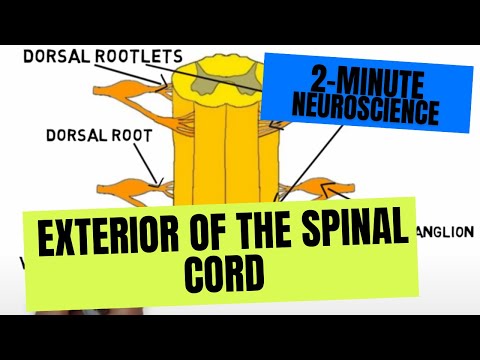
The spinal cord is one of the two main components of the central nervous system (the other being the brain). It receives all of the sensory information from the periphery of the body and carries it to the brain, and also contains motor neurons that supply the muscles we use to move around. Neurons carrying sensory information enter the back of the spinal cord as dorsal rootlets, which all emerge from a single dorsal root. The cell bodies of these sensory neurons are contained in a cluster of cell bodies called a dorsal root ganglion. Motor nerves leave the front of the cord as ventral rootlets and come together to form a ventral root; the dorsal and ventral roots merge together to form the spinal nerves.
The spinal cord is divided into segments, each of which gives rise to a pair of spinal nerves. There are 31 segments, named for their location: there are 8 cervical segments, 12 thoracic segments, 5 lumbar segments, 5 sacral segments, and 1 coccygeal segment.
The spinal cord leaves the skull through an opening called the foramen magnum and travels down the vertebral column, ending in a pointed structure called the conus medullaris. The cord is surrounded by a membrane called the dural tube, but the cord ends before the dural tube and the spinal nerves that leave the cord from some lumbar and sacral segments have to travel down through the dural tube to reach their site of exit from the vertebral canal. This leaves a portion of the dural tube that does not contain the spinal cord but is filled with spinal nerves. It is from this area--known as the lumbar cistern--that cerebrospinal fluid is sampled as part of a lumbar puncture or spinal tap; it is done here because there is less chance the cord could be damaged by the insertion of a needle. The nerves in the lumbar cistern fan out like a horses tail and are resultantly called the cauda equina (which means horse’s tail in Latin). There are two locations along the cord that are slightly enlarged - the cervical and lumbar enlargements - because these parts of the cord contain more neurons in order to supply the limbs. An extension of the meninges called the filum terminale extends from the end of the cord to the tailbone to anchor the cord in place. [1]
- Nolte J. The Human Brain: An Introduction to its Functional Anatomy. 6th ed. Philadelphia, PA. Elsevier; 2009.