- Subjects: Neurosciences
- |
- Contributor:
- Neuroscientifically Challenged


- nervous system
- human brain
- spinal cord
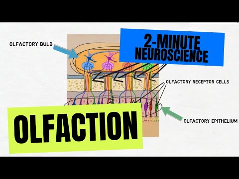
When you look at the spinal cord in cross-section at any level you will see a butterfly shaped region of grey matter surrounded by white matter. The grey matter is made up of cell bodies of neurons while the white matter consists of axons that travel up the spinal cord to the brain and down the spinal cord to the body. There is a small groove called the posterolateral sulcus where dorsal roots enter the cord carrying sensory information. There is another groove that is not very distinct on the front of the cord called the anterolateral sulcus. Ventral roots leave the cord from the anterolateral sulcus to carry motor information to the muscles.
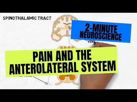
The grey matter is divided into three regions. The posterior horn contains interneurons that make connections within the spinal cord and neurons that enter ascending pathways carrying sensory information to the brain. There is a section of the posterior horn called the substantia gelatinosa that contains neurons that specifically carry pain and temperature sensations to the brain.
The anterior horn contains the cell bodies of motor neurons that activate skeletal muscle. These neurons, called alpha motor neurons, leave the cord in the ventral roots and represent the way the nervous system enacts voluntary and involuntary movements.
The intermediate grey matter has some characteristics of the areas surrounding it, but it also contains neurons involved in autonomic functions, or functions that are automatic and occur without conscious control like heart rate and respiration
The white matter of the spinal cord consists of bundles of ascending and descending fibers that carry sensory information to the brain and motor information to the body, respectively. These bundles of fibers are called funiculi. The back of the spinal cord contains the posterior funiculi, which contain important pathways that carry information about touch and limb position to the brain. The lateral funiculi are found in the lateral portion of the cord; important pain pathways are found here as well as important descending pathways that are responsible for causing movement. The anterior funiculi contain various ascending and descending pathways. [1]
- Nolte J. The Human Brain: An Introduction to its Functional Anatomy. 6th ed. Philadelphia, PA. Elsevier; 2009.