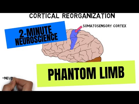
- Subjects: Neuroimaging
- |
- Contributor:
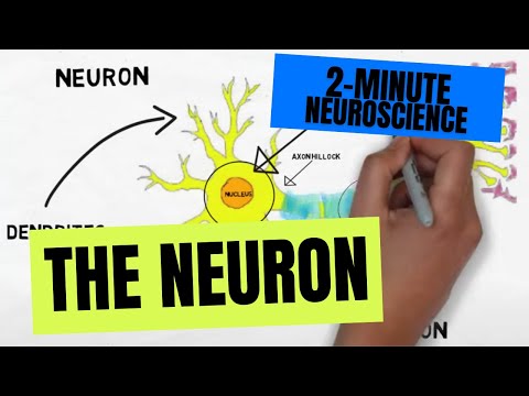
- Neuroscientifically Challenged


- neuroimaging
- Functional magnetic resonance imaging
- brain activity
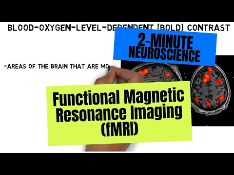
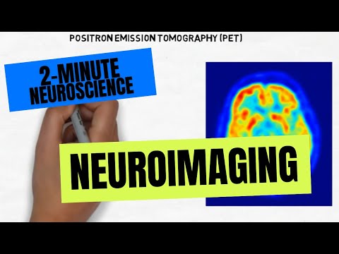
Functional magnetic resonance imaging, or fMRI, is a type of neuroimaging used to obtain images of brain activity. fMRI involves exposing the brain to multiple magnetic fields, and relies on the observation that protons in the nuclei of hydrogen atoms respond to this procedure by emitting an electromagnetic signal that can be detected by the fMRI scanner. The fMRI scanner is capable of determining some of the properties of the tissue the signal came from, and can use this information to reconstruct a high-resolution image of the brain. Additionally, the fMRI scanner can detect differences in the magnetic properties of oxygenated vs. deoxygenated blood, and thus can identify changes in levels of oxygenated blood in different regions of the brain using a method called blood-oxygen-level-dependent, or BOLD, contrast.
BOLD is typically what enables us to identify which brain areas are most active in fMRI. Areas of the brain that are more active tend to receive higher levels of oxygenated blood. Thus, higher levels of oxygenated blood in a particular brain region are believed to correspond to higher neural activity in that region. On a typical fMRI image, color-coding is used to represent differences in the level of oxygenated blood and thus differences in activity. Activity in those areas can then be associated with whatever task was performed at the time of the scan.
fMRI was developed in the early 1990s and since has become a very popular neuroimaging method. Nevertheless, in its short history fMRI research has been plagued by issues like small sample sizes, the use of methods that lead to a high number of false positives, and a small proportion of study results that have been independently reproduced. As the field has become more aware of these problems, many researchers have begun to adjust their approach to address them. [1][2][3]
MRI image at :40 courtesy of DrOONeil on Wikimedia Commons: https://commons.wikimedia.org/wiki/File:FMRI_Brain_Scan.jpg
- Poldrack RA, Baker CI, Durnez J, Gorgolewski KJ, Matthews PM, Munafò MR, Nichols TE, Poline JB, Vul E, Yarkoni T. Scanning the horizon: towards transparent and reproducible neuroimaging research. Nature Reviews Neuroscience. 2017 Feb;18(2):115-126. doi: 10.1038/nrn.2016.167.
- Small SA, Heeger DJ. Functional Imaging of Cognition. In: Kandel ER, Schwartz JH, Jessell TM, eds. Principles of Neural Science, 5th ed. New York: McGraw-Hill; 2013.
- Stamatakis EA, Orfanidou E, Papanicolaou AC. Functional Magnetic Resonance Imaging. In: Papanicolaou AC, ed. The Oxford Handbook of Functional Brain Imaging in Neuropsychology and Cognitive Neurosciences. New York: Oxford University Press; 2014.