| Version | Summary | Created by | Modification | Content Size | Created at | Operation |
|---|---|---|---|---|---|---|
| 1 | Alicja Kmiecik | + 2398 word(s) | 2398 | 2021-08-11 08:38:52 | | | |
| 2 | Dean Liu | Meta information modification | 2398 | 2021-09-28 06:21:04 | | |
Video Upload Options
Nucleobindin 2 (NUCB2) was first described in 1994 in KM3 acute lymphoblastic leukemia cell line as a DNA binding/EF-hand/acidic-amino acid-rich protein. It has been extensively studied since Oh-I et al. identified nesftain-1 as a NUCB2 cleavage product. Several reports indicate that NUCB2/NESF-1 is also expressed in many organs and tissues (e.g., in the stomach, pancreas, heart, reproductive organs, and adipose tissue).
1. Introduction
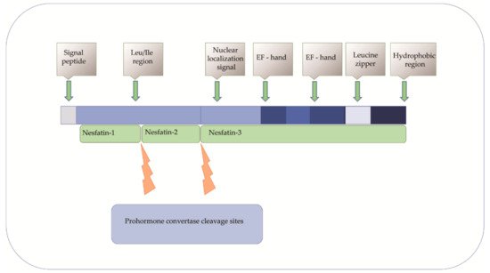
Nucleobindin 2 (NUCB2) was first described in 1994 in KM3 acute lymphoblastic leukemia cell line as a DNA binding/EF-hand/acidic-amino acid-rich protein [1][2]. It has been extensively studied since Oh-I et al. identified nesftain-1 as a NUCB2 cleavage product [3]. Several reports indicate that NUCB2/NESF-1 is also expressed in many organs and tissues (e.g., in the stomach, pancreas, heart, reproductive organs, and adipose tissue) [4][5][6][7][8]. Nucleobindin-2/Nnesfatin-1 is also a secretory component of various body fluids, including saliva, synovial fluid, milk, and plasma or serum [9][10][11][12][13]. As NUCB2 and nesfatin-1 are colocalized, these two names are used interchangeably. The NUCB2/NESF-1 gene in humans is located on chromosome 11 and consists of 14 exons spanning 54 785 nucleotides [14]. Nucleobindin-2 is a 396-amino acid protein preceded by a 24-amino acid signal peptide [15]. The protein is proteolytically cleaved by the prohormone convertase. As a result, N-terminal nesfatin-1, nesfatin-2, and the C–terminal nesfatin-3 are formed [16]. The functions of nesfatin-2 and the C–terminal nesfatin-3 have not been understood yet [17]. Nucleobindin-2 has characteristic functional domains, such as a signal peptide, a Leu ⁄ Ile rich region, two Ca2+ binding EF-hand domains separated by an acidic amino acid-rich region, and a leucine zipper. Therefore it may play a role in many cellular processes [18][19] ( Figure 1 ). Despite the increasing knowledge about the expression and regulation of NUCB2/NESF-1, its role in physiology and pathology is still poorly understood.

2. NUCB2/NESF-1 in Physiology
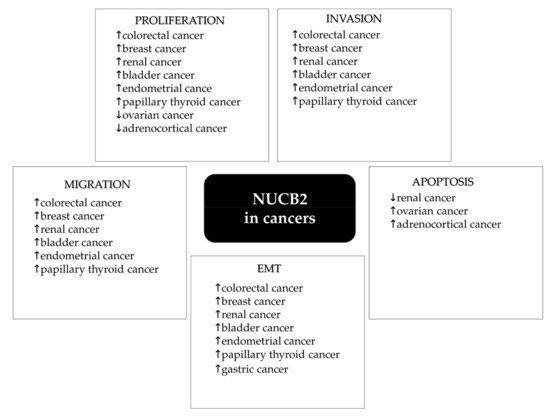
This protein is involved in the regulation of many intracellular processes. Its metabolic function includes food intake, glucose metabolism, and the regulation of immune, cardiovascular, and endocrine systems [20][21][22][23]. Recently, nesfatin-1 has been identified as an anorexigenic neuropeptide that seems to play a crucial role in appetite regulation and energy homeostasis [24]. Oh-I et al. found that intracerebroventricular (IVC) injection of nesfatin-1 decreased food intake in a dose-dependent manner in rats [3]. Furthermore, lower nesfatin-1 concentrations were observed in obese children compared to healthy children [25]. It was also suggested that NUCB2/NESF-1 expressed in the parvocellular region contributed to anorexia by secreting nesfatin-1 [26][27][28]. Nesfatin-1 was reported as a suppressor of gastric emptying and an inhibitor of gastroduodenal motility in mice and rats [21][29]. The expression of NUCB2/NESF-1 was also observed in pancreatic islets, which suggests its role in glucose homeostasis [30]. Su et al. demonstrated that nesfatin-1 reduced blood glucose levels in hyperglycemic db/db mice [31]. Moreover, Gonzales et al. demonstrated that nesfatin-1 enhanced glucose-stimulated insulin secretion in mouse pancreatic β- cells [32]. Nesfatin-1 was found to be an anti-inflammatory and antiapoptotic agent in rats with traumatic brain injury [33]. It was demonstrated that the administration of nesfatin-1 30 min after head trauma could significantly suppress inflammatory-related proteins such as tumor necrosis factor-alpha (TNF-α), IL-1β ( interleukin-1 β), and IL-6 (interleukin-6). Moreover, it was also shown that nesfatin-1 reduced the activity of caspase-3 and the number of apoptotic neuronal cells in traumatic rat brain tissue. It is known that NUCB2/NESF-1 is implicated in the inflammatory response by its involvement in the tumor necrosis factor/tumor necrosis factor receptor 1 (TNF/TNFR1) signaling. Islam et al. revealed that tumor necrosis factor receptor 1 release required the calcium-dependent formation of NUCB2/NESF-1 and aminopeptidase regulator of TNFR1 shedding (ARTS-1) to promote exosome-like vesicle release in human vascular endothelial cells [34]. Feijóo-Bandín et al. showed that mouse and human cardiomyocytes could synthesize and secrete nesfatin-1, which helps regulate glucose metabolism in the hearts of humans and experimental animals [35]. Additionally, the plasma concentration of nesfatin-1 in patients with acute myocardial infarction (AMI) was lower than in the control group, which showed nesfatin-1 to be a protective agent against AMI [36]. On the other hand, it was also found that the infusion of nesfatin-1 to the cerebral cortex and subcutaneous tissue resulted in elevated blood pressure, while intravenous infusion led to vasoconstriction [37]. The role of nesfatin-1 in the cardiovascular system is still poorly understood. There is some evidence that NUCB2/NESF-1 is related to female pubertal maturation. Garcia Galano found that during the female pubertal transition, NUCB2/NESF-1 mRNA and protein levels were significantly increased in the hypothalamus [38]. Intracerebroventricular (ICV) injections of nesfatin-1 induced increased circulating levels of gonadotropin-releasing hormone, luteinizing hormone (LH), and follicle-stimulating hormone (FSH) [39]. Expression of NUCB2/NESF-1 was detected in Leydig cells in rodents and humans. During the pubertal transition, NUCB2/NESF-1 protein level in these cells was significantly increased [38]. To conclude, recent studies have identified nuclebindin-2/nesfatin-1 as a pleiotropic peptide with many physiological functions. Recently, the expression of NUCB2/NESF-1 has been linked to tumor development and metastasis. However, the exact role of NUCB2/NESF-1 in human malignancies is still poorly understood ( Figure 2 ). The current review is the first to present NUCB2/NESF-1 as a potential new prognostic or predictive marker in cancers.

3. NUCB2—A Predictive/Prognostic Biomarker in Cancers?
We evaluated the expression of NUCB2 in breast cancer tissues in our current research (data not published yet). The expression of NUCB2/NESF-1 was localized in the cytoplasm of breast cancer cells ( Figure 3 A). No expression was found in the breast cancer cell nucleus or the tumor stroma ( Figure 3 A). The presence of NUCB2/NESF-1 detected by immunohistochemistry (IHC) was higher in breast carcinoma compared to benign changes in the glandular tissue in the breast (mastopathy; control) ( Figure 3 B).

Additionally, they found a significant positive correlation between nodal metastasis and the clinical stage. Moreover, it was demonstrated that breast cancer patients with high NUCB2 /NESF-1 expression had a significantly poorer overall survival (OS) [40]. Interestingly, the survival analysis with an online analysis tool on 2032 breast cancer cases indicated that a good prognostic effect of high NUCB2 expression was related to longer OS [41]. These conflicting results suggest an important role of NUCB2/NESF-1 in breast cancer progression and highlight a need for further investigation in this area.
Kan et al. were the first to detect NUCB2/NESF-1 expression in colon cancer. The immunofluorescence analysis showed that the expression of NUCB2/NESF-1 was higher compared to non-tumor regions. Additionally, they assessed serum nesfatin-1 levels in colon cancer patients. The obtained results revealed no difference in nesfatin-1 concentration between healthy donors and colon cancer patients [42]. In 2018, Xie et al. found that NUCB2 /NESF-1 mRNA was upregulated in colorectal cancer (CRC) tissues compared to the noncancerous tissue obtained from the same patient. Additionally, immunohistochemistry showed the expression of NUCB2/NESF-1 in 251 colon cancer patients in relation to clinicopathological properties. The protein was predominantly expressed in the cytoplasm and much less in the cancer cell membrane. The results also indicated a positive correlation between NUCB2/NESF-1 expression and lymph node metastasis and the TNM stage. Patients with lymph node metastasis showed a higher expression of NUCB2/NESF-1 compared to those without lymph node metastasis (49.5%, vs. 36.6%, p = 0.043). Moreover, TNM stage III-IV patients had significantly higher NUCB2 /NESF-1 expression compared to TNM stage I-II patients (50.9% vs. 35.0%). No significant association was found between NUCB2/NESF-1 expression and disease-free survival or overall survival in colon cancer patients [43]. To conclude, NUCB2/NESF-1 is suggested to be associated with aggressive progression in colorectal cancer.
The NUCB2 /NESF-1 mRNA level was analyzed in 180 pairs of prostate cancer tissues and the corresponding noncancerous tissue. The analyses revealed that the expression of NUCB2/NESF-1 was significantly higher in cancer cells compared to the noncancerous control. The upregulation of NUCB2/NESF-1 mRNA in prostate cancer was correlated with higher Gleason scores ( p < 0000.1), higher levels of preoperative prostate-specific antigen (PSA) ( p = 0.0004) , positive lymph node metastasis ( p = 0.022) and positive angiolymphatic invasion ( p = 0.0004). No relationship was found between NUCB2/NESF-1 and age, seminal vesicle invasion, pathological stage, or surgical margin status. Additionally, patients with low NUCB2/NESF-1 mRNA levels had significantly longer biochemical recurrence-free survival (BCR-free—the survival time of a person with prostate cancer during which a biochemical marker—PSA does not rise or rises very little) time after radical prostatectomy compared to patients with high NUCB2/NESF-1 mRNA levels. Moreover, multivariate analysis demonstrated that a high NUCB2/NESF-1 mRNA level was an independent predictor of shorter BCR-free survival [44]. In other studies, the same research team showed that high NUCB2/NESF-1 mRNA expression was related to the poor overall survival of patients with prostate cancer. In addition, multivariate Cox analysis indicated that NUCB2/NESF-1 mRNA was an independent prognostic factor for the overall survival of prostate cancer patients [44]. Immunohistochemistry evaluation revealed that NUCB2/NESF-1 expression was significantly higher in prostate cancer compared to benign prostatic hyperplasia ( p < 0000.1) [45]. Furthermore, it was also shown that NUCB2/NESF-1 protein expression was significantly associated with seminal vesicle invasion, higher levels of preoperative PSA, positive lymph node metastasis, positive angiolymphatic invasion, biochemical recurrence, and higher Gleason scores. Multivariate Cox regression analysis revealed that a high NUCB2/NESF-1 protein expression level was an independent prognostic factor for overall survival and BCR-free survival of patients with prostate cancer [14]. To conclude, the above evidence suggests that NUCB2/NESF-1 may be an important biomarker in prostate cancer patients.
4. NUCB2 in Cancer Proliferation, Apoptosis, Migration and Invasion
Proliferation is one of the most important characteristics of cancer. Some proteins are overexpressed in cancer cells and indicated as proliferation markers, which makes them useful in cancer diagnostics. Among the best-evaluated cell proliferation molecules, we classified, e.g., Ki-67, proliferating cell nuclear antigen (PCNA), or minichromosome maintenance (MCM) proteins. Although the proteins have been extensively described in the literature, they also have some limitations when used in the diagnostic process [46]. As a result, there is a need to identify new proliferation-related molecules in cancers. In vitro studies showed that NUCB2/NESF-1 knockdown with siRNA or shRNA in breast cancer (MCF-7, SKBR-3), bladder cancer (T24, 5637), glioblastoma (U251, U87), endometrial cancer (Ishikawa and Sawano), and thyroid cancer (TPC-I, KI) cell lines resulted in the inhibition of cell proliferation [47][48][49][50][51]. Moreover, proliferation-related proteins (Ki-67 and PCNA) were decreased in NUCB2/NESF-1 shRNA-transfected thyroid cancer cells compared to the control. To evaluate the role of NUCB2/NESF-1 related to the growth and metastasis of glioblastoma, NUCB2/NESF-1-silenced cells were injected into nude mice. The findings indicated that the NUCB2/NESF-1 ablation group’s tumor volume was significantly smaller than the control [51]. Additionally, NUCB2/NESF-1 knockdown in the 786-O renal cancer cell line inhibited cell proliferation by arresting the cell cycle at the S phase. Moreover, reduced tumor volume and growth rate were observed in NUCB2/NESF-1-knockdown renal cancer cells in the mice model compared to that in the negative control group [52]. Interestingly, no changes were found in the proliferation properties of NUCB2-knockdown colon cancer cells [42]. Surprisingly, Ramanjaneya et al. found that treatment of H295R adrenocortical cells with recombinant nesfatin-1 resulted in a decreased proliferative capacity of the cells [53]. They put a hypothesis that NUCB2/NESF-1 might be a therapeutic target for adrenal cancer. Similar conclusions were made by Xu et al., who evaluated the treatment of HO-8910 ovarian cancer cells with recombinant human nesfatin-1. It was revealed that nesftain-1 decreased cell proliferation in ovarian cancer in vitro [54]. Surprisingly, treatment of the endometrial cancer cell line (Ishikawa) with recombinant nesfatin-1 promoted cell proliferation [49]. Dysregulation of cell proliferation is known to be related to the mTOR signaling cascade. Takagi et al. revealed that NUCB2/NESF-1 increased mTOR phosphorylation, which resulted in the intense proliferation of the endometrial cancer cell line [49]. Contradictory findings were presented by Xu et al., who reported that NUCB2/NESF-1 decreased mTOR phosphorylation and acted as a tumor suppressor in ovarian cancer [54]. Similarly, Ramanjaneya et al. indicated that NCB2/NESF-1 decreased ERK1/2 phosphorylation, which resulted in decreased proliferation in H295R adrenocortical carcinoma cells [53]. Bearing in mind the above, we may conclude that the role of NUCB2/NESF-1 in cancers is variable and tissue-specific. Additionally, Xu et al. concluded that nesfatin-1 could inhibit the proliferation in human ovarian epithelial carcinoma cell line HO-8910 by inducing apoptosis via the mTOR and RhoA/ROCK signaling pathway [54]. Rho Kinases (ROCKs) are known to be modulators of cell survival and apoptosis. It was reported that NUCB2/nesfatin-1 treatment evoked a marked activation of RhoA, which enhanced apoptosis and inhibited proliferation.
The balance between cell proliferation and apoptosis is crucial for normal development and homeostasis in adults [55]. An imbalance between these two processes causes cancer. Apoptosis is a well-known mechanism of programmed cell death, which plays a crucial role in development and homeostasis. The loss of apoptotic control allows cancer cells to survive and favors tumor progression. One of the major challenges in cancer treatment is identifying factors that terminate the uncontrolled growth of cancer cells [56]. The effects of nesfatin-1 on apoptosis of H295R adrenocortical cells using the DNA fragmentation assay were assessed. The study found that nesfatin-1 induced a concentration-dependent increase in apoptosis of H295R cells compared to control cells. Moreover, the analyses of pro-and-anti-apoptotic gene expression following nesfatin-1 treatment in H295R cells were performed. A significant increase was found in the proapoptotic Bax ( p < 0.05), while a significant decrease was reported in antiapoptotic BCL-XL ( p < 0.01) and BCL-2 ( p < 0.05) mRNA expression following nesfatin-1 stimulation [53]. The study may contribute to cancer prevention and therapy.
To summarize, NUCB2 seems to be an important factor in cancer cell migration and invasion and may affect the expression of MMP-2 and MMP-9. The mechanism underlying this observation is unknown.
The above study confirmed that NUCB-2 might promote EMT via the AMPK/TORC1/ZEB1 pathway in cancer. In addition, a positive correlation was found between the expression of NUCB2/NESF-1 and EMT-related genes such as desmoplakin (DSP) , Integrin Subunit Alpha V (ITGAV) , metalloproteinase 3 ( MMP3) , tetraspanin 13 ( TSPAN13), and Cadherin 2 ( CDH2) in endometrial cancer cells [57].
References
- Skorupska, A.; Bystranowska, D.; Dąbrowska, K.; Ożyhar, A. Calcium ions modulate the structure of the intrinsically disordered Nucleobindin-2 protein. Int. J. Biol. Macromol. 2020, 154, 1091–1104.
- Skorupska, A.; Ożyhar, A.; Bystranowska, D. The physiological role of nucleobindin-2/nesfatin-1 and their potential clinical significance. Postepy Hig. Med. Dosw. 2018, 72, 1084–1096.
- Oh-I, S.; Shimizu, H.; Satoh, T.; Okada, S.; Adachi, S.; Inoue, K.; Eguchi, H.; Yamamoto, M.; Imaki, T.; Hashimoto, K.; et al. Identification of nesfatin-1 as a satiety molecule in the hypothalamus. Nature 2006, 443, 709–712.
- Stengel, A.; Hofmann, T.; Goebel-Stengel, M.; Lembke, V.; Ahnis, A.; Elbelt, U.; Lambrecht, N.W.G.; Ordemann, J.; Klapp, B.F.; Kobelt, P. Ghrelin and NUCB2/nesfatin-1 are expressed in the same gastric cell and differentially correlated with body mass index in obese subjects. Histochem. Cell Biol. 2013, 139, 909–918.
- Foo, K.S.; Brauner, H.; Östenson, C.G.; Broberger, C. Nucleobindin-2/nesfatin in the endocrine pancreas: Distribution and relationship to glycaemic state. J. Endocrinol. 2010, 204, 255–263.
- Angelone, T.; Filice, E.; Pasqua, T.; Amodio, N.; Galluccio, M.; Montesanti, G.; Quintieri, A.M.; Cerra, M.C. Nesfatin-1 as a novel cardiac peptide: Identification, functional characterization, and protection against ischemia/reperfusion injury. Cell. Mol. Life Sci. 2013, 70, 495–509.
- Kim, J.; Yang, H. Nesfatin-1 as a New Potent Regulator in Reproductive System. Dev. Reprod. 2012, 16, 253–264.
- Ramanjaneya, M.; Chen, J.; Brown, J.E.; Tripathi, G.; Hallschmid, M.; Patel, S.; Kern, W.; Hillhouse, E.W.; Lehnert, H.; Tan, B.K.; et al. Identification of nesfatin-1 in human and murine adipose tissue: A novel depot-specific adipokine with increased levels in obesity. Endocrinology 2010, 151, 3169–3180.
- Aydin, S.; Dag, E.; Ozkan, Y.; Erman, F.; Dagli, A.F.; Kilic, N.; Sahin, I.; Karatas, F.; Yoldas, T.; Barim, A.O.; et al. Nesfatin-1 and ghrelin levels in serum and saliva of epileptic patients: Hormonal changes can have a major effect on seizure disorders. Mol. Cell. Biochem. 2009, 328, 49–56.
- Zhang, Y.; Shui, X.; Lian, X. Serum and synovial fluid nesfatin-1 concentration is associated with radiographic severity of knee osteoarthritis. Med Sci. Monit. Int. Med. J. Exp. Clin. Res. 2015, 21, 1078–1082.
- Badillo-suárez, P.A.; Rodríguez-cruz, M.; Nieves-morales, X. Impact of Metabolic Hormones Secreted in Human Breast Milk on Nutritional Programming in Childhood Obesity. J. Mammary Gland. Biol. Neoplasia 2017, 22, 171–191.
- Aydin, S. The presence of the peptides apelin, ghrelin and nesfatin-1 in the human breast milk, and the lowering of their levels in patients with gestational diabetes mellitus. Peptides 2010, 31, 2236–2240.
- Li, Q.C.; Wang, H.Y.; Chen, X.; Guan, H.Z.; Jiang, Z.Y. Fasting plasma levels of nesfatin-1 in patients with type 1 and type 2 diabetes mellitus and the nutrient-related fluctuation of nesfatin-1 level in normal humans. Regul. Pept. 2010, 159, 72–77.
- Zhang, H.; Qi, C.; Wang, A.; Yao, B.; Li, L.; Wang, Y.; Xu, Y. Prognostication of prostate cancer based on NUCB2 protein assessment: NUCB2 in prostate cancer. J. Exp. Clin. Cancer Res. 2013, 32, 1–8.
- Yamada, M.; Horiguchi, K.; Umezawa, R.; Hashimoto, K.; Satoh, T.; Ozawa, A.; Shibusawa, N.; Monden, T.; Okada, S.; Shimizu, H.; et al. Troglitazone, a ligand of peroxisome proliferator-activated receptor-γ, stabilizes NUCB2 (nesfatin) mRNA by activating the ERK1/2 pathway: Isolation and characterization of the human NUCB2 gene. Endocrinology 2010, 151, 2494–2503.
- Nakata, M.; Gantulga, D.; Santoso, P.; Zhang, B.; Masuda, C.; Mori, M.; Okada, T.; Yada, T. Paraventricular NUCB2/nesfatin-1 supports oxytocin and vasopressin neurons to control feeding behavior and fluid balance in male mice. Endocrinology 2016, 157, 2322–2332.
- Ayada, C.; Toru, Ü.; Korkut, Y. Nesfatin-1 and its effects on different systems. Hippokratia 2015, 19, 4–10.
- Miura, K.; Titani, K.; Kurosawa, Y.; Kanai, Y. Molecular cloning of nucleobindin, a novel DNA-binding protein that contains both a signal peptide and a leucine zipper structure. Biochem. Biophys. Res. Commun. 1992, 187, 375–380.
- Taniguchi, N.; Taniura, H.; Niinobe, M.; Takayama, C.; Tominaga-Yoshino, K.; Ogura, A.; Yoshikawa, K. The postmitotic growth suppressor necdin interacts with a calcium-binding protein (NEFA) in neuronal cytoplasm. J. Biol. Chem. 2000, 275, 31674–31681.
- Feijóo-Bandín, S.; Rodríguez-Penas, D.; García-Rúa, V.; Mosquera-Leal, A.; Juanatey, J.R.G.; Lago, F. Nesfatin-1: A new energy-regulating peptide with pleiotropic functions. Implications at cardiovascular level. Endocrine 2015, 52, 11–29.
- Khalili, S.; Khaniani, M.S.; Afkhami, F.; Derakhshan, S.M. NUCB2/Nesfatin-1: A Potent Meal Regulatory Hormone and its Role in Diabetes. Egypt. J. Med. Hum. Genet. 2017, 18, 105–109.
- García-Galiano, D.; Navarro, V.M.; Gaytan, F.; Tena-Sempere, M. Expanding roles of NUCB2/nesfatin-1 in neuroendocrine regulation. J. Mol. Endocrinol. 2010, 45, 281–290.
- Scotece, M.; Conde, J.; Abella, V.; López, V.; Lago, F.; Pino, J.; Gómez-Reino, J.J.; Gualillo, O. NUCB2/nesfatin-1: A New Adipokine Expressed in Human and Murine Chondrocytes with Pro-Inflammatory Properties, An In Vitro Study. J. Orthop. Res. 2014, 32, 653–660.
- Dore, R.; Levata, L.; Lehnert, H.; Schulz, C. Nesfatin-1: Functions and physiology of a novel regulatory peptide. J. Endocrinol. 2017, 232, R45–R65.
- Abaci, A.; Catli, G.; Anik, A.; Kume, T.; Bober, E. The relation of serum nesfatin-1 level with metabolic and clinical parameters in obese and healthy children. Pediatr. Diabetes 2013, 14, 189–195.
- Pałasz, A.; Krzystanek, M.; Worthington, J.; Czajkowska, B.; Kostro, K.; Wiaderkiewicz, R.; Bajor, G. Nesfatin-1, a unique regulatory neuropeptide of the brain. Neuropeptides 2012, 46, 105–112.
- Schalla, M.A.; Stengel, A. Current Understanding of the Role of Nesfatin-1. J. Endocr. Soc. 2018, 2, 1188–1206.
- Wei, Y.; Li, J.; Wang, H.; Wang, G. NUCB2/nesfatin-1: Expression and functions in the regulation of emotion and stress. Prog. Neuro Psychopharmacol. Biol. Psychiatry 2018, 81, 221–227.
- Atsuchi, K.; Asakawa, A.; Ushikai, M.; Ataka, K.; Tsai, M.; Koyama, K.; Sato, Y.; Kato, I.; Fujimiya, M.; Inui, A. Centrally administered nesfatin-1 inhibits feeding behaviour and gastroduodenal motility in mice. Neuroreport 2010, 21, 1008–1011.
- Yang, Y.; Zhang, B.; Nakata, M.; Nakae, J.; Mori, M.; Yada, T. Islet β-cell-produced NUCB2/nesfatin-1 maintains insulin secretion and glycemia along with suppressing UCP-2 in β-cells. J. Physiol. Sci. 2019, 69, 733–739.
- Su, Y.; Zhang, J.; Tang, Y.; Bi, F.; Liu, J.N. The novel function of nesfatin-1: Anti-hyperglycemia. Biochem. Biophys. Res. Commun. 2010, 391, 1039–1042.
- Gonzalez, R.; Reingold, B.K.; Gao, X.; Gaidhu, M.P.; Tsushima, R.G.; Unniappan, S. Nesfatin-1 exerts a direct, glucose-dependent insulinotropic action on mouse islet β- and MIN6 cells. J. Endocrinol. 2011, 208, R9–R16.
- Tang, C.H.; Fu, X.J.; Xu, X.L.; Wei, X.J.; Pan, H.S. The anti-inflammatory and anti-apoptotic effects of nesfatin-1 in the traumatic rat brain. Peptides 2012, 36, 39–45.
- Islam, A.; Adamik, B.; Hawari, F.I.; Ma, G.; Rouhani, F.N.; Zhang, J.; Levine, S.J. Extracellular TNFR1 Release Requires the Calcium-dependent Formation of a Nucleobindin 2-ARTS-1 Complex. J. Biol. Chem. 2006, 281, 6860–6873.
- Feijóo-Bandín, S.; Rodríguez-Penas, D.; García-Rúa, V.; Mosquera-Leal, A.; Otero, M.F.; Pereira, E.; Rubio, J.; Martínez, I.; Seoane, L.M.; Gualillo, O.; et al. Nesfatin-1 in human and murine cardiomyocytes: Synthesis, secretion, and mobilization of GLUT-4. Endocrinology 2013, 154, 4757–4767.
- Dai, H.; Li, X.; He, T.; Wang, Y.; Wang, Z.; Wang, S.; Xing, M.; Sun, W.; Ding, H. Decreased plasma nesfatin-1 levels in patients with acute myocardial infarction. Peptides 2013, 46, 167–171.
- Ayada, C.; Turgut, G.; Turgut, S.; Güçlü, Z. The effect of chronic peripheral nesfatin-1 application on blood pressure in normal and chronic restraint stressed rats: Related with circulating level of blood pressure regulators. Gen. Physiol. Biophys. 2015, 34, 81–88.
- Galiano, D.G.; Pineda, R.; Ilhan, T.; Castellano, J.M.; Ruiz-Pino, F.; Sánchez-Garrido, M.A.; Vazquez, M.J.; Sangiao-Alvarellos, S.; Ruiz, A.R.; Pinilla, L.; et al. Cellular Distribution, Regulated Expression, and Functional Role of the Anorexigenic Peptide, NUCB2/Nesfatin-1, in the Testis. Endocrinology 2012, 153, 1959–1971.
- Prinz, P.; Stengel, A. Expression and regulation of peripheral NUCB2/nesfatin-1. Curr. Opin. Pharmacol. 2016, 31, 25–30.
- Zeng, L.; Zhong, J.; He, G.; Li, F.; Li, J.; Zhou, W.; Liu, W.; Zhang, Y.; Huang, S.; Liu, Z.; et al. Identification of nucleobindin-2 as a potential biomarker for breast cancer metastasis using iTRAQ-based quantitative proteomic analysis. J. Cancer 2017, 8, 3062–3069.
- Györffy, B.; Lanczky, A.; Eklund, A.C.; Denkert, C.; Budczies, J.; Li, Q.; Szallasi, Z. An online survival analysis tool to rapidly assess the effect of 22,277 genes on breast cancer prognosis using microarray data of 1,809 patients. Breast Cancer Res. Treat. 2010, 123, 725–731.
- Kan, J.; Yen, M.C.; Wang, J.Y.; Wu, D.C.; Chiu, Y.J.; Ho, Y.W.; Kuo, P.L. Nesfatin-1/Nucleobindin-2 enhances cell migration, invasion, and epithelial-mesenchymal transition via LKB1/AMPK/TORC1/ZEB1 pathways in colon cancer. Oncotarget 2016, 7, 31336–31349.
- Xie, J.; Chen, L.; Chen, W. High NUCB2 expression level is associated with metastasis and may promote tumor progression in colorectal cancer. Oncol. Lett. 2018, 15, 9188–9194.
- Zhang, H.; Qi, C.; Li, L.; Luo, F.; Xu, Y. Clinical significance of NUCB2 mRNA expression in prostate cancer. J. Exp. Clin. Cancer Res. 2013, 32, 1–6.
- Zhang, H.; Qi, C.; Wang, A.; Li, L.; Xu, Y. High expression of nucleobindin 2 mRNA: An independent prognostic factor for overall survival of patients with prostate cancer. Tumor Biol. 2014, 35, 2025–2028.
- Juríková, M.; Danihel, Ľ.; Polák, Š.; Varga, I. Ki67, PCNA, and MCM proteins: Markers of proliferation in the diagnosis of breast cancer. Acta Histochem. 2016, 118, 544–552.
- Suzuki, S.; Takagi, K.; Miki, Y.; Onodera, Y.; Akahira, J.I.; Ebata, A.; Ishida, T.; Watanabe, M.; Sasano, H.; Suzuki, T. Nucleobindin 2 in human breast carcinoma as a potent prognostic factor. Cancer Sci. 2012, 103, 136–143.
- Cho, J.M.; Moon, K.T.; Lee, H.J.; Shin, S.C.; Choi, J.D.; Kang, J.Y.; Yoo, T.K. Nucleobindin 2 expression is an independent prognostic factor for bladder cancer. Medicine 2020, 99, e19597.
- Takagi, K.; Miki, Y.; Tanaka, S.; Hashimoto, C.; Watanabe, M.; Sasano, H.; Ito, K.; Suzuki, T. Nucleobindin 2 (NUCB2) in human endometrial carcinoma: A potent prognostic factor associated with cell proliferation and migration. Endocr. J. 2016, 63, 287–299.
- Zhao, J.; Yun, X.; Ruan, X.; Chi, J.; Yu, Y.; Li, Y.; Zheng, X.; Gao, M. High expression of NUCB2 promotes papillary thyroid cancer cells proliferation and invasion. Onco. Targets. Ther. 2019, 12, 1309–1318.
- Liu, Q.J.; Lv, J.X.; Liu, J.; Zhang, X.B.; Wang, L.B. Nucleobindin-2 promotes the growth and invasion of glioblastoma. Cancer Biother. Radiopharm. 2019, 34, 581–588.
- Tao, R.; Niu, W.B.; Dou, P.H.; Ni, S.B.; Yu, Y.P.; Cai, L.C.; Wang, X.Y.; Li, S.Y.; Zhang, C.; Luo, Z.G. Nucleobindin - 2 enhances the epithelial - mesenchymal transition in renal cell carcinoma. Oncol. Lett. 2020, 19, 3653–3664.
- Ramanjaneya, M.; Tan, B.K.; Rucinski, M.; Kawan, M.; Hu, J.; Kaur, J.; Patel, V.H.; Malendowicz, L.K.; Komarowska, H.; Lehnert, H.; et al. Nesfatin-1 inhibits proliferation and enhances apoptosis of human adrenocortical H295R cells. J. Endocrinol. 2015, 226, 1–11.
- Xu, Y.; Pang, X.; Dong, M.; Wen, F.; Zhang, Y. Nesfatin-1 inhibits ovarian epithelial carcinoma cell proliferation in vitro. Biochem. Biophys. Res. Commun. 2013, 440, 467–472.
- Guo, M.; Hay, B.A. Cell proliferation and apoptosis. Curr. Opin. Cell Biol. 1999, 11, 745–752.
- Letai, A. Apoptosis and cancer. Annu. Rev. Cancer Biol. 2017, 1, 275–294.
- Altan, B.; Kaira, K.; Okada, S.; Saito, T.; Yamada, E.; Bao, H.; Bao, P.; Takahashi, K.; Yokobori, T.; Tetsunari, O.; et al. High expression of nucleobindin 2 is associated with poor prognosis in gastric cancer. Tumor Biol. 2017, 39, 1–7.


