Submitted Successfully!
To reward your contribution, here is a gift for you: A free trial for our video production service.
Thank you for your contribution! You can also upload a video entry or images related to this topic.
Version Summary Created by Modification Content Size Created at Operation
1 -- 1592 2024-03-14 10:30:52 |
2 Revised into English version + 816 word(s) 2408 2024-03-15 08:27:39 | |
3 Revise the content and add references + 625 word(s) 3033 2024-03-16 14:02:49 | |
4 format correct -4 word(s) 3029 2024-03-19 08:24:46 |

Video Upload Options

We provide professional Video Production Services to translate complex research into visually appealing presentations. Would you like to try it?

Confirm

Are you sure to Delete?
Cite
If you have any further questions, please contact Encyclopedia Editorial Office.
Yan, H.; Zhang, S.; Yang, L.; Jiang, M.; Xin, Y.; Liao, X.; Li, Y.; Lu, J. Anticancer Effects of α-Linolenic Acid. Encyclopedia. Available online: https://encyclopedia.pub/entry/56252 (accessed on 19 November 2024).
Yan H, Zhang S, Yang L, Jiang M, Xin Y, Liao X, et al. Anticancer Effects of α-Linolenic Acid. Encyclopedia. Available at: https://encyclopedia.pub/entry/56252. Accessed November 19, 2024.
Yan, Huirong, Senmiao Zhang, Li Yang, Mingjuan Jiang, Yujie Xin, Xuefei Liao, Yanling Li, Jianhong Lu. "Anticancer Effects of α-Linolenic Acid" Encyclopedia, https://encyclopedia.pub/entry/56252 (accessed November 19, 2024).
Yan, H., Zhang, S., Yang, L., Jiang, M., Xin, Y., Liao, X., Li, Y., & Lu, J. (2024, March 14). Anticancer Effects of α-Linolenic Acid. In Encyclopedia. https://encyclopedia.pub/entry/56252
Yan, Huirong, et al. "Anticancer Effects of α-Linolenic Acid." Encyclopedia. Web. 14 March, 2024.
Anticancer Effects of α-Linolenic Acid
Edit

α-linolenic acid (ALA) belongs to the family of n-3 polyunsaturated fatty acids (n-3 PUFAs) and contains a carbon–carbon double bond on the third carbon atom at the methyl end of the carbon chain. This family of essential fatty acids also includes eicosapentaenoic acid (EPA) and docosahexaenoic acid (DHA). ALA has gradually attracted increased attention due to its nutritional and medicinal advantages. Studies have shown that ALA exerts beneficial effects on a variety of diseases, including cancer.

α-linolenic acid anticancer cell proliferation apoptosis inflammatory response tumor metastasis antioxidant

1. Introduction

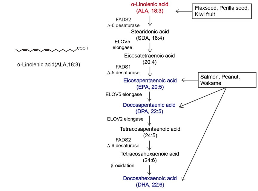
α-linolenic acid (ALA) belongs to the family of n-3 polyunsaturated fatty acids (n-3 PUFAs) and contains a carbon–carbon double bond on the third carbon atom at the methyl end of the carbon chain. This family of essential fatty acids [1] also includes eicosapentaenoic acid (EPA) and docosahexaenoic acid (DHA). ALA is the synthetic precursor of these factors [2][3]. Studies have shown that ALA, DHA, and EPA can be converted into each other via metabolic pathways, as shown in Figure 1, but this conversion is inefficient [4][5]. In the past, attention to ALA was focused mainly on it as a precursor to DHA and EPA, while little was known about ALA itself [6][7]. The most common way to increase the levels of n-3 PUFAs in the body is through dietary intake. ALA can be acquired through the direct consumption of ALA-rich plants, such as flaxseed, perilla seed, chia seed, and rapeseed [8][9], as well as from various ALA preparations available on the market, such as ALA oil, soft capsules, and microcapsules [10]. As components of the phospholipid membrane [11], n-3 PUFAs play diverse roles, including cardiovascular disease prevention [12], anti-inflammatory effects [13], and anticancer effects [14]. However, the effects of ALA, DHA, and EPA on the human body are not consistent. Hemant Poudyal et al. [15] reported that ALA induced different physiological responses compared to DHA or EPA to alleviate the symptoms of metabolic syndrome. Jeong-Eun Choi et al. [16] showed that EPA and DHA exerted antidepressant effects on rats, while ALA did not exert antidepressant effects. Laura E Voorrips [17] showed that ALA was the only n-3 PUFA that was effective at reducing the risk of breast cancer (BC). Based on global medical and nutritional studies [9][18][19][20][21][22][23][24][25][26][27][28][29], ALA can regulate blood lipids, reduce blood viscosity, lower blood pressure, support weight loss, suppress allergic reactions, inhibit inflammation, affect diabetes and bone health, and inhibit cancer occurrence and metastasis. Therefore, it is necessary to distinguish ALA from other n-3 PUFAs.

Figure 1.Overview of α-linolenic acid (ALA). Molecular structure of ALA and the in vivo metabolic pathway by which n-3 polyunsaturated fatty acids (n-3 PUFAs) are generated from ALA. The colors in the figure are intended to emphasize the members of n-3 PUFAs. The vertical arrow represents the metabolism of ALA to other n-3 PUFAs in vivo; Arrows in other directions represent different dietary sources of n-3 PUFAs.

According to the International Agency for Research on Cancer GLOBOCAN 2020 cancer incidence and mortality estimates, in 2020, there were 19.3 million new cancer cases and nearly 10 million deaths worldwide [30], except for melanoma cell cancer. As a populous country, China’s new cancer cases in 2020 accounted for 24% of the world’s new cancer cases [31]. Cancer has surpassed cardiovascular disease as the leading cause of death in China. A prominent feature of tumors is that their growth and proliferation are uncontrolled, and invasion and metastasis are the main problems facing current cancer treatment. Although the etiology of cancer is not yet fully understood, it can be roughly divided into two categories, endogenous and exogenous, and nutrients are the factors most closely related to daily life [32]. Nutrient intake can regulate the tumor microenvironment, thereby affecting cancer cell proliferation, apoptosis, and invasion. Current cancer treatment strategies, including surgery, radiotherapy, and chemotherapy, reduce the quality of life of patients, and diet has gradually become one of the most common treatment methods due to its high acceptance by patients and low toxicity and side effects [13][14]. Most of the initial dietary studies focused on limiting the proliferation of tumor cells by reducing the supply of major nutrients to tumors [33][34][35]. With further research, supplementation with specific nutrients, including histidine and mannose, has also become a strategy for the clinical treatment of cancer [36][37].The n-3 PUFA family has attracted considerable attention for its anticancer effects and use as a dietary supplement. 

Cancer has been a constant threat to human life since its identification, and even when it is treatable, it greatly reduces quality of life. Many studies have shown that ALA exerts significant anticancer effects on multiple cancers [38][39][40][41][42][43][44][45][46][47][48]. In Table 1, a subset of ALA-sensitive cancers is listed, including prostate cancer, BC, hepatocellular carcinoma, colorectal cancer (CRC), and pancreatic cancer. In addition, ALA also exerts effects on many common gastrointestinal tumors and bladder cancer [49][50][51]. As shown in Figure 2, ALA exerts a variety of anticancer effects, including inhibiting proliferation, inducing apoptosis, suppressing tumor metastasis and angiogenesis, and exerting antioxidant effects. To provide a brief introduction to the anticancer effects of ALA, these effects are systematically reviewed, focusing on pharmacological actions and molecular mechanisms.
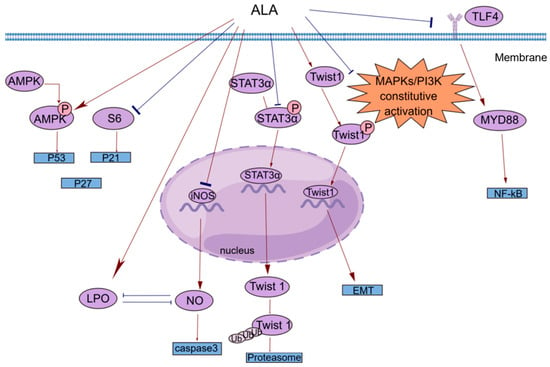
Figure 2. A brief summary of the molecular mechanism of the anticancer effects of ALA. ALA inhibits cell proliferation by regulating the AMPK/S6 axis. ALA can promote cell apoptosis by directly increasing intracellular lipid peroxidation (LPO) or indirectly reducing the accumulation of NO. ALA can suppress tumor metastasis by decreasing the mRNA expression of Twist1 and promoting the degradation of Twist1. The anti-inflammatory effects of ALA may be mediated by blocking the TLR4/MyD88/NF-κB cascade. This figure was constructed with FigDraw (ID: TOUPR3fbb8). There are two kinds of arrows, the flared arrows represent inhibition, and the other is facilitation.
Table 1. The mechanism of the antitumor effects of ALA.
Cancer Effect Effector Molecules Change in Ex-Pression
PCa
(prostate cancer) [52]
anti-inflammatory effect PG/LTs downregulation
BC
(breast cancer) [38][39]
anti-inflammatory effect/inhibition of tumor metastasis COX2/PGE2/Twist 1 downregulation
HCC
(hepatocellular carcinoma) [40][41]
inhibition of proliferation Farnesoid X receptor upregulation
CRC
(colorectal cancer) [42][43]
induction of apoptosis caspase 3 downregulation
PCA
(pancreatic cancer) [44]
anti-inflammatory effect IL-1β/IL-6 downregulation

2. Inhibition of Proliferation

Cell proliferation plays a key role in life. Normal cell proliferation is critical for organismal growth, development, tissue repair, and metabolism. However, the abnormal expression of cancer-related genes in cells caused by various factors can lead to uncontrolled cell proliferation, which is an important part of cancer development.
Esophageal tumors can lead to dysphagia and strongly affect patient quality of life. In clinical practice, small and localized tumors are often surgically removed, but there is no way to perform surgery on larger or non-localized tumors. In addition, the resistance of esophageal cancer to chemotherapy has made the need for new therapies more urgent. Hyun-Seuk Moon’s team [53] reported that dietary ALA with or without oleic acid (OA) could inhibit the proliferation of the esophageal cancer cell lines OE19 and OE33 by regulating the AMPK/S6 axis to treat esophageal tumors. OA and ALA promoted the expression of tumor suppressor genes, such as p53, p21, and p27, by activating AMPK and/or decreasing the phosphorylation of S6. This provides a new idea for cancer treatment involving the consumption of ALA-containing foods for therapeutic purposes. Studies have shown that ALA alone or in combination with other drugs can inhibit cell proliferation in a variety of ways. Peroxisome proliferator-activated receptor-γ (PPAR-γ) is a nuclear receptor that regulates lipid homeostasis. As natural ligands of PPAR-γ, fatty acids can inhibit the growth of cancer cells by activating PPAR-γ [54]. Lijun Yang et al. [55] reported that ALA dose-dependently inhibited the proliferation of renal cell carcinoma (RCC) cells by significantly increasing PPAR-γ activity and gene expression and significantly inhibiting cyclooxygenase-2 (COX-2). COX-2 is an inducible enzyme involved in inflammation. Moreover, when ALA was combined with the PPAR-γ agonist rosiglitazone and the COX-2 inhibitor N-(3-pyridyl) indomethacinamide, its inhibitory effect on the proliferation of the human RCC cell line OS-RC-2 was further increased. ALA inhibited the transformation of cervical cancer cells by reducing the expression of the human papillomavirus oncoproteins E6 and E7, restoring the expression of the tumor suppressor proteins p53 and Rb, and reducing the expression of phosphorylated ERK1/2 and p38. Thus, cell proliferation was inhibited [56]. Consequently, it is possible and promising to use ALA in clinical practice to treat associated tumors by preventing tumor cell proliferation.

3. Induction of Apoptosis

Apoptosis is regulated by genes and is a type of programmed cell death. During embryonic development, certain cell populations undergo apoptosis to eliminate certain cells and complete organogenesis. However, mutagenesis by external factors can cause normal cells to become cancerous. The carcinogenesis of normal cells requires uncontrolled cell proliferation, the dysfunction of cell apoptosis, and the dysregulation of apoptotic regulators. In general, cancerous cells escape apoptosis by upregulating antiapoptotic factors and downregulating proapoptotic factors [57].
Most studies investigating the antiapoptotic effects of n-3 PUFAs have focused on major substances such as EPA and DHA, and little research has been conducted on ALA [58][59][60]. However, the functions of these substances are different. For example, one prospective study separated DHA, EPA, and ALA and found that ALA was the only n-3 PUFA that significantly reduced BC risk [17]. Interestingly, this phenomenon of local generalization is not uncommon. Caspases are a class of cysteine proteases that can mediate apoptosis. The apoptotic effect of ALA is closely related to its ability to increase lipid peroxidation [61]. An increase in lipid peroxides may increase the generation of free radicals, and reactive oxygen species (ROS) can directly activate mitochondrial permeability transition, leading to the loss of mitochondrial membrane potential. This results in cytochrome c (cyt c) release and caspase pathway activation. ALA reduced the mRNA expression of inducible nitric oxide synthase (iNOS) to reduce intracellular levels of NO, which can inhibit lipid peroxidation by scavenging free radicals from lipid peroxidation. ALA also inhibited iNOS-induced NO production in a peroxidation-dependent manner, further activating caspase 3 to induce apoptosis [61]. In addition, studies have shown that ALA can promote apoptosis in a variety of ways, such as by upregulating the expression of the proapoptotic gene Bax, downregulating the expression of the antiapoptotic gene Bcl-2, stabilizing hypoxia-inducible factor-1α (HIF-1α) and downregulating fatty acid synthase (FASN) to promote mitochondrial apoptosis [62][63]. This opens up multiple possibilities for the clinical use of ALA.

4. Anti-Inflammatory Response

The inflammatory response is a double-edged sword. When the normal balance of the body is disrupted, immune activity is increased and typically manifests as inflammation. However, the existence of inflammation itself and the changes in the microenvironment caused by inflammation cause certain pathological symptoms. Cancer patients are prone to secondary inflammatory diseases, and cancer may also develop in response to inflammation [64][65]. For example, patients with inflammatory bowel diseases such as ulcerative colitis and Crohn’s disease have an increased risk of CRC and a higher mortality rate than patients with sporadic CRC [66]. The main reason may be the recurrent chronic inflammatory response, which continuously damages the intestinal mucosa. The mucosa is in a state of long-term repair accompanied by intestinal microbial heterotopia and atypical hyperplasia, which can eventually lead to cancer [67]. ALA has been shown to exert powerful anti-inflammatory effects [68], and different epidemiological studies have shown that ALA is inversely correlated with plasma levels of inflammatory factors, including C-reactive protein (CRP), interleukin-6 (IL-6), interleukin-1β (IL-1β), interferon γ (IFN-γ), tumor necrosis factor-α (TNF-α), and E-selectin [25][26]. The potential anti-inflammatory mechanisms of ALA are diverse and include reducing the level of arachidonic acid (AA) in the blood and downregulating COX-2 [69].
A common clinical phenomenon in alcoholic hepatitis patients is the leakage of bacterial endotoxins through the damaged intestinal barrier into the portal vein. The endotoxins then bind to lipopolysaccharide-binding protein (LBP), triggering the TLR4/MyD88/NF-κB inflammatory cascade in the liver. ALA can inhibit the lipopolysaccharide (LPS)-induced inflammatory response by blocking this cascade [70]. N-6 polyunsaturated fatty acids, such as linolenic acid (LA), can be metabolized to AA, which is the precursor of many potent proinflammatory mediators, including prostaglandins (PGs) and leukotrienes (LTs) [71]. ALA intake can reduce blood levels of AA because ALA competes with LA for the enzyme delta-6-desaturase. Moreover, ALA is the preferred substrate of this enzyme. Increasing ALA intake can limit the conversion of LA to AA, thereby reducing the biosynthesis of proinflammatory eicosanoid acid and further exerting anticancer effects [52][72]. COX-2 enzymes are involved in the synthesis of proinflammatory prostaglandins, and one possible mechanism by which ALA inhibits cancer is that it inhibits inflammation through the downregulation of COX-2 [73]. NF-κB plays an important role in the inflammatory and immune responses. COX-2 is a downstream target of NF-κB activation. ALA suppresses tumors by reducing the expression of NF-κB and its target genes in tumor cells [56].

5. Inhibition of Tumor Metastasis

Tumors can spread in vivo to local normal tissues, to nearby lymph nodes, tissues, and organs, or to distant tissues through fluid transport, which is a feature that has made them a leading cause of cancer-related death [74]. Cancer metastasis can be divided into the following steps: tumor growth in situ, angiogenesis, epithelial–mesenchymal transition (EMT), invasion, intravasation, survival in the blood circulation, extravasation, dormancy, and metastatic tumor growth [75].
In vivo and in vitro experiments have shown that ALA inhibits metastasis in various cancers to varying degrees. Marianela Vara-Messler et al. [76] fed BALB/c mice a chia seed oil diet rich in ALA and corn oil rich in LA as a control and found that ALA could significantly reduce the incidence of BC and the number of metastatic lesions in mice. Mechanistically, ALA can alter signaling in BC cells by altering the cell membrane structure, which increases fatty acid unsaturation and exerts anticancer effects. In vitro experiments were performed to explore the mechanisms by which ALA inhibits cancer metastasis, including the EMT and tumor angiogenesis. Twist1 is required to initiate EMT and promote tumor metastasis and is regulated by signal transducer and activator of transcription 3α (STAT3α) and mitogen-activated protein kinases (MAPKs). Shih-Chung Wang et al. [39] studied the effect of ALA on Twist1 and Twist1-mediated TNBC cell migration and reported that ALA could reduce the mRNA expression of Twist1, reduce the accumulation of STAT3α in the nucleus, and reduce the protein and phosphorylation levels of Twist1. ALA promoted Twist1 degradation, thereby eliminating EMT and inhibiting Twist1-mediated migration in TNBC cells. One of the hallmark features of EMT is the loss of epithelial integrity due to the degradation of adherens junctions, which maintain epithelial cell contacts. A major driver of this degradation is proteolytic digestion by matrix metalloproteinases (MMPs). In tumors, MMP2 and MMP9 are involved in connective tissue degradation, tumor-induced angiogenesis, and cell migration. Another mechanism by which ALA inhibits tumor metastasis is by reducing vascular endothelial growth factor (VEGF), MMP-2 and MMP-9 protein expression [56]. As an important vasodilator, NO can stimulate VEGF production and participate in every step of VEGF-mediated tumor angiogenesis. NO can be generated by the enzyme iNOS in vivo. ALA not only reduces NO levels by reducing the mRNA expression of iNOS but also increases lipid peroxidation. The accumulation of peroxides increases the generation of free radicals; thus, the inactivation and reduction of NO decrease tumor angiogenesis and inhibit tumor metastasis [56][61].

6. Antioxidant Effect

Oxidative stress (OS) refers to the breakdown of the balance between ROS production and elimination in the body and is mainly characterized by the excessive production of highly reactive molecules such as ROS and reactive nitrogen species (RNS). OS is closely related to the occurrence and development of tumors [77][78]. Rashmi Deshpande et al. [61]’s study revealed that ALA could inhibit cancer by stimulating ROS production to induce apoptosis. However, this study was performed in a relatively simple in vitro experimental environment, and there are more complex and diverse mechanisms in vivo that can counteract this effect. Leslie Couedelo et al. [79] showed that ALA intake induced vitamin E depletion. Since vitamin E is a potent antioxidant, it can be used to capture the free radicals produced without producing OS. In addition, Jin Hyang Song and Teruo Miyazawa reported that excessive incorporation of n-3 PUFAs into cell membranes can have adverse effects by enhancing membrane sensitivity to lipid peroxidation (LPO) and inducing OS, but these studies were based on a high-fat diet [80]. In conclusion, when ALA is added to the diet, its effect on the body as a whole should be considered, and the amount of intake should also be considered.
Flaxseed oil (FO) is rich in ALA and is often used as a dietary supplement to treat cancer and improve health. Jyoti Sharma et al. [81] examined mice with skin cancer induced by 7, 12-dimethylbenzo-[a] thane (DMBA) combined with croton oil and showed that FO scavenged free radicals by increasing the levels of enzymatic and nonenzymatic antioxidants, including superoxide dismutase (SOD), catalase (CAT), glutathione peroxidase (GPx), and glutathione (GSH), in the skin and liver. SOD, CAT, and GPx are important antioxidant enzymes that work in concert to prevent excessive levels of intracellular ROS. The main role of SOD is to accelerate the dismutation of superoxide anions to hydrogen peroxide, after which CAT and GPx convert the resulting hydrogen peroxide into harmless substances [82]. The free radical scavenging effect of GSH is mediated by the active sulfhydryl-SH groups in its structure, which are easily dehydrogenated through oxidation. In addition, GSH can bind to GPx and requires different secondary enzymes and cofactors, including nicotinamide–adenine dinucleotide phosphate (NADPH), to exert its effects [81]. NADPH oxidase, which is the major source of ROS in the vasculature, is composed of a catalytic subunit and a regulatory cytosolic subunit. Hao Han [83] examined atherosclerosis and showed that FO could inhibit NADPH oxidase by reducing the mRNA and protein expression of the NADPH oxidase catalytic subunit, thereby regulating cytoplasmic subunit expression, reducing malondialdehyde levels and increasing GSH levels to exert antioxidant effects. ALA-rich FO is consumed mainly as a cooking oil, and there is undoubtedly a close association between this nutrient and the gut microbiota. Xiaoyan Sun et al. [84] used an FO diet to study the antioxidant capacity of colon epithelial cells in aged rats, and the results showed that the antioxidant levels of aged rats increased significantly after FO administration. Mechanistically, the intake of FO may have changed some intestinal bacteria. OS can play a role in the entire process of tumor development, and ALA intake can slow the toxic effects on the body when administered at different stages of tumor development.

References

  1. Arthur A. Spector; Hee-Yong Kim; Discovery of essential fatty acids. J. Lipid Res.. 2015, 56, 11-21.
  2. Toshimi Ogawa; Kento Sawane; Kouta Ookoshi; Ryuta Kawashima; Supplementation with Flaxseed Oil Rich in Alpha-Linolenic Acid Improves Verbal Fluency in Healthy Older Adults. Nutr.. 2023, 15, 1499.
  3. Marija Takic; Biljana Pokimica; Gordana Petrovic-Oggiano; Tamara Popovic; Effects of Dietary α-Linolenic Acid Treatment and the Efficiency of Its Conversion to Eicosapentaenoic and Docosahexaenoic Acids in Obesity and Related Diseases. Mol.. 2022, 27, 4471.
  4. Isabelle M. Berquin; Iris J. Edwards; Yong Q. Chen; Multi-targeted therapy of cancer by omega-3 fatty acids. Cancer Lett.. 2008, 269, 363-377.
  5. Kyu-Bong Kim; Yoon A. Nam; Hyung Sik Kim; A. Wallace Hayes; Byung-Mu Lee; α-Linolenic acid: Nutraceutical, pharmacological and toxicological evaluation. Food Chem. Toxicol.. 2014, 70, 163-178.
  6. Burdge, G.C.; Calder, P.C. Conversion of alpha-linolenic acid to longer-chain polyunsaturated fatty acids in human adults. Reprod. Nutr. Dev.. 2005, 45, 581–597.
  7. Aliza H. Stark; Ram Reifen; Michael A. Crawford; Past and Present Insights on Alpha-linolenic Acid and the Omega-3 Fatty Acid Family. Crit. Rev. Food Sci. Nutr.. 2015, 56, 2261-2267.
  8. Fereidoon Shahidi; Priyatharini Ambigaipalan; Omega-3 Polyunsaturated Fatty Acids and Their Health Benefits. Annu. Rev. Food Sci. Technol.. 2018, 9, 345-381.
  9. Qianghua Yuan; Fan Xie; Wei Huang; Mei Hu; Qilu Yan; Zemou Chen; Yan Zheng; Li Liu; The review of alpha‐linolenic acid: Sources, metabolism, and pharmacology. Phytotherapy Res.. 2021, 36, 164-188.
  10. John Parker; Amanda N. Schellenberger; Amy L. Roe; Hellen Oketch-Rabah; Angela I. Calderón; Therapeutic Perspectives on Chia Seed and Its Oil: A Review. Planta Medica. 2018, 84, 606-612.
  11. Natacha Porta; Béatrice Bourgois; Claude Galabert; Cécile Lecointe; Pierre Cappy; Régis Bordet; Louis Vallée; Stéphane Auvin; Anticonvulsant effects of linolenic acid are unrelated to brain phospholipid cell membrane compositions. Epilepsia. 2009, 50, 65-71.
  12. Federica Laguzzi; Agneta Åkesson; Matti Marklund; Frank Qian; Bruna Gigante; Traci M. Bartz; Julie K. Bassett; Anna Birukov; Hannia Campos; Yoichiro Hirakawa; Fumiaki Imamura; Susanne Jäger; Maria Lankinen; Rachel A. Murphy; Mackenzie Senn; Toshiko Tanaka; Nathan Tintle; Jyrki K. Virtanen; Kazumasa Yamagishi; Matthew Allison; Ingeborg A. Brouwer; Ulf De Faire; Gudny. Eiriksdottir; Luigi Ferrucci; Nita G. Forouhi; Johanna M. Geleijnse; Allison M Hodge; Hitomi Kimura; Markku Laakso; Ulf Risérus; Anniek C. van Westing; Stefania Bandinelli; Ana Baylin; Graham G. Giles; Vilmundur Gudnason; Hiroyasu Iso; Rozenn N. Lemaitre; Toshiharu Ninomiya; Wendy S. Post; Bruce M. Psaty; Jukka T. Salonen; Matthias B. Schulze; Michael Y. Tsai; Matti Uusitupa; Nicholas J. Wareham; Seung-Won Oh; Alexis C. Wood; William S. Harris; David Siscovick; Dariush Mozaffarian; Karin Leander; Fatty Acids and Outcomes Research Consortium (FORCE); Role of Polyunsaturated Fat in Modifying Cardiovascular Risk Associated With Family History of Cardiovascular Disease: Pooled De Novo Results From 15 Observational Studies. Circ.. 2024, 149, 305-316.
  13. Evan C. Lien; Matthew G. Vander Heiden; A framework for examining how diet impacts tumour metabolism. Nat. Rev. Cancer. 2019, 19, 651-661.
  14. Susan E. Steck; E. Angela Murphy; Dietary patterns and cancer risk. Nat. Rev. Cancer. 2019, 20, 125-138.
  15. Hemant Poudyal; Sunil K. Panchal; Leigh C. Ward; Lindsay Brown; Effects of ALA, EPA and DHA in high-carbohydrate, high-fat diet-induced metabolic syndrome in rats. J. Nutr. Biochem.. 2013, 24, 1041-1052.
  16. Jeong-Eun Choi; Yongsoon Park; EPA and DHA, but not ALA, have antidepressant effects with 17β-estradiol injection via regulation of a neurobiological system in ovariectomized rats. J. Nutr. Biochem.. 2017, 49, 101-109.
  17. Laura E Voorrips; Henny Am Brants; Alwine Fm Kardinaal; Gerrit J Hiddink; Piet A van Den Brandt; R Alexandra Goldbohm; Intake of conjugated linoleic acid, fat, and other fatty acids in relation to postmenopausal breast cancer: the Netherlands Cohort Study on Diet and Cancer. Am. J. Clin. Nutr.. 2002, 76, 873-882.
  18. Asmaa S Abdelhamid; Tracey J Brown; Julii S Brainard; Priti Biswas; Gabrielle C Thorpe; Helen J Moore; Katherine Ho Deane; Fai K AlAbdulghafoor; Carolyn D Summerbell; Helen V Worthington; Fujian Song; Lee Hooper; Omega-3 fatty acids for the primary and secondary prevention of cardiovascular disease. Cochrane Database Syst. Rev.. 2018, 7, CD003177.
  19. B. Dierberger; M. Schäch; I. Anadere; M. Brändle; R. Jacob; Effect of a diet rich in linseed oil on complex viscosity and blood pressure in spontaneously hypertensive rats (SHR). Basic Res. Cardiol.. 1991, 86, 561-566.
  20. Yuanyuan Ding; Yuejin Wang; Chaomei Li; Yongjing Zhang; Shiling Hu; Jiapan Gao; Rui Liu; Hongli An; α-Linolenic acid attenuates pseudo-allergic reactions by inhibiting Lyn kinase activity. Phytomedicine. 2020, 80, 153391.
  21. 21. Hu, F.B.; Stampfer, M.J.; Manson, J.E.; Rimm, E.B.; Wolk, A.; Colditz, G.A.; Hennekens, C.H.; Willett, W.C. Dietary intake of alpha-linolenic acid and risk of fatal ischemic heart disease among women. Am. J. Clin. Nutr.. 1999, 69, 890–897.
  22. Alan A. Hennessy; Paul R. Ross; Gerald F. Fitzgerald; Catherine Stanton; Sources and Bioactive Properties of Conjugated Dietary Fatty Acids. Lipids. 2016, 51, 377-397.
  23. Youjin Kim; Jasminka Z. Ilich; Implications of dietary α-linolenic acid in bone health. Nutr.. 2011, 27, 1101-1107.
  24. Mihir Parikh; Thane G. Maddaford; J. Alejandro Austria; Michel Aliani; Thomas Netticadan; Grant N. Pierce; Dietary Flaxseed as a Strategy for Improving Human Health. Nutr.. 2019, 11, 1171.
  25. Aleix Sala-Vila; Jennifer Fleming; Penny Kris-Etherton; Emilio Ros; Impact of α-Linolenic Acid, the Vegetable ω-3 Fatty Acid, on Cardiovascular Disease and Cognition. Adv. Nutr. Int. Rev. J.. 2022, 13, 1584-1602.
  26. Aliza H Stark; Michael A Crawford; Ram Reifen; Update on alpha-linolenic acid. Nutr. Rev.. 2008, 66, 326-332.
  27. Yuejin Wang; Yuanyuan Ding; Chaomei Li; Jiapan Gao; Xiaodong Wang; Hongli An; Alpha-linolenic acid inhibits IgE-mediated anaphylaxis by inhibiting Lyn kinase and suppressing mast cell activation. Int. Immunopharmacol.. 2021, 103, 108449.
  28. H M Kaplan; M Deger; K E Erdogan; T Ates; E Demir; Alpha-linolenic acid protects against methotrexate-induced nephrotoxicity in mouse kidney cells.. Eur. Rev. Med. Pharmacol. Sci.. 2023, 27, 11103-11108.
  29. Sana Noreen; Tabussam Tufail; Huma Bader Ul Ain; Chinaza Godswill Awuchi; Pharmacological, nutraceutical, and nutritional properties of flaxseed (Linum usitatissimum): An insight into its functionality and disease mitigation. Food Sci. Nutr.. 2023, 11, 6820-6829.
  30. Hyuna Sung; Jacques Ferlay; Rebecca L. Siegel; Mathieu Laversanne; Isabelle Soerjomataram; Ahmedin Jemal; Freddie Bray; Msc Freddie Bray Bsc; Jacques Ferlay Me; Msc Isabelle Soerjomataram Md; Lindsey A. Torre Msph; DVM Ahmedin Jemal PhD; Global Cancer Statistics 2020: GLOBOCAN Estimates of Incidence and Mortality Worldwide for 36 Cancers in 185 Countries. CA: A Cancer J. Clin.. 2021, 71, 209-249.
  31. Wei Cao; Hong-Da Chen; Yi-Wen Yu; Ni Li; Wan-Qing Chen; Changing profiles of cancer burden worldwide and in China: a secondary analysis of the global cancer statistics 2020. Chin. Med J.. 2021, 134, 783-791.
  32. Maria Chiara Mentella; Franco Scaldaferri; Caterina Ricci; Antonio Gasbarrini; Giacinto Abele Donato Miggiano; Cancer and Mediterranean Diet: A Review. Nutr.. 2019, 11, 2059.
  33. Emeline Dierge; Yvan Larondelle; Olivier Feron; Cancer diets for cancer patients: Lessons from mouse studies and new insights from the study of fatty acid metabolism in tumors. Biochim.. 2020, 178, 56-68.
  34. Irene Caffa; Vanessa Spagnolo; Claudio Vernieri; Francesca Valdemarin; Pamela Becherini; Min Wei; Sebastian Brandhorst; Chiara Zucal; Else Driehuis; Lorenzo Ferrando; Francesco Piacente; Alberto Tagliafico; Michele Cilli; Luca Mastracci; Valerio G. Vellone; Silvano Piazza; Anna Laura Cremonini; Raffaella Gradaschi; Carolina Mantero; Mario Passalacqua; Alberto Ballestrero; Gabriele Zoppoli; Michele Cea; Annalisa Arrighi; Patrizio Odetti; Fiammetta Monacelli; Giulia Salvadori; Salvatore Cortellino; Hans Clevers; Filippo De Braud; Samir G. Sukkar; Alessandro Provenzani; Valter D. Longo; Alessio Nencioni; Fasting-mimicking diet and hormone therapy induce breast cancer regression. Nat.. 2020, 583, 620-624.
  35. Salvatore Cortellino; Alessandro Raveane; Claudia Chiodoni; Gloria Delfanti; Federica Pisati; Vanessa Spagnolo; Euplio Visco; Giuseppe Fragale; Federica Ferrante; Serena Magni; Fabio Iannelli; Federica Zanardi; Giulia Casorati; Francesco Bertolini; Paolo Dellabona; Mario P. Colombo; Claudio Tripodo; Valter D. Longo; Fasting renders immunotherapy effective against low-immunogenic breast cancer while reducing side effects. Cell Rep.. 2022, 40, 111256.
  36. Naama Kanarek; Heather R. Keys; Jason R. Cantor; Caroline A. Lewis; Sze Ham Chan; Tenzin Kunchok; Monther Abu-Remaileh; Elizaveta Freinkman; Lawrence D. Schweitzer; David M. Sabatini; Histidine catabolism is a major determinant of methotrexate sensitivity. Nat.. 2018, 559, 632-636.
  37. Pablo Sierra Gonzalez; James O’prey; Simone Cardaci; Valentin J. A. Barthet; Jun-Ichi Sakamaki; Florian Beaumatin; Antonia Roseweir; David M. Gay; Gillian Mackay; Gaurav Malviya; Elżbieta Kania; Shona Ritchie; Alice D. Baudot; Barbara Zunino; Agata Mrowinska; Colin Nixon; Darren Ennis; Aoisha Hoyle; David Millan; Iain A. McNeish; Owen J. Sansom; Joanne Edwards; Kevin M. Ryan; Mannose impairs tumour growth and enhances chemotherapy. Nat.. 2018, 563, 719-723.
  38. 38. Horia, E.; Watkins, B.A. Comparison of stearidonic acid and alpha-linolenic acid on PGE2 production and COX-2 protein levels in MDA-MB-231 breast cancer cell cultures. J. Nutr. Biochem. . 2005, 16, 184–192.
  39. Shih-Chung Wang; Hai-Lun Sun; Yi-Hsuan Hsu; Shu-Hui Liu; Chong-Kuei Lii; Chia-Han Tsai; Kai-Li Liu; Chin-Shiu Huang; Chien-Chun Li; α-Linolenic acid inhibits the migration of human triple-negative breast cancer cells by attenuating Twist1 expression and suppressing Twist1-mediated epithelial-mesenchymal transition. Biochem. Pharmacol.. 2020, 180, 114152.
  40. Masataka Okuno; Takuji Tanaka; Chihito Komaki; Seisuke Nagase; Yoshimune Shiratori; Yasutoshi Muto; Kenta Kajiwara; Toshio Maki; Hisataka Moriwaki; Suppressive effect of low amounts of safflower and perilla oils on diethylnitrosamine‐induced hepatocarcinogenesis in male F344 rats. Nutr. Cancer. 1998, 30, 186-193.
  41. Shu Feng; Xingming Xie; Chaochun Chen; Shi Zuo; Xueke Zhao; Haiyang Li; Alpha-linolenic acid inhibits hepatocellular carcinoma cell growth through Farnesoid X receptor/β-catenin signaling pathway. Nutr. Metab.. 2022, 19, 1-10.
  42. John P. Chamberland; Hyun-Seuk Moon; Down-regulation of malignant potential by alpha linolenic acid in human and mouse colon cancer cells. Fam. Cancer. 2014, 14, 25-30.
  43. MarÍA JosÉ GonzÁLez-FernÁNdez; Ignacio Ortea; José Luis Guil-Guerrero; α-Linolenic and γ-linolenic acids exercise differential antitumor effects on HT-29 human colorectal cancer cells. Toxicol. Res.. 2020, 9, 474-483.
  44. Kyung Suk Park; Joo Weon Lim; Hyeyoung Kim; Inhibitory Mechanism of Omega‐3 Fatty Acids in Pancreatic Inflammation and Apoptosis. Ann. New York Acad. Sci.. 2009, 1171, 421-427.
  45. Ze-Bin Dai; Xiao-Li Ren; Yi-Lang Xue; Ya Tian; Bing-Bing He; Chang-Long Xu; Bo Yang; Association of Dietary Intake and Biomarker of α-Linolenic Acid With Incident Colorectal Cancer: A Dose-Response Meta-Analysis of Prospective Cohort Studies. Front. Nutr.. 2022, 9, 948604.
  46. Ana Calado; Pedro Miguel Neves; Teresa Santos; Paula Ravasco; The Effect of Flaxseed in Breast Cancer: A Literature Review. Front. Nutr.. 2018, 5, 4.
  47. Idris Adewale Ahmed; Maryam Abimbola Mikail; Mohammad Rais Mustafa; Muhammad Ibrahim; Rozana Othman; Lifestyle interventions for non-alcoholic fatty liver disease. Saudi J. Biol. Sci.. 2019, 26, 1519-1524.
  48. Michael A. Tsoukas; Byung-Joon Ko; Theodore R. Witte; Fadime Dincer; W. Elaine Hardman; Christos S. Mantzoros; Dietary walnut suppression of colorectal cancer in mice: Mediation by miRNA patterns and fatty acid incorporation. J. Nutr. Biochem.. 2015, 26, 776-783.
  49. Maree T. Brinkman; Margaret R. Karagas; Michael S. Zens; Alan R. Schned; Raoul C. Reulen; Maurice P. Zeegers; Intake of α-linolenic acid and other fatty acids in relation to the risk of bladder cancer: results from the New Hampshire case–control study. Br. J. Nutr.. 2011, 106, 1070-1077.
  50. Jinfeng Dai; Junhui Shen; Wensheng Pan; Shengrong Shen; Undurti N Das; Effects of polyunsaturated fatty acids on the growth of gastric cancer cells in vitro. Lipids Heal. Dis.. 2013, 12, 71-15.
  51. Ji Hoon Yu; Sin‐Gun Kang; Un‐Young Jung; Chul‐Ho Jun; Hyeyoung Kim; Effects of Omega‐3 Fatty Acids on Apoptosis of Human Gastric Epithelial Cells Exposed to Silica‐Immobilized Glucose Oxidase. Ann. New York Acad. Sci.. 2009, 1171, 359-364.
  52. Jingjing Li; Zhennan Gu; Yong Pan; Shunhe Wang; Haiqin Chen; Hao Zhang; Wei Chen; Yong Q. Chen; Dietary supplementation of α-linolenic acid induced conversion of n-3 LCPUFAs and reduced prostate cancer growth in a mouse model. Lipids Heal. Dis.. 2017, 16, 1-9.
  53. Hyun-Seuk Moon; Saime Batirel; Christos S. Mantzoros; Alpha linolenic acid and oleic acid additively down-regulate malignant potential and positively cross-regulate AMPK/S6 axis in OE19 and OE33 esophageal cancer cells. Metab.. 2014, 63, 1447-1454.
  54. 54. Kliewer, S.A.; Sundseth, S.S.; Jones, S.A.; Brown, P.J.; Wisely, G.B.; Koble, C.S.; Devchand, P.; Wahli, W.; Willson, T.M.; Len-hard, J.M.; et al. Fatty acids and eicosanoids regulate gene expression through direct interactions with peroxisome proliferator-activated re-ceptors alpha and gamma. Proc. Natl. Acad. Sci. USA. 1997, 94, 4318–4323.
  55. Lijun Yang; Jianlin Yuan; Liwen Liu; Changhong Shi; Longxin Wang; Feng Tian; Fei Liu; He Wang; Chen Shao; Qiang Zhang; Zhinan Chen; Weijun Qin; Weihong Wen; α-linolenic acid inhibits human renal cell carcinoma cell proliferation through PPAR-γ activation and COX-2 inhibition. Oncol. Lett.. 2013, 6, 197-202.
  56. Rashmi Deshpande; Prakash Mansara; Ruchika Kaul-Ghanekar; Alpha-linolenic acid regulates Cox2/VEGF/MAP kinase pathway and decreases the expression of HPV oncoproteins E6/E7 through restoration of p53 and Rb expression in human cervical cancer cell lines. Tumor Biol.. 2015, 37, 3295-3305.
  57. Mayra Montecillo-Aguado; Belen Tirado-Rodriguez; Sara Huerta-Yepez; The Involvement of Polyunsaturated Fatty Acids in Apoptosis Mechanisms and Their Implications in Cancer. Int. J. Mol. Sci.. 2023, 24, 11691.
  58. Z.Y. Chen; N.W. Istfan; Docosahexaenoic acid is a potent inducer of apoptosis in HT-29 colon cancer cells. Prostaglandins, Leukot. Essent. Fat. Acids. 2000, 63, 301-308.
  59. Sun En Lee; Joo Weon Lim; Hyeyoung Kim; Activator Protein‐1 Mediates Docosahexaenoic Acid‐induced Apoptosis of Human Gastric Cancer Cells. Ann. New York Acad. Sci.. 2009, 1171, 163-169.
  60. Hilde Heimli; Camilla Giske; Soheil Naderi; Christian A. Drevon; Kristin Hollung; Eicosapentaenoic acid promotes apoptosis in Ramos cells via activation of caspase‐3 and ‐9. Lipids. 2002, 37, 797-802.
  61. 62. Deshpande, R.; Mansara, P.; Suryavanshi, S.; Kaul-Ghanekar, R. Alpha-linolenic acid regulates the growth of breast and cervical cancer cell lines through regulation of NO release and in-duction of lipid peroxidation. J. Mol. Biochem. 2013, 2, 6–17.
  62. Subhadeep Roy; Atul Kumar Rawat; Shreesh Raj Sammi; Uma Devi; Manjari Singh; Swetlana Gautam; Rajnish Kumar Yadav; Jitendra Kumar Rawat; Lakhveer Singh; Mohd. Nazam Ansari; Abdulaziz S. Saeedan; Rakesh Pandey; Dinesh Kumar; Gaurav Kaithwas; Alpha-linolenic acid stabilizes HIF-1 α and downregulates FASN to promote mitochondrial apoptosis for mammary gland chemoprevention. Oncotarget. 2017, 8, 70049-70071.
  63. Ji‐Yoon Kim; Han Deuk Park; Eunju Park; Jeong‐Woo Chon; Yoo Kyoung Park; Growth‐Inhibitory and Proapoptotic Effects of Alpha‐Linolenic Acid on Estrogen‐Positive Breast Cancer Cells. Ann. New York Acad. Sci.. 2009, 1171, 190-195.
  64. Alberto Mantovani; Paola Allavena; Antonio Sica; Frances Balkwill; Cancer-related inflammation. Nat.. 2008, 454, 436-444.
  65. Eran Elinav; Roni Nowarski; Christoph A. Thaiss; Bo Hu; Chengcheng Jin; Richard A. Flavell; Inflammation-induced cancer: crosstalk between tumours, immune cells and microorganisms. Nat. Rev. Cancer. 2013, 13, 759-771.
  66. Muhammad Shahid Nadeem; Vikas Kumar; Fahad A. Al-Abbasi; Mohammad Amjad Kamal; Firoz Anwar; Risk of colorectal cancer in inflammatory bowel diseases. Semin. Cancer Biol.. 2019, 64, 51-60.
  67. Ana Elisa Valencise Quaglio; Thais Gagno Grillo; Ellen Cristina Souza De Oliveira; Luiz Claudio Di Stasi; Ligia Yukie Sassaki; Gut microbiota, inflammatory bowel disease and colorectal cancer. World J. Gastroenterol.. 2022, 28, 4053-4060.
  68. Nuoxi Fan; Jennifer L. Fusco; Daniel W. Rosenberg; Antioxidant and Anti-Inflammatory Properties of Walnut Constituents: Focus on Personalized Cancer Prevention and the Microbiome. Antioxidants. 2023, 12, 982.
  69. Maria Azrad; Chelsea Turgeon; Wendy Demark-Wahnefried; Current Evidence Linking Polyunsaturated Fatty Acids with Cancer Risk and Progression. Front. Oncol.. 2013, 3, 224.
  70. Meng Wang; Xiao-Jing Zhang; Chunyan Yan; Chengwei He; Peng Li; Meiwan Chen; Huanxing Su; Jian-Bo Wan; Preventive effect of α-linolenic acid-rich flaxseed oil against ethanol-induced liver injury is associated with ameliorating gut-derived endotoxin-mediated inflammation in mice. J. Funct. Foods. 2016, 23, 532-541.
  71. Jacqueline K. Innes; Philip C. Calder; Omega-6 fatty acids and inflammation. Prostaglandins, Leukot. Essent. Fat. Acids. 2018, 132, 41-48.
  72. Tse-Hung Huang; Pei-Wen Wang; Shih-Chun Yang; Wei-Ling Chou; Jia-You Fang; Cosmetic and Therapeutic Applications of Fish Oil’s Fatty Acids on the Skin. Mar. Drugs. 2018, 16, 256.
  73. D. Williams; M. Verghese; L.T. Walker; J. Boateng; L. Shackelford; C.B. Chawan; Flax seed oil and flax seed meal reduce the formation of aberrant crypt foci (ACF) in azoxymethane-induced colon cancer in Fisher 344 male rats. Food Chem. Toxicol.. 2007, 45, 153-159.
  74. Patricia S Steeg; Tumor metastasis: mechanistic insights and clinical challenges. Nat. Med.. 2006, 12, 895-904.
  75. Scott Valastyan; Robert A. Weinberg; Tumor Metastasis: Molecular Insights and Evolving Paradigms. Cell. 2011, 147, 275-292.
  76. Marianela Vara-Messler; Maria E. Pasqualini; Andrea Comba; Renata Silva; Carola Buccellati; Annalisa Trenti; Lucia Trevisi; Aldo R. Eynard; Angelo Sala; Chiara Bolego; Mirta A. Valentich; Increased dietary levels of α-linoleic acid inhibit mammary tumor growth and metastasis. Eur. J. Nutr.. 2015, 56, 509-519.
  77. MarijaDragan Jelic; AljosaD Mandic; SlobodanM Maricic; BranislavaU Srdjenovic; Oxidative stress and its role in cancer. J. Cancer Res. Ther.. 2021, 17, 22-28.
  78. Simone Reuter; Subash C. Gupta; Madan M. Chaturvedi; Bharat B. Aggarwal; Oxidative stress, inflammation, and cancer: How are they linked?. Free. Radic. Biol. Med.. 2010, 49, 1603-1616.
  79. Leslie Couëdelo; Benjamin Buaud; Hélène Abrous; Ikram Chamekh-Coelho; Didier Majou; Carole Boué-Vaysse; Effect of increased levels of dietary α-linolenic acid on the n-3 PUFA bioavailability and oxidative stress in rat. Br. J. Nutr.. 2021, 127, 1320-1333.
  80. Jin Hyang Song; Teruo Miyazawa; Enhanced level of n-3 fatty acid in membrane phospholipids induces lipid peroxidation in rats fed dietary docosahexaenoic acid oil. Atheroscler.. 2001, 155, 9-18.
  81. Jyoti Sharma; Ritu Singh; P. K. Goyal; Chemomodulatory Potential of Flaxseed Oil Against DMBA/Croton Oil–Induced Skin Carcinogenesis in Mice. Integr. Cancer Ther.. 2015, 15, 358-367.
  82. Joël R. Drevet; The antioxidant glutathione peroxidase family and spermatozoa: A complex story. Mol. Cell. Endocrinol.. 2006, 250, 70-79.
  83. Hao Han; Fubin Qiu; Haifeng Zhao; Haiying Tang; Xiuhua Li; Dongxing Shi; Dietary flaxseed oil improved western-type diet-induced atherosclerosis in apolipoprotein-E knockout mice. J. Funct. Foods. 2018, 40, 417-425.
  84. Xiaoyan Sun; Juntong Yu; Yong Wang; Jianming Luo; Guangwen Zhang; Xichun Peng; Flaxseed oil ameliorates aging in d‐galactose induced rats via altering gut microbiota and mitigating oxidative damage. J. Sci. Food Agric.. 2022, 102, 6432-6442.
More
Information
Contributors MDPI registered users' name will be linked to their SciProfiles pages. To register with us, please refer to https://encyclopedia.pub/register : , , , , , , ,
View Times: 249
Revisions: 4 times (View History)
Update Date: 19 Mar 2024
1000/1000
ScholarVision Creations