Submitted Successfully!
To reward your contribution, here is a gift for you: A free trial for our video production service.
Thank you for your contribution! You can also upload a video entry or images related to this topic.
Version Summary Created by Modification Content Size Created at Operation
1 -- 4835 2022-10-14 13:58:27 |
2 format correct + 19 word(s) 4854 2022-10-18 07:39:38 | |
3 format correct Meta information modification 4854 2022-10-18 07:44:36 |

Video Upload Options

Do you have a full video?

Confirm

Are you sure to Delete?
Cite
If you have any further questions, please contact Encyclopedia Editorial Office.
Masanetz, R.K.;  Winkler, J.;  Winner, B.;  Günther, C.;  Süß, P. The Gut-Immune-Brain Axis in Inflammatory Bowel Disease. Encyclopedia. Available online: https://encyclopedia.pub/entry/29560 (accessed on 02 July 2024).
Masanetz RK,  Winkler J,  Winner B,  Günther C,  Süß P. The Gut-Immune-Brain Axis in Inflammatory Bowel Disease. Encyclopedia. Available at: https://encyclopedia.pub/entry/29560. Accessed July 02, 2024.
Masanetz, Rebecca Katharina, Jürgen Winkler, Beate Winner, Claudia Günther, Patrick Süß. "The Gut-Immune-Brain Axis in Inflammatory Bowel Disease" Encyclopedia, https://encyclopedia.pub/entry/29560 (accessed July 02, 2024).
Masanetz, R.K.,  Winkler, J.,  Winner, B.,  Günther, C., & Süß, P. (2022, October 17). The Gut-Immune-Brain Axis in Inflammatory Bowel Disease. In Encyclopedia. https://encyclopedia.pub/entry/29560
Masanetz, Rebecca Katharina, et al. "The Gut-Immune-Brain Axis in Inflammatory Bowel Disease." Encyclopedia. Web. 17 October, 2022.
The Gut-Immune-Brain Axis in Inflammatory Bowel Disease
Edit

Inflammatory bowel disease (IBD) is a chronic inflammatory disease comprising two major clinical entities—Crohn’s disease (CD) and ulcerative colitis (UC). IBD incidence remains constantly high in industrialized countries and continuously rises in emerging economies. Importantly, IBD is associated with neuropsychiatric symptoms that strongly worsen IBD disease burden. Mounting evidence indicates that chronic gut inflammation induces a systemic immune response that might cause the CNS manifestation in IBD. In line with this, biologicals targeting inflammatory circuits exerted robust positive effects on depressive symptoms in many autoimmune diseases, and in IBD in particular. Therefore, research in recent years increasingly focused on the characterization of local and systemic immune reactions in IBD, and on entry routes of inflammatory cells and molecules into the CNS. The ultimate aim is to understand how the changes in the neuroimmune landscape impair the function of neurons to cause neuropsychiatric symptoms. In addition, the role of intestinal microbiota in the gut–immune–brain axis in IBD will be discussed. 

inflammatory bowel disease neuroinflammation gut microbiota Crohn's disease ulcerative colitis systemic inflammation depression gut-brain axis

1. Routes from Peripheral Inflammation to the Central Nervous System in Inflammatory Bowel Disease

Chronic gut-derived systemic inflammation is able to take different anatomical routes connecting the gastrointestinal tract and the bloodstream with the  central nervous system (CNS) in order to provoke an inflammatory response within the brain (Figure 1).
Figure 1. Alterations at immune-to-brain interfaces during Inflammatory bowel disease (IBD). (1) At the blood–brain barrier (BBB), downregulation of tight junction proteins and endothelial permeability mediate influx of inflammatory molecules and immune cells. Additional non-disruptive BBB changes comprise endothelial upregulation of adhesion proteins for the interaction and transmigration of circulating immune cells, as well as the release of mediators by endothelial cells and perivascular macrophages that modulate microglial and neuronal function. (2) At the choroid plexus (CP), decreased expression of tight junction proteins and increased fenestration followed by transient closure of the vascular barrier are observed, whereas permeability of the epithelial barrier is transiently enhanced. Numbers of stromal monocytes and neutrophils are increased during intestinal inflammation. (3) In the meninges, infiltration of gut-derived immune cells as well as activation of meningeal immune cells and the NLRP3 inflammasome can be detected. (4) Neural communication pathways linking gut inflammation and the brain include the afferent input via the vagal nerve to the nucleus tractus solitarii (NTS), and information from sensory afferent neurons stimulated in the periphery that provoke neural activation in different brain regions. Gut-derived immune cells (2) can enter the CSF via the CP or the meninges. CP: choroid plexus; CSF: cerebrospinal fluid; SCFA: short chain fatty acids; Tnf: tumor necrosis factor; Il-1β: interleukin-1β; NTS: nucleus tractus solitarii. Figure created with BioRender.com (accessed on 15 September 2022).

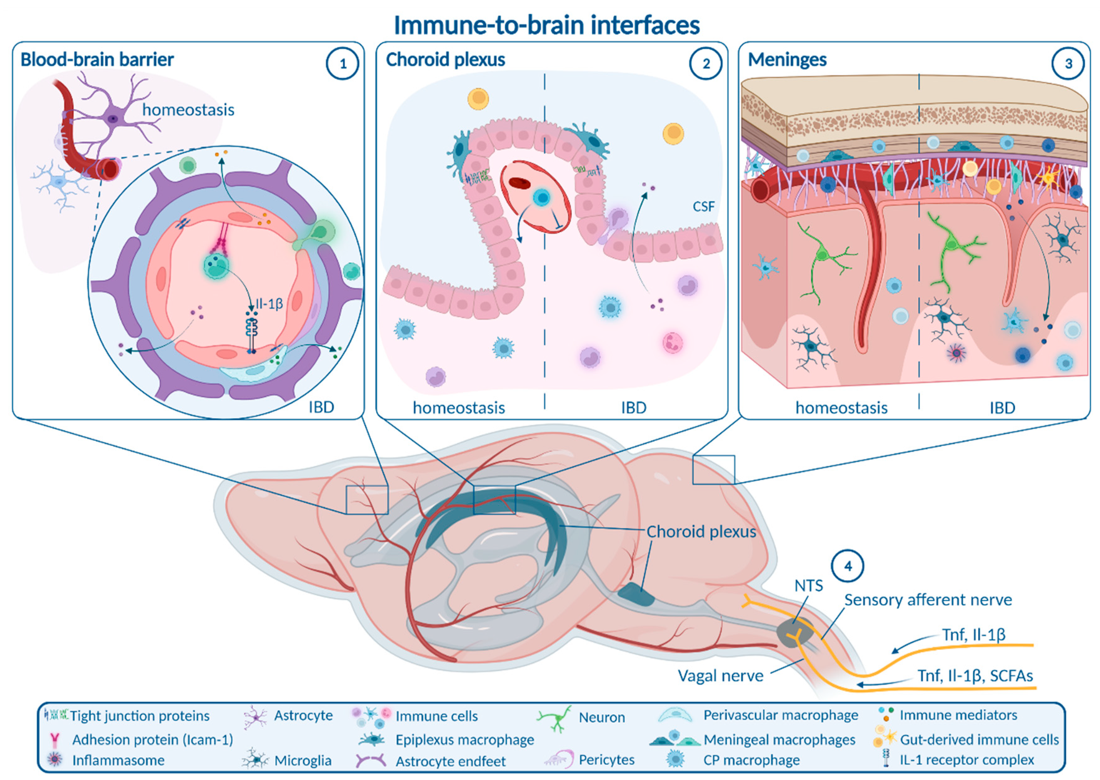
1.1. Enteric, Autonomic and Sensory Nervous System Signaling

The peripheral (i.e., enteric, autonomic) and central nervous systems are tightly connected to form a bidirectional neural communication highway between the gastrointestinal tract and the brain. This route can be employed by inflammatory signaling towards the CNS directly from the gut, without requiring transition into the bloodstream [1]. A major transit route of gut-derived signals is the vagal nerve. Under homeostasis, vagal nerve afferents in the gut monitor key physiological parameters [2] and microbial metabolites like the SCFA butyrate, a surrogate for nutritional state [3]. These afferents project to the nucleus tractus solitarii (NTS) in the brainstem, directly activating neurons in the efferent dorsal motor nucleus of the vagal nerve to signal back to the gut, but also project to many other brain regions [4]. Under inflammatory conditions, vagal sensory neurons are able to sense Tnf and Il-1β, and signal inflammatory cues to the NTS by cytokine-specific electrophysiological patterns [5]. Moreover, early studies implicated the vagal nerve in CNS transmission of peripherally derived Il-1β in fever induction that was abrogated in rats by vagotomy [6]. The role of the vagal nerve in transmitting chronic gut inflammation in IBD towards the brain is not well elucidated and requires further investigation. In chronic mild gut inflammation caused by infection of mice with the parasite Trichuris muris (T. muris), anxiety-like behavior was reduced by systemic anti-inflammatory treatment, but not altered by vagotomy [7]. Besides a potential role of the vagal nerve in signaling of inflammatory cues to the brain, cholinergic efferent vagal nerve fibers can ameliorate gastrointestinal inflammation in TNBS-induced colitis [8]. Moreover, vagal nerve stimulation was reported to be beneficial both in depression and IBD, again pointing to the 10th cranial nerve as a major regulator of the gut–immune–brain axis [1][9].
A second neural route potentially involved in immune communication from the gut to the brain is the nociceptive system. IBD is associated with severe abdominal pain. In vitro data suggested that TNF in the supernatant of colonic biopsies from UC patients activated nociceptors on dorsal root ganglia neurons [10]. In line with this, IBD patients receiving TNF antagonists show a rapid and profound reduction in blood oxygen level dependent (BOLD) brain activity in functional magnetic resonance imaging (fMRI) after application of painful stimuli [11]. Strikingly, this CNS-mediated treatment response was present prior to the resolution of gastrointestinal inflammation [11]. These data suggest TNF-mediated upward signaling of gastrointestinal inflammation via the nociceptive afferent system contributes to functional CNS alterations in IBD. Therefore, neural communication pathways like the vagal nerve and the nociceptive system transmit gut inflammation to the brain.

1.2. Blood–Brain Barrier

The blood–brain barrier (BBB) controls the passage of circulating molecules and cells from the blood into the brain parenchyma. It consists of vascular endothelial cells connected by tight junctions and situated on a vascular basement membrane. The brain parenchyma is bordered by a layer of astrocytic end feet and their corresponding basement membrane. The perivascular space is located between the two basement membranes and contains perivascular macrophages and pericytes [12][13]. In most brain regions, the BBB prevents passive diffusion of hydrophilic molecules larger than 500 Da into the brain [14]. Thus, the BBB is by far less permeable than the GVB. However, several small regions located around the third and fourth ventricle, the so-called circumventricular organs (CVOs), exhibit more permeable endothelia to allow bidirectional interaction between the brain and the periphery, e.g., for endocrine signaling.
Systemic inflammation can modulate BBB function by different mechanisms [15]. Disruption of BBB tight junctions facilitates paracellular influx of inflammatory molecules and immune cells. In animal models of IBD, BBB disruption was investigated based on expression of tight junction markers and dye permeability assays, leading to heterogenous results. Reduction in the tight junction markers occludin and claudin-5 in the brains of mice with acute DSS-induced colitis was reported in one study [16], while this reduction required additional hypoxia in a second study [17]. Similarly, the permeability of Evans Blue-binding albumin (molecular weight 69 kDa) into the brain parenchyma of mice with acute DSS-induced colitis was increased [18] or unchanged [16], respectively. A recent time course analysis in mice treated with DSS for 3 days followed by 2 days of drinking water revealed a reduction in vascular zonula occludens-1 (Zo-1) after 3 days and a normalization after 5 days [19]. Strikingly, these mice showed an initial increase followed by a significant decrease in permeability for systemically administered fluorescent dextran (molecular weight 70 kDa), which also normalized after 5 days [19], indicating rapid dynamics of BBB regulation including a transient closure. Transient alterations of BBB permeability were also observed in TNBS-induced colitis showing enhanced permeation of Evans Blue-albumin after 1 day and a normalization at later time points [20]. Moreover, TNBS-induced colitis caused higher leakage of fluorescein (molecular weight 376 Da) into the brain parenchyma and different circumventricular organs [21][22]. Overall, these data indicate that disruptive changes in the BBB during the course of colitis may be of transient and highly dynamic nature. While changes in BBB permeability during the phase of acute colitis were demonstrated, data on chronic preclinical IBD are largely lacking.
Besides disruption and increased permeability of the BBB, systemic inflammation is able to induce so-called non-disruptive BBB alterations. This includes the upregulation of cellular adhesion molecules like intercellular adhesion molecule-1 (Icam-1) and vascular adhesion molecule-1 (Vcam-1) on BBB endothelia, facilitating the evasion of immune cells via the intact BBB [15]. Such mechanisms were barely addressed in experimental colitis models, except for one study reporting increased Vcam-1 mRNA in the brains of DSS-treated mice [23]. Infiltration of blood-derived immune cells contributes to parenchymal CNS inflammation due to either disruptive or non-disruptive BBB changes in distinct experimental models of IBD. Increased numbers of blood-derived monocytes and macrophages were observed in acute DSS-induced colitis [17][23][24], and to an even larger extent in chronic DSS-induced colitis [23]. Brain infiltration of neutrophils in acute DSS-induced colitis was reported either increased [23][24] or reduced [17]. In T cell transfer colitis, brain infiltration of monocytes and T cells was proposed [23]. However, many of these data must be interpreted with caution, as they are solely based on flow cytometry experiments and might be confounded by cells residing in the vascular lumen, the perivascular space, the meninges or the choroid plexus (CP). Future detailed structural studies must decipher the precise path of peripheral-derived immune cell localization and entry route.
Finally, systemic inflammation may result in direct inflammatory activation of BBB endothelial cells, which are able to secrete inflammatory mediators to modulate microglial and neuronal function [25]. During aging, upregulation of Vcam-1 on BBB endothelia inhibits neural progenitor cell (NPC) activity, mediates microglial activation, and induces an age-related impairment in hippocampal-dependent learning and memory [26]. These alterations were reversed by anti-Vcam1 antibody treatment [26]. The role of endothelia, rather as active neuroimmune players than a passive bystander, has not been addressed in the context of mucosal inflammation or experimental colitis.
Collectively, the permeability of the BBB during acute colitis is dynamically regulated, while insights on the BBB in chronic colitis as well as non-disruptive changes and immune activation of endothelial cells and perivascular macrophages during IBD need to be further analyzed in future studies.

1.3. Choroid Plexus and Blood–CSF Barrier

Besides alterations in BBB integrity, the CP has been identified as a gateway for pathogens, cells, and molecule transport into the CNS via the cerebrospinal fluid (CSF) [19][27][28]. The CP contains blood vessels with fenestrated endothelia as well as a layer of epithelial cells connected by tight junctions separating the CP from the CSF. Together, CP endothelia and epithelia form the blood–cerebrospinal fluid barrier (BCSFB). In addition, the CP contains a heterogeneous pool of immune cells, including T cells, B cells, dendritic cells, natural killer cells, lymphocytes, and two types of resident macrophages, one of which resembles brain parenchymal microglia [29].
In recent years, several findings shed light on the CP and the BCSFB as an important immune interface between the systemic circulation and the CNS. During aging and systemic inflammation, type I-interferon signaling in the CP becomes upregulated, which mediates glial cell activation and cognitive impairment [25][30]. Moreover, lipopolysaccharide (LPS)-induced systemic inflammation has the potential to trigger the release of extracellular vesicles by the CP, which propagate inflammation towards the brain [28].
Recently, the BCSFB has been thoroughly characterized in acute colitis induced by application of DSS for 3 days [19]. Strikingly, increased permeation of the CP vasculature after 1 day was followed by a transient closure of the vascular CP barrier after 3 days, whereas the tight junction marker Zo-1 was reduced between CP epithelial cells [19]. Together, these changes lead to a decrease in fluorescent tracer permeation into the CSF. Transient closure of the CP vascular barrier was orchestrated by the Wnt-β-catenin-pathway in endothelial cells and contributed to anxiety-like behavior and memory impairment [19]. These findings challenge the concept of general vascular barrier disruption by systemic inflammation, and suggest a contribution of the open CP vascular barrier to cognitive processing. Apart from the BCSFB, the role of the diverse CP immune cells during IBD and related CNS comorbidity is not well understood. The closure of CP vasculature in DSS-treated mice was accompanied by reduced numbers of CP macrophages and a rapid reduction in neutrophil infiltration [19]. Overall, acute DSS-induced colitis led to increased numbers of CD45+ leukocytes in the CP, but a further characterization of cell types was not performed [31]. Future studies focusing on the contribution of CP immune cell subsets and the BCSFB, especially in chronic experimental colitis models, are necessary.
In summary, the CP and the BCSFB may display a promising target for the treatment of IBD-associated neuropsychiatric comorbidity. This is further suggested by recent imaging data drawing a possible link between CP volume, BBB and BCSFB closure, and neuroinflammation in patients with depression [27].

1.4. Meninges

The meninges, covering the CNS surface, are divided into the leptomeninges (pia mater and arachnoidea mater) and the dura mater, both of which contain resident macrophage populations as well as diverse other immune cells under homeostasis [29]. A growing body of evidence underpins the relevance for meningeal immune signaling for CNS homeostasis. T cells in the meninges regulate neural activity and social behavior through IFN-γ that directly activates γ-aminobutyric-acid (GABA)-ergic neurons [32]. Additionally, meningeal γδT cells were recently shown to signal to glutamatergic neurons via Il-17a inducing anxiety-like behavior [33]. Interestingly, immunological interaction between the gut and the meninges was observed in stroke, where gut-derived CD11c+ myeloid cells were found to migrate to the meninges and CNS [34]. Moreover, T cells expressing gut-homing receptors have been shown to circulate in the CSF of patients with non-inflammatory neurological disease [35]. These cells might enter the CSF via the meninges or the CP.
In mice with chronic DSS-induced colitis, activation of the NACHT, LRR, and pyrin domain containing protein 3 (NLRP3) inflammasome in the meninges was proposed and linked to an increased infiltration of gut-derived T cells [36]. These findings indicate that the meninges may act as a gut-to-brain immune interface in IBD.
Overall, distinct anatomical interaction pathways may contribute to the propagation of gut-derived systemic inflammation towards the CNS, subsequently triggering neuroinflammation.

2. How Neuroinflammation Is Linked to Depression and Anxiety

CNS immune activation is a well-known phenomenon in depression and anxiety. Recent findings indicate that immune processes represent a key pathogenic driver rather than a pure epi-phenomenon in both conditions. This is supported by the notion that brain-resident immune cells including microglia and brain-resident CD4+ T cells are essential for neuronal homeostasis and physiological behavior [37][38][39]. Moreover, microglia were reported to mediate behavioral deficits in models of depression and anxiety induced by chronic unpredictable stress or early-life inflammation induced by intraperitoneal LPS injection [40][41][42]. Involved inflammatory pathways being potential therapeutic targets include microglia–astrocyte crosstalk via glutaminase-1 [43], the NLRP3 inflammasome [44][45] and the clearance of reactive oxygen species (ROS) via silent information regulator 2 homolog 1 (Sirt1)—nuclear factor erythroid 2-related factor 2 (Nrf2)—hemoxygenase 1 (Ho-1)—signaling [46][47].
Interestingly, modulation of the BBB is also implicated in the etiology of stress-induced depression and anxiety in chronic social defeat stress. In particular, stress-susceptibility and depression-like behaviors were dependent on downregulation of the tight junction marker claudin-5 (Cldn5) in the hippocampus and nucleus accumbens, which was orchestrated via Tnf and histone deacetylase 1 (Hdac1) and facilitated vascular influx of Il-6 into the brain parenchyma [48][49]. As chronic gastrointestinal inflammation was reported to cause BBB tight junction downregulation comparable to social stress, it might also represent a trigger for BBB-mediated depressive-like behavior. Furthermore, non-disruptive BBB alterations may contribute to anxiety-like behavior. Social stress-induced anxiety was mediated by Il1-receptor 1 producing endothelial cells, which were activated by Il-1β-expressing monocytes attracted to the BBB by microglia [50]. These findings suggest that endothelia closely interact with myeloid cells and may act as a gatekeeper to further develop neuropsychiatric symptoms. A better understanding of non-disruptive BBB changes in IBD is necessary to explore if such mechanisms are present during gastrointestinal inflammation.
Though these and other studies strongly imply the relevance of neuroinflammation in depression and anxiety, they do not precisely delineate how neuroinflammation alter the function of neuronal circuits involved in behavioral and emotional regulation to ultimately trigger psychiatric symptoms. In the context of IBD, this might be mediated by several pathways (Figure 2).
Figure 2. Impaired neuronal functions in inflammatory gut-to-brain communication. Microglia, peripheral immune cells and endothelia contribute to immune-mediated impairment of neuronal functions in IBD by different mechanisms. First, reduced neurotrophic signaling by BDNF is induced by neuroinflammation and observed in IBD. Second, inflammation interferes with neurotransmitter metabolism, e.g., resulting in reduced availability of serotonin. Third, electrophysiological properties of distinct neuronal populations are modified, and synaptic plasticity is reduced. Fourth, adult hippocampal neurogenesis is impaired. Fifth, increased microglial engulfment of synapses may lead to aberrant synaptic degradation. Finally, inflammatory signaling could induce neuronal cell death. Intestinal microbiota essentially contribute to neuronal alterations in IBD, either by augmenting inflammatory activation of immune cells or by direct influence on neurons. Disturbed neuronal function is the pathological correlate of behavioral changes and neuropsychiatric comorbidity in IBD. BDNF: brain derived neurotrophic factor; ↑: increased; ↓: decreased. Figure created with BioRender.com (accessed on 18 August 2022).
First, CNS immune activation may compromise neuronal activity and synaptic transmission in key regions involved in anxiety and depression. Microglial activation was recently shown to reduce neuronal excitability in the dorsal striatum in a prostaglandin-dependent manner. Thus, targeting cyclooxygenase-1 (Cox-1)-mediated prostaglandin synthesis in microglia may alleviate depressive symptoms [40]. In the basolateral amygdala, inflammation induced by peripheral LPS application leads to increased glutamate release and projection neuron excitability, which was linked to depressive and anxiety-like behavior [51]. In line with these findings, TNBS-induced colitis reduced synaptic plasticity and elicited enhanced synaptic transmission in hippocampal glutamatergic neurons [52]. Interestingly, manganese-enhanced MRI indicated reduced hippocampal activity in chronic DSS-induced colitis [18]. Recently, inflammation-induced dysregulation of neuronal circuits was proposed to diminish inhibitory input from the prefrontal cortex and hippocampus on hypothalamic corticotropin releasing hormone (CRH) secretion. This, in turn, might aggravate both colitis and depressive-like behavior [53]. Together, electrophysiological properties and synaptic transmission of defined neuronal subtypes may be impaired during IBD-related neuroinflammation.
Additionally, CNS immune activation can shift neurotransmitter metabolism, in particular resulting in reduced availability of serotonin. Impaired serotoninergic signaling is involved in depression and is a major target for antidepressant treatment. In IBD, peripheral serotonin may act as a double-edged sword by augmenting mucosal inflammation [54], but protecting the enteric nervous system [55]. Of note, antidepressant serotonergic treatment positively influenced the disease course among patients with CD and UC, decreasing systemic pro-inflammatory cytokine levels [56]. Activated microglia express indoleamine 2,3-dioxygenase (IDO) to catabolize tryptophan into kynurenine instead of serotonin. Kynurenine is further processed into excitotoxic metabolites [57]. In acute DSS-induced colitis, IDO expression was increased in the prefrontal cortex [58]. Moreover, chronic colitis induced by infection with T. muris was accompanied by increased serum kynurenine levels [7]. These data suggest impaired tryptophan-serotonin metabolism in IBD, but a direct link between IBD-related neuroinflammation, impaired serotonergic signaling, and behavioral deficits has not yet been drawn.
Additionally, microglia can engulf and prune synapses. Microglial synaptic pruning is essential for proper brain development, but is aberrantly upregulated during neurodegeneration [59]. Intriguingly, the complement system, which is essentially involved in synaptic pruning, was recently implicated in stress-induced depressive-like behavior [60]. Moreover, microglia-synapse interactions were altered in models of depression in a spatiotemporally distinct manner. Early-life LPS-induced inflammation enhanced microglial engulfment of glutamatergic neuronal spines in the anterior cingulate cortex and thereby elicited depressive-like behavior in adolescence [41]. Depression and anxiety provoked by chronic unpredictable stress were linked to upregulation of microglial phagocytosis by neuronal colony-stimulating factor (CSF) 1, leading to reduced dendritic spine density on pyramidal neurons in the medial prefrontal cortex [42]. In contrast, early-life stress disturbed microglial engulfment of excitatory synapses in stress-sensitive CRH-expressing neurons in the paraventricular nucleus (PVN) of the hypothalamus [61]. The resulting activation of the hypothalamo–pituitary–adrenal axis impaired behavioral stress response. In IBD, synaptic clearance and involved pathways like the complement system are yet to be investigated. First insights indicate loss of Map2-positive dendritic nerve fibers in the cortex and hippocampus during chronic DSS-induced colitis in aged mice [36].
Besides structural dynamics of synapses, neuroinflammation in IBD might cause neuronal cell death. In line with this, the number of total neurons in the cortex and hippocampus of aged mice with chronic DSS-induced colitis was reduced [36]. Correspondingly, elevated expression of caspase 3 indicated increased apoptotic cell death in the brain during acute colitis, although this was not yet assigned to particular cell types [16]. Besides apoptosis, other kinds of cell death were not addressed in the CNS during IBD. Altogether, neuroinflammation in IBD could contribute to psychiatric symptoms by inducing structural alterations or degradation of synapses or cell death of neurons.
Impairing neuronal plasticity is another pivotal mechanism by which neuroinflammation might be able to mediate neuropsychiatric symptoms. Adult neurogenesis, the generation of new neurons in the brain throughout adult life, only occurs in few niches including the subgranular zone of the hippocampal dentate gyrus. Adult hippocampal neurogenesis is involved in learning, memory, and pattern separation [62]. However, impaired adult hippocampal neurogenesis is also linked to depression [63][64]. Importantly, there is broad evidence for impaired adult hippocampal neurogenesis during neuroinflammation. While homeostatic microglia maintain adult hippocampal neurogenesis [65], inflammatory cytokines like Tnf [66], as well as Il-1β [67] and peripheral inflammation induced by LPS administration [68], inhibit NPC proliferation and maturation. Intriguingly, elevated blood levels of Ccl11, a chemokine also observed in the serum of CD and UC patients [69][70], decreased adult hippocampal neurogenesis during aging-related peripheral inflammation [71]. In the context of IBD, a direct mechanistic link between neuroinflammation and adult hippocampal neurogenesis was not yet shown, but several studies in acute and chronic DSS-colitis show impaired adult neurogenesis in the hippocampus [72][73][74][75][76]. Noteworthy, acute and chronic DSS-induced colitis altered distinct aspects of adult hippocampal neurogenesis. Acute colitis increased progenitor cell proliferation, but dysregulated cell cycle kinetics, while chronic colitis led to reduced migration and functional integration of newly generated neurons [72]. Collectively, adult hippocampal neurogenesis is vulnerable towards peripheral and cerebral inflammation and may contribute to IBD-linked neuropsychiatric symptoms.
Homeostatic brain functions, including adult neurogenesis, are governed by several trophic factors. One pivotal factor is the brain derived neurotrophic factor (BDNF), which signals via its receptor tyrosine receptor kinase b (Trkb). BDNF supports the release of neurotransmitters as well as the expression and function of neurotransmitter receptors and ion channels [77]. Moreover, BDNF augments synaptic plasticity and adult neurogenesis [77]. Interestingly, reduced BDNF levels were linked to depression [44], and a major mode of action of antidepressant drugs was recently revealed to be the amplification of BDNF-Trkb-signaling [78]. Of note, there is evidence for reduced BDNF signaling during neuroinflammation. The expression of BDNF is suppressed by Il-1β [79]. Furthermore, astrocyte-derived Il-33 was reported to reduce BDNF levels in the amygdala, which was linked to impaired signaling of GABAergic neurons [80]. In line with this, BDNF levels in the brain were reduced in acute and chronic DSS-induced as well as in DNBS-induced colitis [24][74][75][81]. Treatment with liver hydrolysate rescued neuroinflammation and depressive-like behavior in acute DSS-induced colitis, putatively by inducing BDNF expression via adenosine monophosphate-activated protein kinase (AMPK) [75]. Though neuroinflammation and reduced BDNF levels were only coincident and not causally linked in IBD models, impaired BDNF signaling might be triggered by neuroinflammation and contribute to depression and anxiety in IBD.
In summary, neuroinflammation can trigger neuronal dysfunction via a plethora of distinct mechanisms, thereby mediating neuropsychiatric comorbidity in IBD. Though many of these potential mechanisms were described in animal models for IBD, a major limitation of most studies is the lack of a causal relation between coinciding neuroinflammation and depressive-like behavior. Only marginal data supporting this causality were generated in pharmacological studies. Inhibition of the DAMP S100a9 alleviated DSS-induced colitis, neuroinflammation, and behavioral impairment [24]. However, these effects might be explained solely by the reduction in colitis rather than interference with immune gut-to-brain communication. Interestingly, systemic inhibition of RNS did not affect TNBS-induced colitis but diminished hippocampal Tnf levels and reversed depressive-like behavior [82]. Moreover, local intracerebroventricular administration of the antibiotic and immune modulatory drug minocycline reduced microglial activation and normalized synaptic plasticity in TNBS-induced colitis [52]. Though these findings link neuroinflammation and neuronal dysfunction in the context of IBD, the applied treatment paradigms are unspecific. Thus, more specific approaches are required to investigate the causal link between individual immune cell types, neuronal dysfunction and neuropsychiatric symptoms in IBD.

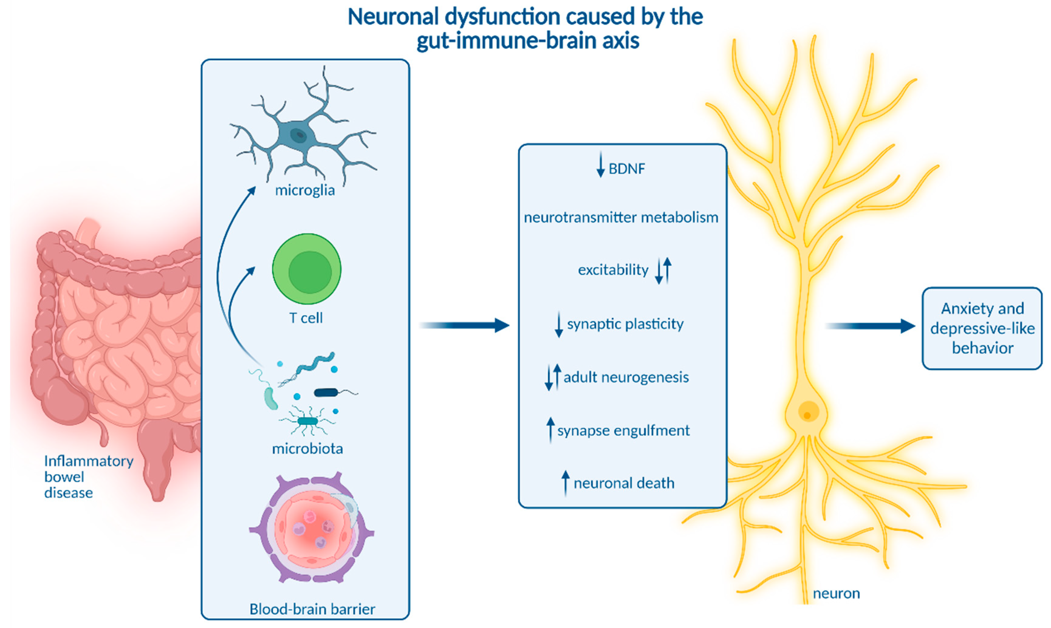
3. Impact of Microbiota on Neuroinflammation and Neuropsychiatric Disease

Having highlighted different routes of transmission from chronic gastrointestinal inflammation into the systemic circulation and into the CNS as well as consecutive neuronal dysfunction and behavioral impairment, researchers will shed some light on the role of gut microbiota in the gut-immune-brain interplay. The intestinal microbiota is substantially involved in gastrointestinal inflammation and neuropsychiatric diseases [83][84][85][86]. Therefore, microbiota and their metabolites are emerging key players in the gut-immune brain axis during IBD. This notion has encouraged several probiotic treatment approaches. Indeed, application of probiotic bacterial strains alleviated DSS-induced colitis, reduced systemic and CNS cytokine levels, induced micro-RNA expression related to restoring inflammation-associated microbiota dysbiosis, and improved depressive and anxiety-like behavior [87][88][89][90]. However, it is unclear whether effects of probiotic treatments were solely indirect based on the reduction in gut inflammation or also directly interfered with inflammatory gut-to-brain communication or neurons. It is important to note, that changes in the gut microbiota may be cause or consequence of intestinal inflammation and modulate neuropsychiatric symptoms via affecting neuroinflammation, but also exert direct effects on neurons. Researchers will therefore highlight different modes of action which affect the CNS during IBD-related dysbiosis.
First, microbiota and microbiota-derived molecules actively shape the CNS immune landscape, and the presence of a complex microbiota is essential for microglia activation and function [89][91][92][93]. Among other CNS-associated myeloid cell types, commensal microbiota strongly influence CP macrophages, while their impact on perivascular and meningeal macrophages is moderate [89]. Different microbial metabolites were described to modulate microglia. First, microbiota-derived SCFAs signaling via the free fatty acid receptor 2 (Ffar2) were implicated in microglial maturation and function [92]. A recent differential analysis of distinct SCFAs revealed acetate to be a key regulator of microglial metabolism and phagocytosis [92]. In the context of multiple sclerosis, the SCFA propionate was shown to induce regulatory Treg activation, whereas pro-inflammatory Th1 and Th17 responses were diminished [94]. Moreover, bacterial metabolites of dietary tryptophan signal via the aryl hydrocarbon receptor to reduce microglial inflammatory activation of astrocytes [95]. In the context of IBD, UC patients with comorbid depression or anxiety showed a distinct intestinal bacterial profile linked to reduced blood levels of the metabolites 2ʹ-deoxy-D-ribose and L-pipecolic acid [83]. Intriguingly, substitution of these metabolites in mice alleviated DSS-induced colitis as well as cytokine levels in the blood and brain, but also ameliorated anxiety and depressive-like behavior [83]. These findings suggest a role of microbial metabolites in gut-immune-brain communication during IBD. Future studies addressing the modulation of neuroinflammation and behavior in mouse models for experimental colitis by the above-mentioned metabolites will improve people's understanding of microbial influence on IBD-related CNS morbidity.
In addition to gut bacteria, mucosal fungi were shown to promote Th17 cell activation, which promotes social behavior by direct signaling to neurons via Il-17 [96]. Collectively, intestinal microbiota are able to modulate systemic and CNS immune responses in IBD and could thereby contribute to IBD-related CNS comorbidity.
Besides indirect immune-mediated effects of microbiota and derived metabolites, they have the potential to exert immune-independent effects on neurons via different pathways. Gut bacteria-derived outer membrane vesicles (OMVs) can cross the intestinal barrier and even the BBB, enabling a shuttled transfer of microbial bioactive molecules to the brain [97]. Interestingly, bacterial OMVs were differentially taken up by neurons with a regionally distinct affinity [97]. Though their influence on neuronal function is unknown, bacteria-derived OMV cargo uptake may be enhanced in IBD due to gut barrier and BBB dysfunction and may be a complementary part of the gut-to-brain communication.
Moreover, intestinal microbiota are involved in neurotransmitter metabolism. Expansion of Bacteroides species contributes to depressive-like behavior and impaired hippocampal neurogenesis by regulating tryptophan and neurotransmitter metabolism [86]. Interestingly, Bacteroides species were also linked to the development of colitis [98]. Apart from that, gut microbiota are a major source of the neurotransmitter serotonin. Reduced serotonin abundance as a potential pathogenic mechanism driving depression may be caused by microbial dysbiosis and impaired serotonin production in the gastrointestinal tract [99].
In addition, gut microbiota influence the expression of micro-RNA (miRNA) in the gut and in different brain regions, which is associated with depression and anxiety in mice [100][101]. Interestingly, differential miRNA expression associated with microbiota dysbiosis distinguishes IBD patients and healthy individuals. MiRNAs are thus suggested as biomarkers and promising targets to treat intestinal inflammation [102].
Compromised gut microbiota in IBD might therefore contribute to the pathophysiology of concomitant anxiety and depression via affecting neurotransmitter homeostasis and miRNA expression.
Besides prototypical neurotransmitters and miRNA, microbial metabolites have the potential to actively signal to neurons. The phenolic compounds phenyl sulfate, pyrocatechol sulfate, and 3-(3-sulfooxyphenyl)propanoic acid as well as indoxyl sulfate were implicated in synaptic remodeling and fear extinction learning [103]. Moreover, δ-valerobetaine produced by diverse bacterial species modulates inhibitory synaptic transmission and neuronal network activity independent of microglia, preventing age-related decline in cognitive function [104]. SCFAs are able to act locally in the ENS and enhance enteric neuronal survival and neurogenesis potentially acting via the 5-hydroxytryptamine type 4 (5-HT4) receptor [105]. SCFAs were also found in the brain, but their levels were not altered during chronic DSS-induced colitis [18].
In acute DSS-induced colitis, bacteria of the Lachnospiraceae, Ruminococcaceae and Muribaculaceae families were observed to be associated with anxiety and depressive-like behavior. Fecal microbial transfer of colitis-characteristic gut microbiota composition into germ free or antibiotic-treated mice was sufficient to transmit behavioral abnormalities without inducing neuroinflammation, suggesting that immune-independent mechanisms promote microbiota-mediated behavioral alterations in IBD [84].
Altogether, gut microbiota likely contribute to the development of neuroinflammation and neuropsychiatric symptoms in IBD. Future studies will need to identify essential bacterial strains and the mechanisms they employ to alter neuroinflammation or directly modify neuronal function specifically during chronic gastrointestinal inflammation.

References

  1. Breit, S.; Kupferberg, A.; Rogler, G.; Hasler, G. Vagus Nerve as Modulator of the Brain–Gut Axis in Psychiatric and Inflammatory Disorders. Front. Psychiatry 2018, 9, 44.
  2. Ichiki, T.; Wang, T.; Kennedy, A.; Pool, A.-H.; Ebisu, H.; Anderson, D.J.; Oka, Y. Sensory representation and detection mechanisms of gut osmolality change. Nature 2022, 602, 468–474.
  3. Goswami, C.; Iwasaki, Y.; Yada, T. Short-chain fatty acids suppress food intake by activating vagal afferent neurons. J. Nutr. Biochem. 2018, 57, 130–135.
  4. Browning, K.N.; Verheijden, S.; Boeckxstaens, G.E. The Vagus Nerve in Appetite Regulation, Mood, and Intestinal Inflammation. Gastroenterology 2017, 152, 730–744.
  5. Steinberg, B.E.; Silverman, H.A.; Robbiati, S.; Gunasekaran, M.K.; Tsaava, T.; Battinelli, E.; Stiegler, A.; Bouton, C.E.; Chavan, S.S.; Tracey, K.J.; et al. Cytokine-specific Neurograms in the Sensory Vagus Nerve. Bioelectron. Med. 2016, 3, 7–17.
  6. Watkins, L.R.; Goehler, L.E.; Relton, J.K.; Tartaglia, N.; Silbert, L.; Martin, D.; Maier, S.F. Blockade of interleukin-1 induced hyperthermia by subdiaphragmatic vagotomy: Evidence for vagal mediation of immune-brain communication. Neurosci. Lett. 1995, 183, 27–31.
  7. Bercik, P.; Verdu, E.F.; Foster, J.A.; Macri, J.; Potter, M.; Huang, X.; Malinowski, P.; Jackson, W.; Blennerhassett, P.; Neufeld, K.A.; et al. Chronic Gastrointestinal Inflammation Induces Anxiety-Like Behavior and Alters Central Nervous System Biochemistry in Mice. Gastroenterology 2010, 139, 2102–2112.e1.
  8. Sun, P.; Zhou, K.; Wang, S.; Li, P.; Chen, S.; Lin, G.; Zhao, Y.; Wang, T. Involvement of MAPK/NF-κB signaling in the activation of the cholinergic anti-inflammatory pathway in experimental colitis by chronic vagus nerve stimulation. PLoS ONE 2013, 8, e69424.
  9. Sinniger, V.; Pellissier, S.; Fauvelle, F.; Trocmé, C.; Hoffmann, D.; Vercueil, L.; Cracowski, J.L.; David, O.; Bonaz, B. A 12-month pilot study outcomes of vagus nerve stimulation in Crohn’s disease. Neurogastroenterol. Motil. 2020, 32, e13911.
  10. Ibeakanma, C.; Vanner, S.J. TNI± is a key mediator of the pronociceptive effects of mucosal supernatant from human ulcerative colitis on colonic DRG neurons. Gut 2010, 59, 612–621.
  11. Hess, A.; Roesch, J.; Saake, M.; Sergeeva, M.; Hirschmann, S.; Neumann, H.; Dörfler, A.; Neurath, M.F.; Atreya, R. Functional Brain Imaging Reveals Rapid Blockade of Abdominal Pain Response Upon Anti-TNF Therapy in Crohn’s Disease. Gastroenterology 2015, 149, 864–866.
  12. Engelhardt, B.; Vajkoczy, P.; Weller, R.O. The movers and shapers in immune privilege of the CNS. Nat. Immunol. 2017, 18, 123–131.
  13. Galea, I. The blood–brain barrier in systemic infection and inflammation. Cell. Mol. Immunol. 2021, 18, 2489–2501.
  14. Spadoni, I.; Zagato, E.; Bertocchi, A.; Paolinelli, R.; Hot, E.; Di Sabatino, A.; Caprioli, F.; Bottiglieri, L.; Oldani, A.; Viale, G.; et al. A gut-vascular barrier controls the systemic dissemination of bacteria. Science 2015, 350, 830–834.
  15. Varatharaj, A.; Galea, I. The blood-brain barrier in systemic inflammation. Brain Behav. Immun. 2017, 60, 1–12.
  16. Han, Y.; Zhao, T.; Cheng, X.; Zhao, M.; Gong, S.-H.; Zhao, Y.-Q.; Wu, H.-T.; Fan, M.; Zhu, L.-L. Cortical Inflammation is Increased in a DSS-Induced Colitis Mouse Model. Neurosci. Bull. 2018, 34, 1058–1066.
  17. Han, Y.; Ding, L.; Cheng, X.; Zhao, M.; Zhao, T.; Guo, L.; Li, X.; Geng, Y.; Fan, M.; Liao, H.; et al. Hypoxia Augments Cerebral Inflammation in a Dextran Sulfate Sodium-Induced Colitis Mouse Model. Front. Cell. Neurosci. 2020, 14, 611764.
  18. Mitchell, J.; Kim, S.J.; Howe, C.; Lee, S.; Her, J.Y.; Patel, M.; Kim, G.; Lee, J.; Im, E.; Rhee, S.H. Chronic Intestinal Inflammation Suppresses Brain Activity by Inducing Neuroinflammation in Mice. Am. J. Pathol. 2022, 192, 72–86.
  19. Carloni, S.; Bertocchi, A.; Mancinelli, S.; Bellini, M.; Erreni, M.; Borreca, A.; Braga, D.; Giugliano, S.; Mozzarelli, A.M.; Manganaro, D.; et al. Identification of a choroid plexus vascular barrier closing during intestinal inflammation. Science 2021, 374, 439–448.
  20. Zhao, X.; Liang, P.; Liu, J.; Jiang, H.; Fan, X.; Chen, G.; Zhou, C. Elevation of arachidonoylethanolamide levels by activation of the endocannabinoid system protects against colitis and ameliorates remote organ lesions in mice. Exp. Ther. Med. 2017, 14, 5664–5670.
  21. Hathaway, C.A.; Appleyard, C.B.; Percy, W.H.; Williams, J.L. Experimental colitis increases blood-brain barrier permeability in rabbits. Am. J. Physiol.-Gastrointest. Liver Physiol. 1999, 276, G1174–G1180.
  22. Natah, S.S.; Mouihate, A.; Pittman, Q.J.; Sharkey, K.A. Disruption of the blood-brain barrier during TNBS colitis. Neurogastroenterol. Motil. 2005, 17, 433–446.
  23. Barnes, S.E.; Zera, K.A.; Ivison, G.T.; Buckwalter, M.S.; Engleman, E.G. Brain profiling in murine colitis and human epilepsy reveals neutrophils and TNFα as mediators of neuronal hyperexcitability. J. Neuroinflamm. 2021, 18, 199.
  24. Talley, S.; Valiauga, R.; Anderson, L.; Cannon, A.R.; Choudhry, M.A.; Campbell, E.M. DSS-induced inflammation in the colon drives a proinflammatory signature in the brain that is ameliorated by prophylactic treatment with the S100A9 inhibitor paquinimod. J. Neuroinflamm. 2021, 18, 263.
  25. Blank, T.; Detje, C.N.; Spieß, A.; Hagemeyer, N.; Brendecke, S.M.; Wolfart, J.; Staszewski, O.; Zöller, T.; Papageorgiou, I.; Schneider, J.; et al. Brain Endothelial- and Epithelial-Specific Interferon Receptor Chain 1 Drives Virus-Induced Sickness Behavior and Cognitive Impairment. Immunity 2016, 44, 901–912.
  26. Yousef, H.; Czupalla, C.J.; Lee, D.; Chen, M.B.; Burke, A.N.; Zera, K.A.; Zandstra, J.; Berber, E.; Lehallier, B.; Mathur, V.; et al. Aged blood impairs hippocampal neural precursor activity and activates microglia via brain endothelial cell VCAM1. Nat. Med. 2019, 25, 988–1000.
  27. Althubaity, N.; Schubert, J.; Martins, D.; Yousaf, T.; Nettis, M.A.; Mondelli, V.; Pariante, C.; Harrison, N.A.; Bullmore, E.T.; Dima, D.; et al. Choroid plexus enlargement is associated with neuroinflammation and reduction of blood brain barrier permeability in depression. NeuroImage Clin. 2022, 33, 102926.
  28. Balusu, S.; Van Wonterghem, E.; De Rycke, R.; Raemdonck, K.; Stremersch, S.; Gevaert, K.; Brkic, M.; Demeestere, D.; Vanhooren, V.; Hendrix, A.; et al. Identification of a novel mechanism of blood–brain communication during peripheral inflammation via choroid plexus—derived extracellular vesicles. EMBO Mol. Med. 2016, 8, 1162–1183.
  29. Van Hove, H.; Martens, L.; Scheyltjens, I.; De Vlaminck, K.; Antunes, A.R.P.; De Prijck, S.; Vandamme, N.; De Schepper, S.; Van Isterdael, G.; Scott, C.L.; et al. A single-cell atlas of mouse brain macrophages reveals unique transcriptional identities shaped by ontogeny and tissue environment. Nat. Neurosci. 2019, 22, 1021–1035.
  30. Baruch, K.; Deczkowska, A.; David, E.; Castellano, J.M.; Miller, O.; Kertser, A.; Berkutzki, T.; Barnett-Itzhaki, Z.; Bezalel, D.; Wyss-Coray, T.; et al. Aging-induced type I interferon response at the choroid plexus negatively affects brain function. Science 2014, 346, 89–93.
  31. Dempsey, E.; Abautret-Daly, Á.; Docherty, N.G.; Medina, C.; Harkin, A. Persistent central inflammation and region specific cellular activation accompany depression- and anxiety-like behaviours during the resolution phase of experimental colitis. Brain Behav. Immun. 2019, 80, 616–632.
  32. Filiano, A.J.; Xu, Y.; Tustison, N.; Marsh, R.L.; Baker, W.; Smirnov, I.; Overall, C.C.; Gadani, S.P.; Turner, S.; Weng, Z.; et al. Unexpected role of interferon-γ in regulating neuronal connectivity and social behaviour. Nature 2016, 535, 425–429.
  33. De Lima, K.A.; Rustenhoven, J.; Da Mesquita, S.; Wall, M.; Salvador, A.F.; Smirnov, I.; Cebinelli, G.M.; Mamuladze, T.; Baker, W.; Papadopoulos, Z.; et al. Meningeal γδ T cells regulate anxiety-like behavior via IL-17a signaling in neurons. Nat. Immunol. 2020, 21, 1421–1429.
  34. Brea, D.; Poon, C.; Benakis, C.; Lubitz, G.; Murphy, M.; Iadecola, C.; Anrather, J. Stroke affects intestinal immune cell trafficking to the central nervous system. Brain Behav. Immun. 2021, 96, 295–302.
  35. Kivisäkk, P.; Tucky, B.; Wei, T.; Campbell, J.J.; Ransohoff, R.M. Human cerebrospinal fluid contains CD4+ memory T cells expressing gut- or skin-specific trafficking determinants: Relevance for immunotherapy. BMC Immunol. 2006, 7, 14.
  36. He, X.-F.; Li, L.-L.; Xian, W.-B.; Li, M.-Y.; Zhang, L.-Y.; Xu, J.-H.; Pei, Z.; Zheng, H.-Q.; Hu, X.-Q. Chronic colitis exacerbates NLRP3-dependent neuroinflammation and cognitive impairment in middle-aged brain. J. Neuroinflamm. 2021, 18, 153.
  37. Badimon, A.; Strasburger, H.J.; Ayata, P.; Chen, X.; Nair, A.; Ikegami, A.; Hwang, P.; Chan, A.T.; Graves, S.M.; Uweru, J.O.; et al. Negative feedback control of neuronal activity by microglia. Nature 2020, 586, 417–423.
  38. Parkhurst, C.N.; Yang, G.; Ninan, I.; Savas, J.N.; Yates, J.R., 3rd; Lafaille, J.J.; Hempstead, B.L.; Littman, D.R.; Gan, W.B. Microglia promote learning-dependent synapse formation through brain-derived neurotrophic factor. Cell 2013, 155, 1596–1609.
  39. Pasciuto, E.; Burton, O.T.; Roca, C.P.; Lagou, V.; Rajan, W.D.; Theys, T.; Mancuso, R.; Tito, R.Y.; Kouser, L.; Callaerts-Vegh, Z.; et al. Microglia Require CD4 T Cells to Complete the Fetal-to-Adult Transition. Cell 2020, 182, 625–640.e24.
  40. Klawonn, A.M.; Fritz, M.; Castany, S.; Pignatelli, M.; Canal, C.; Similä, F.; Tejeda, H.A.; Levinsson, J.; Jaarola, M.; Jakobsson, J.; et al. Microglial activation elicits a negative affective state through prostaglandin-mediated modulation of striatal neurons. Immunity 2021, 54, 225–234.e6.
  41. Cao, P.; Chen, C.; Liu, A.; Shan, Q.; Zhu, X.; Jia, C.; Peng, X.; Zhang, M.; Farzinpour, Z.; Zhou, W.; et al. Early-life inflammation promotes depressive symptoms in adolescence via microglial engulfment of dendritic spines. Neuron 2021, 109, 2573–2589.e9.
  42. Wohleb, E.S.; Terwilliger, R.; Duman, C.H.; Duman, R.S. Stress-Induced Neuronal Colony Stimulating Factor 1 Provokes Microglia-Mediated Neuronal Remodeling and Depressive-like Behavior. Biol. Psychiatry 2018, 83, 38–49.
  43. Ji, C.; Tang, Y.; Zhang, Y.; Li, C.; Liang, H.; Ding, L.; Xia, X.; Xiong, L.; Qi, X.-R.; Zheng, J.C. Microglial glutaminase 1 deficiency mitigates neuroinflammation associated depression. Brain Behav. Immun. 2022, 99, 231–245.
  44. Li, W.; Ali, T.; He, K.; Liu, Z.; Shah, F.A.; Ren, Q.; Liu, Y.; Jiang, A.; Li, S. Ibrutinib alleviates LPS-induced neuroinflammation and synaptic defects in a mouse model of depression. Brain Behav. Immun. 2021, 92, 10–24.
  45. Kaufmann, F.N.; Costa, A.P.; Ghisleni, G.; Diaz, A.; Rodrigues, A.L.; Peluffo, H.; Kaster, M.P. NLRP3 inflammasome-driven pathways in depression: Clinical and preclinical findings. Brain Behav. Immun. 2017, 64, 367–383.
  46. Dang, R.; Wang, M.; Li, X.; Wang, H.; Liu, L.; Wu, Q.; Zhao, J.; Ji, P.; Zhong, L.; Licinio, J.; et al. Edaravone ameliorates depressive and anxiety-like behaviors via Sirt1/Nrf2/HO-1/Gpx4 pathway. J. Neuroinflamm. 2022, 19, 41.
  47. Ali, T.; Hao, Q.; Ullah, N.; Rahman, S.U.; Shah, F.A.; He, K.; Zheng, C.; Li, W.; Murtaza, I.; Li, Y.; et al. Melatonin Act as an Antidepressant via Attenuation of Neuroinflammation by Targeting Sirt1/Nrf2/HO-1 Signaling. Front. Mol. Neurosci. 2020, 13, 96.
  48. Dudek, K.A.; Dion-Albert, L.; Lebel, M.; LeClair, K.; Labrecque, S.; Tuck, E.; Perez, C.F.; Golden, S.A.; Tamminga, C.; Turecki, G.; et al. Molecular adaptations of the blood-brain barrier promote stress resilience vs. depression. Proc. Natl. Acad. Sci. USA 2020, 117, 3326–3336.
  49. Menard, C.; Pfau, M.L.; Hodes, G.E.; Kana, V.; Wang, V.X.; Bouchard, S.; Takahashi, A.; Flanigan, M.E.; Aleyasin, H.; LeClair, K.B.; et al. Social stress induces neurovascular pathology promoting depression. Nat. Neurosci. 2017, 20, 1752–1760.
  50. McKim, D.B.; Weber, M.D.; Niraula, A.; Sawicki, C.M.; Liu, X.; Jarrett, B.L.; Ramirez-Chan, K.; Wang, Y.; Roeth, R.M.; Sucaldito, A.D.; et al. Microglial recruitment of IL-1β-producing monocytes to brain endothelium causes stress-induced anxiety. Mol. Psychiatry 2018, 23, 1421–1431.
  51. Zheng, Z.-H.; Tu, J.-L.; Li, X.-H.; Hua, Q.; Liu, W.-Z.; Liu, Y.; Pan, B.-X.; Hu, P.; Zhang, W.-H. Neuroinflammation induces anxiety- and depressive-like behavior by modulating neuronal plasticity in the basolateral amygdala. Brain Behav. Immun. 2021, 91, 505–518.
  52. Riazi, K.; Galic, M.A.; Kuzmiski, J.B.; Ho, W.; Sharkey, K.A.; Pittman, Q.J. Microglial activation and TNFalpha production mediate altered CNS excitability following peripheral inflammation. Proc. Natl. Acad. Sci. USA 2008, 105, 17151–17156.
  53. Craig, C.F.; Filippone, R.T.; Stavely, R.; Bornstein, J.C.; Apostolopoulos, V.; Nurgali, K. Neuroinflammation as an etiological trigger for depression comorbid with inflammatory bowel disease. J. Neuroinflamm. 2022, 19, 4.
  54. Ghia, J.E.; Li, N.; Wang, H.; Collins, M.; Deng, Y.; El-Sharkawy, R.T.; Côté, F.; Mallet, J.; Khan, W.I. Serotonin has a key role in pathogenesis of experimental colitis. Gastroenterology 2009, 137, 1649–1660.
  55. Shajib, M.S.; Baranov, A.; Khan, W.I. Diverse Effects of Gut-Derived Serotonin in Intestinal Inflammation. ACS Chem. Neurosci. 2017, 8, 920–931.
  56. Kristensen, M.S.; Kjærulff, T.M.; Ersbøll, A.K.; Green, A.; Hallas, J.; Thygesen, L.C. The Influence of Antidepressants on the Disease Course among Patients with Crohn’s Disease and Ulcerative Colitis-A Danish Nationwide Register-Based Cohort Study. Inflamm. Bowel Dis. 2019, 25, 886–893.
  57. Chen, L.-M.; Bao, C.-H.; Wu, Y.; Liang, S.-H.; Wang, D.; Wu, L.-Y.; Huang, Y.; Liu, H.-R.; Wu, H.-G. Tryptophan-kynurenine metabolism: A link between the gut and brain for depression in inflammatory bowel disease. J. Neuroinflamm. 2021, 18, 135.
  58. Sroor, H.M.; Hassan, A.M.; Zenz, G.; Valadez-Cosmes, P.; Farzi, A.; Holzer, P.; El-Sharif, A.; Gomaa, F.A.-Z.M.; Kargl, J.; Reichmann, F. Experimental colitis reduces microglial cell activation in the mouse brain without affecting microglial cell numbers. Sci. Rep. 2019, 9, 20217.
  59. Süß, P.; Schlachetzki, J.C.M. Microglia in Alzheimer’s Disease. Curr. Alzheimer Res. 2020, 17, 29–43.
  60. Crider, A.; Feng, T.; Pandya, C.D.; Davis, T.; Nair, A.; Ahmed, A.O.; Baban, B.; Turecki, G.; Pillai, A. Complement component 3a receptor deficiency attenuates chronic stress-induced monocyte infiltration and depressive-like behavior. Brain Behav. Immun. 2018, 70, 246–256.
  61. Bolton, J.L.; Short, A.K.; Othy, S.; Kooiker, C.L.; Shao, M.; Gunn, B.G.; Beck, J.; Bai, X.; Law, S.M.; Savage, J.C.; et al. Early stress-induced impaired microglial pruning of excitatory synapses on immature CRH-expressing neurons provokes aberrant adult stress responses. Cell Rep. 2022, 38, 110600.
  62. Vadodaria, K.C.; Gage, F.H. SnapShot: Adult Hippocampal Neurogenesis. Cell 2014, 156, 1114–1114.e1.
  63. Jacobs, B.L.; van Praag, H.; Gage, F.H. Adult brain neurogenesis and psychiatry: A novel theory of depression. Mol. Psychiatry 2000, 5, 262–269.
  64. Toda, T.; Parylak, S.L.; Linker, S.B.; Gage, F.H. The role of adult hippocampal neurogenesis in brain health and disease. Mol. Psychiatry 2019, 24, 67–87.
  65. Sierra, A.; Encinas, J.M.; Deudero, J.J.P.; Chancey, J.; Enikolopov, G.; Wadiche, L.; Tsirka, S.E.; Maletic-Savatic, M. Microglia Shape Adult Hippocampal Neurogenesis through Apoptosis-Coupled Phagocytosis. Cell Stem Cell 2010, 7, 483–495.
  66. Iosif, R.E.; Ekdahl, C.T.; Ahlenius, H.; Pronk, C.J.H.; Bonde, S.; Kokaia, Z.; Jacobsen, S.E.W.; Lindvall, O. Tumor Necrosis Factor Receptor 1 Is a Negative Regulator of Progenitor Proliferation in Adult Hippocampal Neurogenesis. J. Neurosci. 2006, 26, 9703–9712.
  67. Goshen, I.; Kreisel, T.; Ben-Menachem-Zidon, O.; Licht, T.; Weidenfeld, J.; Ben-Hur, T.; Yirmiya, R. Brain interleukin-1 mediates chronic stress-induced depression in mice via adrenocortical activation and hippocampal neurogenesis suppression. Mol. Psychiatry 2007, 13, 717–728.
  68. Monje, M.L.; Toda, H.; Palmer, T.D. Inflammatory Blockade Restores Adult Hippocampal Neurogenesis. Science 2003, 302, 1760–1765.
  69. Korolkova, O.Y.; Myers, J.N.; Pellom, S.T.; Wang, L.; M’Koma, A.E. Characterization of Serum Cytokine Profile in Predominantly Colonic Inflammatory Bowel Disease to Delineate Ulcerative and Crohn’s Colitides. Clin. Med. Insights Gastroenterol. 2015, 8, 29–44.
  70. Tatsuki, M.; Hatori, R.; Nakazawa, T.; Ishige, T.; Hara, T.; Kagimoto, S.; Tomomasa, T.; Arakawa, H.; Takizawa, T. Serological cytokine signature in paediatric patients with inflammatory bowel disease impacts diagnosis. Sci. Rep. 2020, 10, 14638.
  71. Villeda, S.A.; Luo, J.; Mosher, K.I.; Zou, B.; Britschgi, M.; Bieri, G.; Stan, T.M.; Fainberg, N.; Ding, Z.; Eggel, A.; et al. The ageing systemic milieu negatively regulates neurogenesis and cognitive function. Nature 2011, 477, 90–94.
  72. Gampierakis, I.-A.; Koutmani, Y.; Semitekolou, M.; Morianos, I.; Polissidis, A.; Katsouda, A.; Charalampopoulos, I.; Xanthou, G.; Gravanis, A.; Karalis, K.P. Hippocampal neural stem cells and microglia response to experimental inflammatory bowel disease (IBD). Mol. Psychiatry 2021, 26, 1248–1263.
  73. Salvo, E.; Stokes, P.; Keogh, C.E.; Brust-Mascher, I.; Hennessey, C.; Knotts, T.A.; Sladek, J.A.; Rude, K.M.; Swedek, M.; Rabasa, G.; et al. A murine model of pediatric inflammatory bowel disease causes microbiota-gut-brain axis deficits in adulthood. Am. J. Physiol. Gastrointest. Liver Physiol. 2020, 319, G361–G374.
  74. Zonis, S.; Pechnick, R.N.; Ljubimov, V.A.; Mahgerefteh, M.; Wawrowsky, K.; Michelsen, K.S.; Chesnokova, V. Chronic intestinal inflammation alters hippocampal neurogenesis. J. Neuroinflamm. 2015, 12, 65.
  75. Nakagawasai, O.; Yamada, K.; Takahashi, K.; Odaira, T.; Sakuma, W.; Ishizawa, D.; Takahashi, N.; Onuma, K.; Hozumi, C.; Nemoto, W.; et al. Liver hydrolysate prevents depressive-like behavior in an animal model of colitis: Involvement of hippocampal neurogenesis via the AMPK/BDNF pathway. Behav. Brain Res. 2020, 390, 112640.
  76. Takahashi, K.; Kurokawa, K.; Miyagawa, K.; Mochida-Saito, A.; Nemoto, Y.; Iwasa, H.; Nakagawasai, O.; Tadano, T.; Takeda, H.; Tsuji, M. Antidementia effects of Enterococcus faecalis 2001 are associated with enhancement of hippocampal neurogenesis via the ERK-CREB-BDNF pathway in olfactory bulbectomized mice. Physiol. Behav. 2020, 223, 112997.
  77. Park, H.; Poo, M.-M. Neurotrophin regulation of neural circuit development and function. Nat. Rev. Neurosci. 2013, 14, 7–23.
  78. Casarotto, P.C.; Girych, M.; Fred, S.M.; Kovaleva, V.; Moliner, R.; Enkavi, G.; Biojone, C.; Cannarozzo, C.; Sahu, M.P.; Kaurinkoski, K.; et al. Antidepressant drugs act by directly binding to TRKB neurotrophin receptors. Cell 2021, 184, 1299–1313.e19.
  79. Barrientos, R.; Sprunger, D.; Campeau, S.; Higgins, E.; Watkins, L.; Rudy, J.; Maier, S. Brain-derived neurotrophic factor mRNA downregulation produced by social isolation is blocked by intrahippocampal interleukin-1 receptor antagonist. Neuroscience 2003, 121, 847–853.
  80. Zhuang, X.; Zhan, B.; Jia, Y.; Li, C.; Wu, N.; Zhao, M.; Chen, N.; Guo, Y.; Du, Y.; Zhang, Y.; et al. IL-33 in the basolateral amygdala integrates neuroinflammation into anxiogenic circuits via modulating BDNF expression. Brain Behav. Immun. 2022, 102, 98–109.
  81. Haj-Mirzaian, A.; Amiri, S.; Amini-Khoei, H.; Hosseini, M.-J.; Haj-Mirzaian, A.; Momeny, M.; Rahimi-Balaei, M.; Dehpour, A.R. Anxiety- and Depressive-Like Behaviors are Associated with Altered Hippocampal Energy and Inflammatory Status in a Mouse Model of Crohn’s Disease. Neuroscience 2017, 366, 124–137.
  82. Heydarpour, P.; Rahimian, R.; Fakhfouri, G.; Khoshkish, S.; Fakhraei, N.; Salehi-Sadaghiani, M.; Wang, H.; Abbasi, A.; Dehpour, A.R.; Ghia, J.E. Behavioral despair associated with a mouse model of Crohn’s disease: Role of nitric oxide pathway. Prog. Neuropsychopharmacol. Biol. Psychiatry 2016, 64, 131–141.
  83. Yuan, X.; Chen, B.; Duan, Z.; Xia, Z.; Ding, Y.; Chen, T.; Liu, H.; Wang, B.; Yang, B.; Wang, X.; et al. Depression and anxiety in patients with active ulcerative colitis: Crosstalk of gut microbiota, metabolomics and proteomics. Gut Microbes 2021, 13, 1987779.
  84. Vicentini, F.A.; Szamosi, J.C.; Rossi, L.; Griffin, L.; Nieves, K.; Bihan, D.; Lewis, I.A.; Pittman, Q.J.; Swain, M.G.; Surette, M.G.; et al. Colitis-associated microbiota drives changes in behaviour in male mice in the absence of inflammation. Brain Behav. Immun. 2022, 102, 266–278.
  85. Valles-Colomer, M.; Falony, G.; Darzi, Y.; Tigchelaar, E.F.; Wang, J.; Tito, R.Y.; Schiweck, C.; Kurilshikov, A.; Joossens, M.; Wijmenga, C.; et al. The neuroactive potential of the human gut microbiota in quality of life and depression. Nat. Microbiol. 2019, 4, 623–632.
  86. Zhang, Y.; Fan, Q.; Hou, Y.; Zhang, X.; Yin, Z.; Cai, X.; Wei, W.; Wang, J.; He, D.; Wang, G.; et al. Bacteroides species differentially modulate depression-like behavior via gut-brain metabolic signaling. Brain Behav. Immun. 2022, 102, 11–22.
  87. Takahashi, K.; Nakagawasai, O.; Nemoto, W.; Odaira, T.; Sakuma, W.; Onogi, H.; Nishijima, H.; Furihata, R.; Nemoto, Y.; Iwasa, H.; et al. Effect of Enterococcus faecalis 2001 on colitis and depressive-like behavior in dextran sulfate sodium-treated mice: Involvement of the brain–gut axis. J. Neuroinflamm. 2019, 16, 201.
  88. Emge, J.R.; Huynh, K.; Miller, E.N.; Kaur, M.; Reardon, C.; Barrett, K.E.; Gareau, M.G. Modulation of the microbiota-gut-brain axis by probiotics in a murine model of inflammatory bowel disease. Am. J. Physiol. Liver Physiol. 2016, 310, G989–G998.
  89. Sankowski, R.; Ahmari, J.; Mezö, C.; de Angelis, A.L.H.; Fuchs, V.; Utermöhlen, O.; Buch, T.; Blank, T.; de Agüero, M.G.; Macpherson, A.J.; et al. Commensal microbiota divergently affect myeloid subsets in the mammalian central nervous system during homeostasis and disease. EMBO J. 2021, 40, e108605.
  90. Rodríguez-Nogales, A.; Algieri, F.; Garrido-Mesa, J.; Vezza, T.; Utrilla, M.P.; Chueca, N.; García, F.; Rodríguez-Cabezas, M.E.; Gálvez, J. Intestinal anti-inflammatory effect of the probiotic Saccharomyces boulardii in DSS-induced colitis in mice: Impact on microRNAs expression and gut microbiota composition. J. Nutr. Biochem. 2018, 61, 129–139.
  91. Erny, D.; Dokalis, N.; Mezö, C.; Castoldi, A.; Mossad, O.; Staszewski, O.; Frosch, M.; Villa, M.; Fuchs, V.; Mayer, A.; et al. Microbiota-derived acetate enables the metabolic fitness of the brain innate immune system during health and disease. Cell Metab. 2021, 33, 2260–2276.e7.
  92. Erny, D.; Hrabě de Angelis, A.L.; Jaitin, D.; Wieghofer, P.; Staszewski, O.; David, E.; Keren-Shaul, H.; Mahlakoiv, T.; Jakobshagen, K.; Buch, T.; et al. Host microbiota constantly control maturation and function of microglia in the CNS. Nat. Neurosci. 2015, 18, 965–977.
  93. Mossad, O.; Batut, B.; Yilmaz, B.; Dokalis, N.; Mezö, C.; Nent, E.; Nabavi, L.S.; Mayer, M.; Maron, F.J.M.; Buescher, J.M.; et al. Gut microbiota drives age-related oxidative stress and mitochondrial damage in microglia via the metabolite N6-carboxymethyllysine. Nat. Neurosci. 2022, 25, 295–305.
  94. Duscha, A.; Gisevius, B.; Hirschberg, S.; Yissachar, N.; Stangl, G.I.; Eilers, E.; Bader, V.; Haase, S.; Kaisler, J.; David, C.; et al. Propionic Acid Shapes the Multiple Sclerosis Disease Course by an Immunomodulatory Mechanism. Cell 2020, 180, 1067–1080.e16.
  95. Rothhammer, V.; Borucki, D.M.; Tjon, E.C.; Takenaka, M.C.; Chao, C.C.; Ardura-Fabregat, A.; de Lima, K.A.; Gutiérrez-Vázquez, C.; Hewson, P.; Staszewski, O.; et al. Microglial control of astrocytes in response to microbial metabolites. Nature 2018, 557, 724–728.
  96. Leonardi, I.; Gao, I.H.; Lin, W.-Y.; Allen, M.; Li, X.V.; Fiers, W.D.; De Celie, M.B.; Putzel, G.G.; Yantiss, R.K.; Johncilla, M.; et al. Mucosal fungi promote gut barrier function and social behavior via Type 17 immunity. Cell 2022, 185, 831–846.e14.
  97. Bittel, M.; Reichert, P.; Sarfati, I.; Dressel, A.; Leikam, S.; Uderhardt, S.; Stolzer, I.; Phu, T.A.; Ng, M.; Vu, N.K.; et al. Visualizing transfer of microbial biomolecules by outer membrane vesicles in microbe-host-communication in vivo. J. Extracell. Vesicles 2021, 10, e12159.
  98. Bloom, S.M.; Bijanki, V.N.; Nava, G.M.; Sun, L.; Malvin, N.P.; Donermeyer, D.L.; Dunne, W.M.; Allen, P.M.; Stappenbeck, T.S. Commensal Bacteroides Species Induce Colitis in Host-Genotype-Specific Fashion in a Mouse Model of Inflammatory Bowel Disease. Cell Host Microbe 2011, 9, 390–403.
  99. Dicks, L.M.T.; Hurn, D.; Hermanus, D. Gut Bacteria and Neuropsychiatric Disorders. Microorganisms 2021, 9, 2583.
  100. Chen, J.-J.; Zeng, B.-H.; Li, W.-W.; Zhou, C.-J.; Fan, S.-H.; Cheng, K.; Zeng, L.; Zheng, P.; Fang, L.; Wei, H.; et al. Effects of gut microbiota on the microRNA and mRNA expression in the hippocampus of mice. Behav. Brain Res. 2017, 322, 34–41.
  101. Hoban, A.E.; Stilling, R.M.; Moloney, G.; Shanahan, F.; Dinan, T.G.; Clarke, G.; Cryan, J.F. The microbiome regulates amygdala-dependent fear recall. Mol. Psychiatry 2018, 23, 1134–1144.
  102. Casado-Bedmar, M.; Viennois, E. MicroRNA and Gut Microbiota: Tiny but Mighty—Novel Insights into Their Cross-talk in Inflammatory Bowel Disease Pathogenesis and Therapeutics. J. Crohn’s Colitis 2021, 16, 992–1005.
  103. Chu, C.; Murdock, M.H.; Jing, D.; Won, T.H.; Chung, H.; Kressel, A.; Tsaava, T.; Addorisio, M.E.; Putzel, G.G.; Zhou, L.; et al. The microbiota regulate neuronal function and fear extinction learning. Nature 2019, 574, 543–548.
  104. Mossad, O.; Nent, E.; Woltemate, S.; Folschweiller, S.; Buescher, J.M.; Schnepf, D.; Erny, D.; Staeheli, P.; Bartos, M.; Szalay, A.; et al. Microbiota-dependent increase in δ-valerobetaine alters neuronal function and is responsible for age-related cognitive decline. Nat. Aging 2021, 1, 1127–1136.
  105. Vicentini, F.A.; Keenan, C.M.; Wallace, L.E.; Woods, C.; Cavin, J.-B.; Flockton, A.R.; Macklin, W.B.; Belkind-Gerson, J.; Hirota, S.A.; Sharkey, K.A. Intestinal microbiota shapes gut physiology and regulates enteric neurons and glia. Microbiome 2021, 9, 210.
More
Information
Subjects: Neurosciences
Contributors MDPI registered users' name will be linked to their SciProfiles pages. To register with us, please refer to https://encyclopedia.pub/register : , , , ,
View Times: 624
Entry Collection: Gastrointestinal Disease
Revisions: 3 times (View History)
Update Date: 18 Oct 2022
1000/1000
Video Production Service