Submitted Successfully!
To reward your contribution, here is a gift for you: A free trial for our video production service.
Thank you for your contribution! You can also upload a video entry or images related to this topic.
Version Summary Created by Modification Content Size Created at Operation
1 -- 2877 2022-05-27 10:29:21 |
2 format correct Meta information modification 2877 2022-05-27 11:00:30 |

Video Upload Options

Do you have a full video?

Confirm

Are you sure to Delete?
Cite
If you have any further questions, please contact Encyclopedia Editorial Office.
Vicario, M.; Martinez, C.; , . Eosinophils in the Gastrointestinal Tract. Encyclopedia. Available online: https://encyclopedia.pub/entry/23484 (accessed on 29 March 2024).
Vicario M, Martinez C,  . Eosinophils in the Gastrointestinal Tract. Encyclopedia. Available at: https://encyclopedia.pub/entry/23484. Accessed March 29, 2024.
Vicario, María, Cristina Martinez,  . "Eosinophils in the Gastrointestinal Tract" Encyclopedia, https://encyclopedia.pub/entry/23484 (accessed March 29, 2024).
Vicario, M., Martinez, C., & , . (2022, May 27). Eosinophils in the Gastrointestinal Tract. In Encyclopedia. https://encyclopedia.pub/entry/23484
Vicario, María, et al. "Eosinophils in the Gastrointestinal Tract." Encyclopedia. Web. 27 May, 2022.
Eosinophils in the Gastrointestinal Tract
Edit

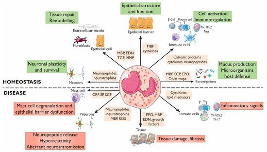
Eosinophils are innate immune granulocytes actively involved in defensive responses and in local and systemic inflammatory processes. Beyond these effector roles, eosinophils are fundamental to maintaining homeostasis in the tissues they reside. Gastrointestinal eosinophils modulate barrier function and mucosal immunity and promote tissue development through their direct communication with almost every cellular component. This is possible thanks to the variety of receptors they express and the bioactive molecules they store and release, including cytotoxic proteins, cytokines, growth factors, and neuropeptides and neurotrophines. A growing body of evidence points to the eosinophil as a key neuro-immune player in the regulation of gastrointestinal function, with potential implications in pathophysiological processes. Eosinophil–neuron interactions are facilitated by chemotaxis and adhesion molecules, and the mediators released may have excitatory or inhibitory effects on each cell type, with physiological consequences dependent on the type of innervation involved. Of special interest are the disorders of the brain–gut interaction (DBGIs), mainly functional dyspepsia (FD) and irritable bowel syndrome (IBS), in which mucosal eosinophilia and eosinophil activation have been identified. 

intestinal eosinophils neuro-immune interaction disorders of brain–gut interaction

1. Eosinophils Regulate the Intestinal Barrier Function

Although the impact of eosinophils on the intestinal epithelium in in vivo human studies is difficult to decipher, data gained from in vitro and pre-clinical models have shown a significant influence of eosinophils on mucosal homeostasis (Figure 1).
Figure 1. Roles of eosinophils in gastrointestinal homeostasis and disease. The interactions between eosinophils and other constituents of the intestinal mucosa (nerves, immune cells, endothelium, stroma, and luminal factors) mediate barrier integrity maintenance or tissue damage through the release of granule mediators. Major basic protein and eosinophil-derived cytokines contribute to barrier function through prostaglandin and ion secretion and mucus production. Eosinophils also impact tissue immuno-regulation and inflammation through cytokines and growth factors that promote adaptive and innate immunity, including a direct effect on mast cells in inflammatory or under stress conditions. Eosinophils also prime B cells for antigen-specific IgM production and sustain long-lived plasma cells. They participate in protection against infections through the release of DNA traps and the stimulation of mucus production through cationic proteins. In addition, eosinophils are also a source of a varied range of proteins and cytokines involved in fibrogenesis and angiogenesis that promote tissue remodeling, repair, and fibrosis. Eosinophils can regulate the function, survival, and development of nerve cells through the release of eosinophil-derived neuropeptides and neurotrophines, MBP, and ROS, and can promote hyperreactivity and aberrant neurotransmission in pathological conditions. CRF, Corticotropin releasing factor; ECP, Eosinophil cationic protein; EDN, Eosinophil-derived neurotoxin; EPO, Eosinophil peroxidase; MBP, Major basic protein; MMP, Matrix metalloprotease; ROS, Reactive oxygen species; SCF, Stem cell factor; SP, Substance P; TGF, Transforming growth factor.
The intestinal epithelium exerts a broad variety of functions ranging from simply being a physical barrier to luminal content to directly promoting active defensive immunity, achieved thanks to the existence of bi-directional communication between intestinal epithelial cells and the whole plethora of mucosal resident immune cells. Eosinophil-induced promotion of intestinal barrier protection seems to depend on the abundance of eosinophils, a low eosinophil–epithelial cell ratio being associated with enhanced barrier integrity [1][2][3], while a total depletion of intestinal eosinophils is associated with barrier disruption, as experimentally demonstrated by a high-fat diet treatment [4]. A remarkable exception to this pattern is the case of the healthy esophagus, where increased eosinophil numbers in EoE are linked to chronic inflammation and barrier disruption [5]. Notably, the close proximity of eosinophils and intestinal epithelial cells facilitates the crosstalk between these two cell types which may evoke a wide range of effects, ultimately contributing to maintenance of homeostasis or barrier dysfunction.
The interplay between eosinophils and the airway epithelium has been extensively studied, and similar interactions may be expected in the intestinal mucosa. Indeed, a large number of studies have demonstrated that, similar to the airways, intestinal epithelial cells are potent sources of immune-modulating cytokines, chemokines, and growth factors, with large effects on eosinophil recruitment, survival, and activation [6][7]. On the other hand, eosinophils can signal to epithelial cells through the generation of reactive oxygen species (ROS) and the release of leukotrienes and other granule-stored mediators, thus augmenting local inflammation and promoting epithelial proliferation [8][9]. In EoE, eosinophil-derived mediators have been shown to target esophageal epithelial integrity. More specifically, IL-13 down-regulates the intercellular adhesion molecule desmoglein-1, but not desmoglein-3, leading to altered epithelial barrier function and the promotion of inflammatory responses [10][11]. Eosinophils also modulate the secretion of mucus and bactericidal substances contributing to the reinforcement of epithelial barrier protection against infection. In fact, eosinophil-deficient mice show significantly reduced numbers of mucus-secreting goblet cells in the small bowel [12]. Moreover, supernatants derived from activated eosinophils increase mucin levels in human airway epithelial cells [13][14], and blocking the eotaxin receptor with anti-CCR3 monoclonal antibody in a mouse model of asthma reduces lung eosinophil recruitment and mucus overproduction [15]. Additionally, eosinophils have the ability to synthesize and rapidly release a web-like complex meshwork of DNA fibers and granule proteins called eosinophil extracellular traps (EETs) in a ROS-dependent manner but independently of eosinophil death, creating a second physical barrier that limits bacterial invasion [16]. However, EETs could also have deleterious effects on intestinal health due to increased production of mucus secretions [17]. Indeed, EETs have been associated with respiratory diseases and a number of other inflammation-associated diseases affecting the skin (atopic dermatitis, urticaria) and the gastrointestinal tract (IBD and EoE) [17].
Additional to the direct effect on the epithelium, eosinophils also contribute to barrier maintenance by promoting tissue remodeling/repair after tissue damage induced by pathogens, toxins, or cell death. A dual role of eosinophils in tissue degradation and repair has been described. In fact, eosinophils release eosinophil-derived neurotoxin (EDN), which promotes fibroblast proliferation [18], and major basic protein (MBP), which synergizes with IL-5 and transforming growth factor beta (TGF-β) to enhance fibroblast expression and secretion of IL-6 and IL-11 [19]. In addition, MBP and EDN are potent heparanase inhibitors, which suggests that eosinophils may be involved in proteoglycan degradation prevention and contribute, therefore, to proteoglycan accumulation in fibrotic tissues [20]. Tissue degradation can be sensed by eosinophils which react to promote healing by facilitating epithelial proliferation and acting on the vasculature through the release of vascular endothelial growth factor, fibroblast growth factor, and transforming growth factor-β1 (TGF-β1) [21] in order to recover tissue structure and promote homeostasis.

2. Eosinophils Modulate Intestinal Immune Responses

Eosinophils are a major cellular element of the intestinal mucosa and participate in the regulation of immunity and in the promotion of inflammatory responses (Figure 2). They are omnipresent at sites of Th2-mediated inflammation, where they can precede or be rapidly recruited to tissue, independently of adaptive immune responses [22]. This suggests that eosinophils themselves can modulate and/or sustain the Th2 character of the local tissue immune microenvironment and also promote Th2 memory cell functions by, for example, releasing cytokines [23]. Beyond Th2 immunity, preformed eosinophil mediators are also related to other immune responses; these include cytokines associated with Th1 (IFN-γ, IL-12), T-regulatory (IL-10, TGF-β), and Th17 [24][25] activities. In addition, eosinophils have the capacity to initiate and polarize adaptive immune responses. Unlike their blood counterparts, intestinal eosinophils constitutively express on their cell surface antigen presentation markers, including MHC class II, CD80, and activating receptor FcγRIII, suggesting that gut eosinophils may be primed for antigen presentation [26][27][28]. In addition, eosinophils also promote the initiation of adaptive immune responses through the granule proteins EDN and EPO which are involved in dendritic cell migration, activation, and maturation, leading, therefore, to enhanced antigen-specific Th2 responses [29]. Notably, eosinophils contribute to immunoglobulin production by plasma cells, as shown in animal studies by mediating adjuvant-elicited priming of B cells and optimal antigen-specific early IgM through IL-4 [30]. Eosinophils are crucial for class-switch generation and the maintenance of IgA plasma cells in the lamina propria, mainly through the expression of the A proliferation-inducing ligand (APRIL) and other cytokines, as identified in models of eosinophil-depleted mice [31].
Eosinophil-derived cytokines and chemokines also have effects on innate immune cells, especially on mast cells. Eosinophil–mast cell interactions have been the focus of many hypotheses trying to explain mucosal immune responses (reviewed in [32]). The large amount of evidence of eosinophil and mast cell contribution in the same scenarios, along with the important role mast cells play in allergic and inflammatory diseases also related to the brain–gut axis, suggest an additional eosinophil-derived effector function in immunoregulation within this axis. This potential contribution should no longer be interpreted as a minor interaction based on the release of eosinophil proteins and their inflammatory activity. In fact, eosinophils can promote mast cell growth, survival, and activation by several cytokines and by granule-derived proteins, such as MBP, stem cell factor (SCF), and nerve growth factor (NGF) [32][33]. Additionally, mast cells secrete mediators needed for reciprocally activating and promoting the survival of eosinophils [33]. However, mast cells may not be necessarily required for eosinophil survival, as in a mast cell-deficient mouse model of EoE the number of eosinophils remain unaffected [34].

3. Eosinophil–Neuron Interactions

3.1. Innervation of the Gastrointestinal Tract

The gastrointestinal tract is densely populated by two complex networks of neurons (intrinsic innervation) and immune cells that have co-evolved mechanisms to sense and rapidly adapt to the highly dynamic environmental challenges taking place at the intestinal mucosa. The first network is the submucosal plexus or Meissner’s plexus, located in the submucosal region between the circular smooth muscle and the submucosa, which controls glandular secretions, regulates local blood flow, and controls water secretion into the lumen. Fundamental research in different species, such as guinea pigs, rats, and humans, has shown that secretomotor neurons release acetylcholine (ACh) and vasoactive intestinal polypeptide (VIP) which stimulate chloride (Cl-) and water secretion. Nevertheless, the findings in mice are contradictory. In this animal species, the activation of submucosal neurons by electrical field stimulation involves in part the release of Ach and the activation of muscarinic receptors; however, the pharmacological stimulation of these neurons with veratridine does not involve the release of Ach [35]. The second network, called the myenteric plexus or Auerbach plexus, is located between the circular and the longitudinal smooth muscle layers and plays a crucial role in controlling gastrointestinal motility. Excitatory motor neurons synthetize and release ACh as the main neurotransmitter, along with substance P (SP) and other tachykinins. Inhibitory motor neurons synthetize nitric oxide, the main inhibitory neurotransmitter in the upper gastrointestinal tract, and adenosine triphosphate (ATP), the main inhibitory neurotransmitter in the lower gastrointestinal tract. Both plexi of the enteric nervous system (ENS) operate independently but are in turn modulated by the autonomic nervous system via efferent sympathetic and parasympathetic innervation (extrinsic innervation). The cell bodies of these nerves are located in the celiac, superior, and inferior mesenteric ganglions of the sympathetic chain and in the brain, respectively. The central nervous system (CNS) processes sensory information from the different layers of the gastrointestinal tract via the vagal afferent (non-painful physiological stimuli) and spinal afferent nerves (nociceptive stimuli—pain). Gastrointestinal symptoms are triggered basically through the stimulation of chemosensitive nociceptors present in spinal afferent nerves that innervate the lamina propria and through the activation of mechanosensitive nociceptors present in the longitudinal and circular smooth muscle [36]. The activation of spinal afferent nerves induces the release of sensory neuropeptides, such as SP, CGRP, and NKA.
How the different branches of the extrinsic and intrinsic nervous system crosstalk with innate and adaptive immune cells residing in the gut to jointly coordinate critical physiological functions and responses to challenges has been a matter of intense research. Indeed, functional neuro-immune interactions have been described as playing fundamental roles in intestinal health and disease [37] and there are excellent reviews discussing neuronal crosstalk in several gut immune populations (mainly mast cells, macrophages, and T- and B-cells) [38][39]
Like other gut resident immune cells, eosinophils express a wide range of neuropeptides and their receptors that confer on them abilities of interacting with the nervous system, either directly by cell-to-cell contact or indirectly through eosinophil crosstalk with other immune cells residing in the lamina propria [25][37]. This eosinophil–neuron communication has been shown to have a bi-directional nature (Figure 2); eosinophils can be either the source or the target of the interactions. Indeed, neurons can recruit and activate eosinophils, while eosinophils have been described to show trophic, stimulatory, and inhibitory effects on neurons.
Figure 2. Eosinophil–neuron interaction. Eosinophils and neurons communicate bidirectionally, modulating functions in both cell types. Nerve cells can recruit eosinophils through the release of neuropeptides, cytokines, and chemokines, and activate degranulation through nerve contact facilitated by adhesion molecules (intercellular and neural cell adhesion molecules (ICAMs and NCAMs). The release of bioactive mediators by eosinophils, such as cationic proteins, ROS, and neuropeptides/neurotrophines, results in a variety of neuronal effects, including nerve growth, nerve damage, neuropeptide synthesis and release, and activation and sensitization, leading to hyperreactivity and aberrant neuropeptide release in pathological conditions. A1/A2a/A3, Adenosine receptor; AR, Adrenergic receptor; B1/B2, Bradykinin receptor; CB2, Cannabinoid receptor 2; CGRP, Calcitonin gene-related peptide; CRTH2, Chemoattractant receptor-homologous molecule expressed on Th2 cells; H1/H2/H4R, Histamine receptor H1, H2, H4; ICAM, Intercellular cell adhesion molecule; M2/M3R, Muscarinic receptor 2, 3; nAChRs α-3/-α4/α-7, Nicotinic acetylcholine receptor α-3, -α4, and α-7; NK-1/NK-2/NK-3, Tachykinin receptor 1, 2, 3; P2Y/P2X, Purinergic receptor 2Y, X; ROS, Reactive oxygen species; TrK, Tropomyosin receptor kinase; VCAM, Vascular cell adhesion molecule; 5-HT1/2A, 5-hydroxytryptamine 1, 2A.

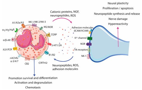
3.2. Neural-Induced Recruitment and Activation of Eosinophils by Extrinsic Nerves

Different in vitro and in vivo studies have shown that airway nerves actively recruit and bind eosinophils, promoting a wide range of effects on both cell types. Direct recruitment of eosinophils can happen through eotaxins constitutively expressed and released by parasympathetic efferent nerves, as occurs in the airways during antigenic challenge [40][41]. In addition, neurotransmitters and sensory neuropeptides released from the peripheral nerve endings of sensory neurons (extrinsic innervation), such as SP, CCK-8, NKA, and CGRP, can also promote eosinophil recruitment [42][43][44][45]. After recruitment, eosinophils adhere to nerves through cell adhesion molecules (CAMs), which leads to eosinophil activation and degranulation [43][45].
After recruitment, eosinophils adhere to nerves through cell adhesion molecules (CAMs), which leads to eosinophil activation and degranulation [43][45]. Two major CAM families are involved in eosinophil–nerve binding: the immunoglobulin superfamily of CAMs, which include vascular, intercellular, and neural cell adhesion molecules (VCAM, ICAM, and NCAM); and integrin CAMs, mainly integrin α4β1/VLA-4 and leukocyte function-associated molecule 1 (LFA1). CAM expression on nerves can be constitutive or inducible by pro-inflammatory cytokines and other mediators. Indeed, ICAM expression is induced by TNF-α and IFN-γ causing increased eosinophil adhesion and switching binding preference from VCAM to ICAM in primary cultures of airway parasympathetic nerves [46], an effect that is prevented by treatment with dexamethasone and NFKB inhibitors [47]. Eosinophil engagement to neural adhesion molecules leads to eosinophil activation and ROS production [48][49]. Indeed, ICAM and VCAM activation concurrently induce neurite retraction via the generation of tyrosine kinase-dependent ROS and by the p38 MAP kinase pathway [50]. At the same time, neural-derived ROS trigger eosinophil degranulation [36], which makes ROS production a shared event in the eosinophil–nerve bi-directional interaction.
Additional neural stimuli, such as ACh released by peripheral efferent nerves, have been linked to eosinophil chemotaxis and degranulation in experimental models [51] and in atopic asthma patients [52]. It is likely that a similar mechanism exists in the gastrointestinal tract, but further investigations are needed to define the eosinophil–nerve interactions and their contributions to organ functions.

3.3. Neural-Induced Recruitment and Activation of Eosinophils by the ENS

Although the anatomy and function of extrinsic nerves are very different, eosinophil–enteric nerve interactions seem to be similar to those described in the parasympathetic efferent innervation of the lungs. Interestingly, eosinophils are in close proximity to terminal varicosities of excitatory motor neurons expressing SP and choline acetyltransferase (ChAT) but not neuronal nitric oxide synthase (nNOS) [53]. In addition, another cell adhesion molecule, NCAM, has been reported to play a role in eosinophil adhesion to myenteric terminal varicosities in the colonic mucosa of rats undergoing a Th2 response caused by parasitic infection [54]. However, the specific mechanisms underlying eosinophil adhesion to excitatory terminal varicosities during intestinal inflammation are still unknown.
In the gastrointestinal mucosa, SP signaling has been shown to trigger the production and release of corticotropin releasing factor (CRF) in vitro and in experimental chronic restraint stress models [55][56]. CRF is a major mediator of stress-induced autonomic, hormonal, and behavioral reflexes that inhibit inflammatory responses at regional levels and influence gut motility and secretion [57] and it has been recently involved in a non-classical non-pro-inflammatory eosinophil activation mechanism, wherein neuromediators selectively induce eosinophil synthesis and release of CRF by piecemeal degranulation [58]. VIP is a key signaling molecule in the neuro-immune network that is secreted by neuronal cells and by different types of immune cells and exerts a wide spectrum of functions. The release of VIP by enteric neurons innervating the intestinal mucosa modulates the epithelial barrier [59]. VIP also regulates the production of both anti- and pro-inflammatory mediators in immune cells [60]. Eosinophils do not express the classical VPAC-1 and VPAC2 but respond to VIP through the Chemoattractant Receptor-Homologous Molecule Expressed on Th2 Cells (CRTH2) [61]. The main effects of VIP on eosinophils are the promotion of chemotaxis and the production of prostaglandin D2, both identified in the context of allergic rhinitis. It may be relevant to determine whether eosinophilia is promoted by VIP released by enteric neurons during intestinal inflammatory conditions, and, additionally, whether eosinophils also impact the intestinal barrier by means of VIP, as it can store and release this neuropeptide [62].

References

  1. Furuta, G.T.; Nieuwenhuis, E.E.S.; Karhausen, J.; Gleich, G.; Blumberg, R.S.; Lee, J.J.; Ackerman, S.J. Eosinophils Alter Colonic Epithelial Barrier Function: Role for Major Basic Protein. Am. J. Physiol.-Gastrointest. Liver Physiol. 2005, 289, G890–G897.
  2. Jacoby, D.B.; Ueki, I.F.; Widdicombe, J.H.; Loegering, D.A.; Gleich, G.J.; Nadel, J.A. Effect of Human Eosinophil Major Basic Protein on Ion Transport in Dog Tracheal Epithelium. Am. Rev. Respir. Dis. 1988, 137, 13–16.
  3. White, S.R.; Sigrist, K.S.; Spaethe, S.M. Prostaglandin Secretion by Guinea Pig Tracheal Epithelial Cells Caused by Eosinophil Major Basic Protein. Am. J. Physiol.-Lung Cell. Mol. Physiol. 1993, 265, L234–L242.
  4. Johnson, A.M.F.; Costanzo, A.; Gareau, M.G.; Armando, A.M.; Quehenberger, O.; Jameson, J.M.; Olefsky, J.M. High Fat Diet Causes Depletion of Intestinal Eosinophils Associated with Intestinal Permeability. PLoS ONE 2015, 10, e0122195.
  5. Furuta, G.T.; Katzka, D.A. Eosinophilic Esophagitis. N. Engl. J. Med. 2015, 373, 1640–1648.
  6. Mishra, A.; Hogan, S.P.; Lee, J.J.; Foster, P.S.; Rothenberg, M.E. Fundamental Signals That Regulate Eosinophil Homing to the Gastrointestinal Tract. J. Clin. Investig. 1999, 103, 1719–1727.
  7. Carlens, J.; Wahl, B.; Ballmaier, M.; Bulfone-Paus, S.; Förster, R.; Pabst, O. Common γ-Chain-Dependent Signals Confer Selective Survival of Eosinophils in the Murine Small Intestine. J. Immunol. 2009, 183, 5600–5607.
  8. Honda, K.; Chihara, J. Eosinophil Activation by Eotaxin--Eotaxin Primes the Production of Reactive Oxygen Species from Eosinophils. Allergy 1999, 54, 1262–1269.
  9. Choe, M.M.; Sporn, P.H.S.; Swartz, M.A. An in vitro Airway Wall Model of Remodeling. Am. J. Physiol.-Lung Cell. Mol. Physiol. 2003, 285, L427–L433.
  10. Sherrill, J.D.; Kc, K.; Wu, D.; Djukic, Z.; Caldwell, J.M.; Stucke, E.M.; Kemme, K.A.; Costello, M.S.; Mingler, M.K.; Blanchard, C.; et al. Desmoglein-1 Regulates Esophageal Epithelial Barrier Function and Immune Responses in Eosinophilic Esophagitis. Mucosal Immunol. 2014, 7, 718–729.
  11. Racca, F.; Pellegatta, G.; Cataldo, G.; Vespa, E.; Carlani, E.; Pelaia, C.; Paoletti, G.; Messina, M.R.; Nappi, E.; Canonica, G.W.; et al. Type 2 Inflammation in Eosinophilic Esophagitis: From Pathophysiology to Therapeutic Targets. Front. Physiol. 2021, 12, 815842.
  12. Jung, Y.; Wen, T.; Mingler, M.K.; Caldwell, J.M.; Wang, Y.H.; Chaplin, D.D.; Lee, E.H.; Jang, M.H.; Woo, S.Y.; Seoh, J.Y.; et al. IL-1β in Eosinophil-Mediated Small Intestinal Homeostasis and IgA Production. Mucosal Immunol. 2015, 8, 930–942.
  13. Cohn, L.; Homer, R.J.; MacLeod, H.; Mohrs, M.; Brombacher, F.; Bottomly, K. Th2-Induced Airway Mucus Production Is Dependent on IL-4Ralpha, but Not on Eosinophils. J. Immunol. 1999, 162, 6178–6183.
  14. Burgel, P.-R.; Lazarus, S.C.; Tam, D.C.-W.; Ueki, I.F.; Atabai, K.; Birch, M.; Nadel, J.A. Human Eosinophils Induce Mucin Production in Airway Epithelial Cells Via Epidermal Growth Factor Receptor Activation. J. Immunol. 2001, 167, 5948–5954.
  15. Shen, H.; Xu, F.; Zhang, G.; Wang, S.; Xu, W. CCR3 Monoclonal Antibody Inhibits Airway Eosinophilic Inflammation and Mucus Overproduction in a Mouse Model of Asthma. Acta Pharmacol. Sin. 2006, 27, 1594–1599.
  16. Yousefi, S.; Gold, J.A.; Andina, N.; Lee, J.J.; Kelly, A.M.; Kozlowski, E.; Schmid, I.; Straumann, A.; Reichenbach, J.; Gleich, G.J.; et al. Catapult-like Release of Mitochondrial DNA by Eosinophils Contributes to Antibacterial Defense. Nat. Med. 2008, 14, 949–953.
  17. Mukherjee, M.; Lacy, P.; Ueki, S. Eosinophil Extracellular Traps and Inflammatory Pathologies—Untangling the Web! Front. Immunol. 2018, 9, 2763.
  18. Puxeddu, I.; Ribatti, D.; Crivellato, E.; Levi-Schaffer, F. Mast Cells and Eosinophils: A Novel Link between Inflammation and Angiogenesis in Allergic Diseases. J. Allergy Clin. Immunol. 2005, 116, 531–536.
  19. Gomes, I.; Mathur, S.K.; Espenshade, B.M.; Mori, Y.; Varga, J.; Ackerman, S.J. Eosinophil-Fibroblast Interactions Induce Fibroblast IL-6 Secretion and Extracellular Matrix Gene Expression: Implications in Fibrogenesis. J. Allergy Clin. Immunol. 2005, 116, 796–804.
  20. Temkin, V.; Aingorn, H.; Puxeddu, I.; Goldshmidt, O.; Zcharia, E.; Gleich, G.J.; Vlodavsky, I.; Levi-Schaffer, F. Eosinophil Major Basic Protein: First Identified Natural Heparanase-Inhibiting Protein. J. Allergy Clin. Immunol. 2004, 113, 703–709.
  21. Lee, J.J.; Jacobsen, E.A.; McGarry, M.P.; Schleimer, R.P.; Lee, N.A. Eosinophils in Health and Disease: The LIAR Hypothesis: Eosinophils and the LIAR Hypothesis. Clin. Exp. Allergy 2010, 40, 563–575.
  22. Shinkai, K.; Mohrs, M.; Locksley, R.M. Helper T Cells Regulate Type-2 Innate Immunity in vivo. Nature 2002, 420, 825–829.
  23. Wang, Y.-H.; Angkasekwinai, P.; Lu, N.; Voo, K.S.; Arima, K.; Hanabuchi, S.; Hippe, A.; Corrigan, C.J.; Dong, C.; Homey, B.; et al. IL-25 Augments Type 2 Immune Responses by Enhancing the Expansion and Functions of TSLP-DC–Activated Th2 Memory Cells. J. Exp. Med. 2007, 204, 1837–1847.
  24. Spencer, L.A.; Szela, C.T.; Perez, S.A.C.; Kirchhoffer, C.L.; Neves, J.S.; Radke, A.L.; Weller, P.F. Human Eosinophils Constitutively Express Multiple Th1, Th2, and Immunoregulatory Cytokines That Are Secreted Rapidly and Differentially. J. Leukoc. Biol. 2008, 85, 117–123.
  25. Akuthota, P.; Wang, H.B.; Spencer, L.A.; Weller, P.F. Immunoregulatory Roles of Eosinophils: A New Look at a Familiar Cell. Clin. Exp. Allergy 2008, 38, 1254–1263.
  26. Xenakis, J.J.; Howard, E.D.; Smith, K.M.; Olbrich, C.L.; Huang, Y.; Anketell, D.; Maldonado, S.; Cornwell, E.W.; Spencer, L.A. Resident Intestinal Eosinophils Constitutively Express Antigen Presentation Markers, and Include Two Phenotypically Distinct Subsets of Eosinophils. Immunology 2018, 154, 298–308.
  27. Farhan, R.K.; Vickers, M.A.; Ghaemmaghami, A.M.; Hall, A.M.; Barker, R.N.; Walsh, G.M. Effective Antigen Presentation to Helper T Cells by Human Eosinophils. Immunology 2016, 149, 413–422.
  28. Smith, K.M.; Rahman, R.S.; Spencer, L.A. Humoral Immunity Provides Resident Intestinal Eosinophils Access to Luminal Antigen via Eosinophil-Expressed Low-Affinity Fcγ Receptors. J. Immunol. 2016, 197, 3716–3724.
  29. Chu, D.K.; Jimenez-Saiz, R.; Verschoor, C.P.; Walker, T.D.; Goncharova, S.; Llop-Guevara, A.; Shen, P.; Gordon, M.E.; Barra, N.G.; Bassett, J.D.; et al. Indigenous Enteric Eosinophils Control DCs to Initiate a Primary Th2 Immune Response in vivo. J. Exp. Med. 2014, 211, 1657–1672.
  30. Wang, H.-B.; Weller, P.F. Pivotal Advance: Eosinophils Mediate Early Alum Adjuvant-Elicited B Cell Priming and IgM Production. J. Leukoc. Biol. 2008, 83, 817–821.
  31. Chu, V.T.; Beller, A.; Rausch, S.; Strandmark, J.; Zänker, M.; Arbach, O.; Kruglov, A.; Berek, C. Eosinophils Promote Generation and Maintenance of Immunoglobulin-A-Expressing Plasma Cells and Contribute to Gut Immune Homeostasis. Immunity 2014, 40, 582–593.
  32. Shakoory, B.; Fitzgerald, S.M.; Lee, S.A.; Chi, D.S.; Krishnaswamy, G. The Role of Human Mast Cell-Derived Cytokines in Eosinophil Biology. J. Interferon Cytokine Res. 2004, 24, 271–281.
  33. Galdiero, M.R.; Varricchi, G.; Seaf, M.; Marone, G.; Levi-Schaffer, F.; Marone, G. Bidirectional Mast Cell–Eosinophil Interactions in Inflammatory Disorders and Cancer. Front. Med. 2017, 4, 103.
  34. Niranjan, R.; Mavi, P.; Rayapudi, M.; Dynda, S.; Mishra, A. Pathogenic Role of Mast Cells in Experimental Eosinophilic Esophagitis. Am. J. Physiol. Gastrointest. Liver Physiol. 2013, 304, G1087–G1094.
  35. Hirota, C.L.; McKay, D.M. Cholinergic Regulation of Epithelial Ion Transport in the Mammalian Intestine. Br. J. Pharmacol. 2006, 149, 463–479.
  36. Brierley, S.M.; Linden, D.R. Neuroplasticity and Dysfunction after Gastrointestinal Inflammation. Nat. Rev. Gastroenterol. Hepatol. 2014, 11, 611–627.
  37. Brinkman, D.J.; ten Hove, A.S.; Vervoordeldonk, M.J.; Luyer, M.D.; de Jonge, W.J. Neuroimmune Interactions in the Gut and Their Significance for Intestinal Immunity. Cells 2019, 8, 670.
  38. Veiga-Fernandes, H.; Mucida, D. Neuro-Immune Interactions at Barrier Surfaces. Cell 2016, 165, 801–811.
  39. Jacobson, A.; Yang, D.; Vella, M.; Chiu, I.M. The Intestinal Neuro-Immune Axis: Crosstalk between Neurons, Immune Cells, and Microbes. Mucosal Immunol. 2021, 14, 555–565.
  40. Chou, D.L.; Daugherty, B.L.; McKenna, E.K.; Hsu, W.M.; Tyler, N.K.; Plopper, C.G.; Hyde, D.M.; Schelegle, E.S.; Gershwin, L.J.; Miller, L.A. Chronic Aeroallergen during Infancy Enhances Eotaxin-3 Expression in Airway Epithelium and Nerves. Am. J. Respir. Cell Mol. Biol. 2005, 33, 1–8.
  41. Fryer, A.D. Neuronal Eotaxin and the Effects of Ccr3 Antagonist on Airway Hyperreactivity and M2 Receptor Dysfunction. J. Clin. Investig. 2005, 116, 228–236.
  42. Numao, T.; Agrawal, D.K. Neuropeptides Modulate Human Eosinophil Chemotaxis. J. Immunol. 1992, 149, 3309–3315.
  43. Jacoby, D.B.; Costello, R.M.; Fryer, A.D. Eosinophil Recruitment to the Airway Nerves. J. Allergy Clin. Immunol. 2001, 107, 211–218.
  44. Dunzendorfer, S.; Meierhofer, C.; Wiedermann, C.J. Signaling in Neuropeptide-Induced Migration of Human Eosinophils. J. Leukoc. Biol. 1998, 64, 828–834.
  45. Kingham, P.J.; McLean, W.G.; Walsh, M.-T.; Fryer, A.D.; Gleich, G.J.; Costello, R.W. Effects of Eosinophils on Nerve Cell Morphology and Development: The Role of Reactive Oxygen Species and P38 MAP Kinase. Am. J. Physiol.-Lung Cell. Mol. Physiol. 2003, 285, L915–L924.
  46. Sawatzky, D.A.; Kingham, P.J.; Court, E.; Kumaravel, B.; Fryer, A.D.; Jacoby, D.B.; McLean, W.G.; Costello, R.W. Eosinophil Adhesion to Cholinergic Nerves via ICAM-1 and VCAM-1 and Associated Eosinophil Degranulation. Am. J. Physiol.-Lung Cell. Mol. Physiol. 2002, 282, L1279–L1288.
  47. Nie, Z.; Nelson, C.S.; Jacoby, D.B.; Fryer, A.D. Expression and Regulation of Intercellular Adhesion Molecule-1 on Airway Parasympathetic Nerves. J. Allergy Clin. Immunol. 2007, 119, 1415–1422.
  48. Walsh, M.-T.; Curran, D.R.; Kingham, P.J.; Morgan, R.K.; Durcan, N.; Gleich, G.J.; McLean, W.G.; Costello, R.W. Effect of Eosinophil Adhesion on Intracellular Signaling in Cholinergic Nerve Cells. Am. J. Respir. Cell Mol. Biol. 2004, 30, 333–341.
  49. Kingham, P.J.; McLean, W.G.; Sawatzky, D.A.; Walsh, M.T.; Costello, R.W. Adhesion-Dependent Interactions between Eosinophils and Cholinergic Nerves. Am. J. Physiol.-Lung Cell. Mol. Physiol. 2002, 282, L1229–L1238.
  50. Taylor-Clark, T.E.; Undem, B.J. Sensing Pulmonary Oxidative Stress by Lung Vagal Afferents. Respir. Physiol. Neurobiol. 2011, 178, 406–413.
  51. Evans, C.M.; Belmonte, K.E.; Costello, R.W.; Jacoby, D.B.; Gleich, G.J.; Fryer, A.D. Substance P-Induced Airway Hyperreactivity Is Mediated by Neuronal M 2 Receptor Dysfunction. Am. J. Physiol.-Lung Cell. Mol. Physiol. 2000, 279, L477–L486.
  52. Raap, M.; Rüdrich, U.; Ständer, S.; Gehring, M.; Kapp, A.; Raap, U. Substance P Activates Human Eosinophils. Exp. Dermatol. 2015, 24, 557–559.
  53. Smyth, C.M.; Akasheh, N.; Woods, S.; Kay, E.; Morgan, R.K.; Thornton, M.A.; O’Grady, A.; Cummins, R.; Sheils, O.; Smyth, P.; et al. Activated Eosinophils in Association with Enteric Nerves in Inflammatory Bowel Disease. PLoS ONE 2013, 8, e64216.
  54. O’Brien, L.M.; Fitzpatrick, E.; Baird, A.W.; Campion, D.P. Eosinophil–Nerve Interactions and Neuronal Plasticity in Rat Gut Associated Lymphoid Tissue (GALT) in Response to Enteric Parasitism. J. Neuroimmunol. 2008, 197, 1–9.
  55. Wallon, C.; Persborn, M.; Jönsson, M.; Wang, A.; Phan, V.; Lampinen, M.; Vicario, M.; Santos, J.; Sherman, P.M.; Carlson, M.; et al. Eosinophils Express Muscarinic Receptors and Corticotropin-Releasing Factor to Disrupt the Mucosal Barrier in Ulcerative Colitis. Gastroenterology 2011, 140, 1597–1607.
  56. Zheng, P.-Y.; Feng, B.-S.; Oluwole, C.; Struiksma, S.; Chen, X.; Li, P.; Tang, S.-G.; Yang, P.-C. Psychological Stress Induces Eosinophils to Produce Corticotrophin Releasing Hormone in the Intestine. Gut 2009, 58, 1473–1479.
  57. Kiank, C.; Taché, Y.; Larauche, M. Stress-Related Modulation of Inflammation in Experimental Models of Bowel Disease and Post-Infectious Irritable Bowel Syndrome: Role of Corticotropin-Releasing Factor Receptors. Brain Behav. Immun. 2010, 24, 41–48.
  58. Salvo-Romero, E.; Martínez, C.; Lobo, B.; Rodiño-Janeiro, B.K.; Pigrau, M.; Sánchez-Chardi, A.D.; González-Castro, A.M.; Fortea, M.; Pardo-Camacho, C.; Nieto, A.; et al. Overexpression of Corticotropin-Releasing Factor in Intestinal Mucosal Eosinophils Is Associated with Clinical Severity in Diarrhea-Predominant Irritable Bowel Syndrome. Sci. Rep. 2020, 10, 20706.
  59. Neunlist, M.; Toumi, F.; Oreschkova, T.; Denis, M.; Leborgne, J.; Laboisse, C.L.; Galmiche, J.P.; Jarry, A. Human ENS regulates the intestinal epithelial barrier permeability and a tight junction-associated protein ZO-1 via VIPergic pathways. Am. J. Physiol. Gastrointest. Liver Physiol. 2003, 285, G1028–G1036.
  60. Verma, A.K.; Manohar, M.; Venkateshaiah, S.U.; Mishra, A. Neuroendocrine cells derived chemokine vasoactive intestinal polypeptide (VIP) in allergic diseases. Cytokine Growth Factor Rev. 2017, 38, 37–48.
  61. El-Shazly, A.E.; Begon, D.Y.; Kustermans, G.; Arafa, M.; Dortu, E.; Henket, M.; Lefebvre, P.P.; Louis, R.; Delvenne, P. Novel Association between Vasoactive Intestinal Peptide and CRTH2 Receptor in Recruiting Eosinophils. J. Biol. Chem. 2013, 288, 1374–1384.
  62. Metwali, A.; Blum, A.M.; Ferraris, L.; Klein, J.S.; Fiocchi, C.; Weinstock, J.V. Eosinophils within the healthy or inflamed human intestine produce substance P and vasoactive intestinal peptide. J. Neuroimmunol. 1994, 52, 69–78.
More
Information
Contributors MDPI registered users' name will be linked to their SciProfiles pages. To register with us, please refer to https://encyclopedia.pub/register : , ,
View Times: 695
Entry Collection: Gastrointestinal Disease
Revisions: 2 times (View History)
Update Date: 27 May 2022
1000/1000