Submitted Successfully!
To reward your contribution, here is a gift for you: A free trial for our video production service.
Thank you for your contribution! You can also upload a video entry or images related to this topic.
Version Summary Created by Modification Content Size Created at Operation
1 + 1150 word(s) 1150 2021-11-29 10:04:21 |
2 format correct Meta information modification 1150 2021-12-13 02:13:53 |

Video Upload Options

Do you have a full video?

Confirm

Are you sure to Delete?
Cite
If you have any further questions, please contact Encyclopedia Editorial Office.
Ziegler, A. Efficient Mucosal Repair Limits Morbidity from Colic. Encyclopedia. Available online: https://encyclopedia.pub/entry/16996 (accessed on 28 March 2024).
Ziegler A. Efficient Mucosal Repair Limits Morbidity from Colic. Encyclopedia. Available at: https://encyclopedia.pub/entry/16996. Accessed March 28, 2024.
Ziegler, Amanda. "Efficient Mucosal Repair Limits Morbidity from Colic" Encyclopedia, https://encyclopedia.pub/entry/16996 (accessed March 28, 2024).
Ziegler, A. (2021, December 10). Efficient Mucosal Repair Limits Morbidity from Colic. In Encyclopedia. https://encyclopedia.pub/entry/16996
Ziegler, Amanda. "Efficient Mucosal Repair Limits Morbidity from Colic." Encyclopedia. Web. 10 December, 2021.
Efficient Mucosal Repair Limits Morbidity from Colic
Edit

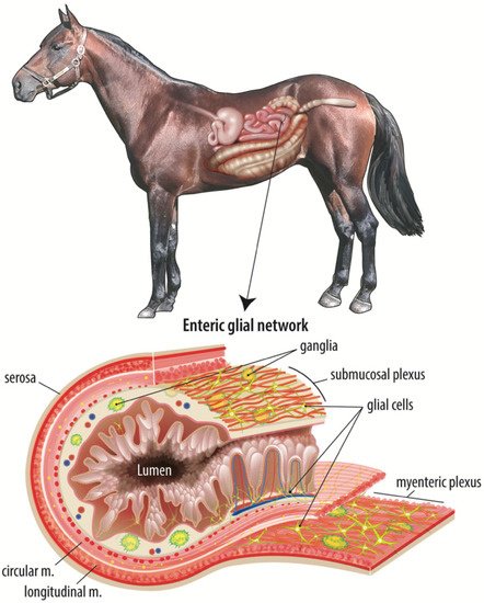
Colic is a leading cause of death in horses, with the most fatal form being strangulating obstruction which directly damages the intestinal barrier. Following surgical intervention, it is imperative that the intestinal barrier rapidly repairs to prevent translocation of gut bacteria and their products and ensure survival of the patient. Age-related disparities in survival have been noted in many species, including horses, humans, and pigs, with younger patients suffering poorer clinical outcomes. Maintenance and repair of the intestinal barrier is regulated by a complex mucosal microenvironment, of which the ENS, and particularly a developing network of subepithelial enteric glial cells, may be of particular importance in neonates with colic. Postnatal development of an immature enteric glial cell network is thought to be driven by the microbial colonization of the gut and therefore modulated by diet-influenced changes in bacterial populations early in life.

horse colic ischemia-reperfusion injury enteric nervous system intestinal barrier

1. Evidence of Age-Dependent Barrier Repair: A Comparative Pig Model

Because clinically recording differences in barrier repair is challenging, studies often rely on animal models of gastrointestinal barrier disruption and repair, particularly in neonatal and early life gastrointestinal research [1][2]. The pig has proven to be a powerful large animal model of gastrointestinal physiology for translation to humans and large animals as it can overcome some limitations of mouse and other rodent models [3]. The pig has been shown to be a viable model for equine gastrointestinal research, where similarities in both anatomy and physiology are of use experimentally [4][5]. The horse and the pig share similar microvascular anatomy within the small intestine, where a single, eccentrically located arteriole and similar branching of the venules within the villus will exhibit comparable epithelial sloughing patterns in response to ischemic injury [6][7][4]. Anatomically, though, there are species differences in the ENS; specifically, differences in the number of layers in the myenteric and submucosal plexuses. These differences have been characterized in the mouse and human and, more recently, in the pig, and it is reasonable to assume that there may be such anatomical differences between the pig and the horse. Though there are minor anatomical differences, functional differences between mammalian species have yet to be fully studied.
Physiologically, the neonatal pig is a good model for ischemia/reperfusion injury in the foal as both species lack intestinal xanthine dehydrogenase, an enzyme involved in the physiologic breakdown of hypoxanthine to xanthine and then to uric acid, at birth, indicating that reperfusion injury may not be as much of a concern in neonates of either species as xanthine dehydrogenase is converted to xanthine oxidase in hypoxic conditions, which produces superoxide during reperfusion [4]. Further, the pig has been established as a powerful model of early life stress, with important implications for the development of lifelong gastrointestinal disease [1][8][9]. One pig study identified a complete defect in ex vivo restitution of the intestinal barrier after ischemic injury in two-week-old piglet jejunum after 30 to 120 min of ischemia [10]. This defect, however, could be rescued by application of a homogenate of ischemic-injured mucosa from weanling-aged animals [10]. This indicates some aspect of the mucosa is not present or is immature in neonates that is apparently present and functional in weanling animals, and research is ongoing to determine the mechanisms responsible for this disparity which is of particular importance to foals suffering from severe colic.

2. A Complex Mucosal Microenvironment Regulates Epithelial Repair

Barrier repair is regulated intensively by complex and coordinated subepithelial cell signaling events. There are many cellular components of the subepithelial microenvironment to consider when investigating age-related differences in intestinal repair. The small intestine is a complex organ comprised of several layers, the outermost being the serosa surrounding the longitudinal and circular muscle layers. The smooth muscle layers facilitate motility, driving segmentation, churning, and forward transit of digesta, while ensuring adequate contact time between the luminal contents and the epithelium to increase absorption of digested nutrients. Between the two layers of smooth muscle is the myenteric plexus of the ENS, a network of intrinsic primary afferent neurons, interneurons, excitatory and inhibitory neurons, and enteric glial cells that regulate smooth muscle activity among many other emerging functions [11][12].
Internal to the smooth muscle layers is the submucosa, a region of loose connective tissue containing vasculature, lymphatic vessels, and the submucosal plexus of the ENS, another network of neurons and enteric glial cells (Figure 1). This region also houses a diverse population of immune cells, mesenchymal cells, and endothelial cells. Intestinal mesenchymal cells consist of fibroblasts, myofibroblasts, pericytes, mesenchymal stem cells, smooth muscle cells, interstitial cells of Cajal, and fibrocytes [13]. These cells function to provide mechanical support, epithelial homeostasis, stem cell maintenance, immune regulation, extracellular matrix maintenance, angiogenesis, and vascular function regulation [13]. Studies have identified a therapeutic effect when inflamed and damaged intestine is treated with mesenchymal stem cells in vitro [14][15][16]. Endothelial cells form the barrier between the intravascular elements and the submucosal microenvironment and are responsible for the induction of inflammation and recruitment of leukocytes through the release of several proinflammatory cytokines and colony-stimulating factors [17]. The innermost layer is the intestinal mucosa, containing subepithelial capillaries and lymphatic vessels, neuronal projections, and yet another network of enteric glial cells with projections extending close enough to directly contact the single-cell thick epithelial barrier. This epithelial population includes a carefully coordinated and organized population of absorptive and secretory enterocytes, enteroendocrine cells, goblet cells, tuft cells, and intestinal stem cells [18].
Figure 1. The adult equine enteric glial network. The intestinal mucosal microenvironment is home to several cell types that all work in a complex, coordinated manner to maintain the epithelial barrier and restore the barrier in response to intestinal injury. Of these cell types, the enteric glial network is thought to act as an intermediary between the enteric neurons and luminal signals, such as nutrients or microbial metabolites. This figure illustrates the complexity and expanse of the enteric glial network, including directly adjacent to the epithelium.
On the opposite side of the epithelial barrier, the small intestinal lumen contains ingesta, mucus, and two microbial populations, one that is suspended within the ingesta and another that is adherent to the mucus layer between the luminal contents and the mucosa. Within these two populations, microbes can be commensal, symbiotic, or pathogenic, and the intestinal microbiota is increasingly implicated broadly in health and disease [19][20]. Mucus is continually secreted by goblet cells in the small intestine and provides both a chemical and physical barrier to the potentially harmful bacteria in the intestinal lumen [21]. Loss of this mucus layer, through ischemic injury for example, increases intestinal permeability and the risk of patient sepsis [22]. Dietary components found in the intestinal lumen also function to stimulate mucus secretion and absorption while other nutrients function to decrease barrier activity. For example, glucose causes intracellular tight junctions to open the paracellular space, allowing nutrients and water to cross the barrier more easily [23]. These components of the luminal microenvironment are equally important to consider when investigating age-dependent mucosal repair following ischemic intestinal injury.

References

  1. Pohl, C.S.; Medland, J.E.; Moeser, A.J. Early-life stress origins of gastrointestinal disease: Animal models, intestinal pathophysiology, and translational implications. Am. J. Physiol. Liver Physiol. 2015, 309, G927–G941.
  2. Smith, F.; Clark, J.E.; Overman, B.L.; Tozel, C.C.; Huang, J.H.; Rivier, J.E.F.; Blisklager, A.T.; Moeser, A.J. Early weaning stress impairs development of mucosal barrier function in the porcine intestine. Am. J. Physiol. Liver Physiol. 2010, 298, G352–G363.
  3. Ziegler, A.; Gonzalez, L.; Blikslager, A. Large Animal Models: The Key to Translational Discovery in Digestive Disease Research. Cell. Mol. Gastroenterol. Hepatol. 2016, 2, 716–724.
  4. Blikslager, A.T.; Roberts, M.C.; Gerard, M.P.; A Argenzio, R. How important is intestinal reperfusion injury in horses? J. Am. Veter. Med. Assoc. 1997, 211, 1387–1389.
  5. Blikslager, A.T.; Roberts, M.C.; Rhoads, J.; A Argenzio, R. Is reperfusion injury an important cause of mucosal damage after porcine intestinal ischemia? Surgery 1997, 121, 526–534.
  6. Granger, D.N.; Holm, L.; Kvietys, P. The Gastrointestinal Circulation: Physiology and Pathophysiology. Compr. Physiol. 2015, 5, 1541–1583.
  7. Bellamy, J.E.C.; Nielsen, N.O.; Latshaw, W.K. The Vascular Architecture of the Porcine Small Intestine. Can. J. Comp. Med. Rev. Can. Med. Comp. 1973, 37, 56–62.
  8. Pohl, C.S.; Medland, J.E.; Mackey, E.; Edwards, L.L.; Bagley, K.D.; Dewilde, M.P.; Williams, K.J.; Moeser, A.J. Early weaning stress induces chronic functional diarrhea, intestinal barrier defects, and increased mast cell activity in a porcine model of early life adversity. Neurogastroenterol. Motil. 2017, 29, e13118.
  9. Medland, J.E.; Pohl, C.S.; Edwards, L.L.; Frandsen, S.; Bagley, K.; Li, Y.; Moeser, A.J. Early life adversity in piglets induces long-term upregulation of the enteric cholinergic nervous system and heightened, sex-specific secretomotor neuron responses. Neurogastroenterol. Motil. 2016, 28, 1317–1329.
  10. Ziegler, A.L.; Pridgen, T.A.; Mills, J.K.; Gonzalez, L.M.; Van Landeghem, L.; Odle, J.; Blikslager, A.T. Epithelial restitution defect in neonatal jejunum is rescued by juvenile mucosal homogenate in a pig model of intestinal ischemic injury and repair. PLoS ONE 2018, 13, e0200674.
  11. McCann, C.; Alves, M.M.; Brosens, E.; Natarajan, D.; Perin, S.; Chapman, C.; Hofstra, R.M.; Burns, A.J.; Thapar, N. Neuronal Development and Onset of Electrical Activity in the Human Enteric Nervous System. Gastroenterology 2019, 156, 1483–1495.e6.
  12. Furness, J.B. The enteric nervous system and neurogastroenterology. Nat. Rev. Gastroenterol. Hepatol. 2012, 9, 286–294.
  13. Koliaraki, V.; Pallangyo, C.K.; Greten, F.R.; Kollias, G. Mesenchymal Cells in Colon Cancer. Gastroenterology 2017, 152, 964–979.
  14. Gong, W.; Guo, M.; Han, Z.; Wang, Y.; Yang, P.; Xu, C.; Wang, Q.; Du, L.; Li, Q.; Zhao, H.; et al. Mesenchymal stem cells stimulate intestinal stem cells to repair radiation-induced intestinal injury. Cell Death Dis. 2016, 7, e2387.
  15. Sémont, A.; Mouiseddine, M.; François, A.; Demarquay, C.; Mathieu, N.; Chapel, A.; Saché, A.; Thierry, D.; Laloi, P.; Gourmelon, P. Mesenchymal stem cells improve small intestinal integrity through regulation of endogenous epithelial cell homeostasis. Cell Death Differ. 2009, 17, 952–961.
  16. Ocansey, D.W.; Wang, L.; Wang, J.; Yan, Y.; Qian, H.; Zhang, X.; Xu, W.; Mao, F. Mesenchymal stem cell–gut microbiota interaction in the repair of inflammatory bowel disease: An enhanced therapeutic effect. Clin. Transl. Med. 2019, 8, 31.
  17. Swerlick, R.A.; Lawley, T.J. Role of Microvascular Endothelial Cells in Inflammation. J. Investig. Dermatol. 1993, 100, S111–S115.
  18. Saps, M.; Miranda, A. Gastrointestinal Pharmacology. Mediat. Drugs Gastrointest. Motil. I 2017, 239, 147–176.
  19. Tamburini, S.; Shen, N.; Wu, H.C.; Clemente, S.T.N.S.J.C. The microbiome in early life: Implications for health outcomes. Nat. Med. 2016, 22, 713–722.
  20. Sampson, T.R.; Debelius, J.W.; Thron, T.; Janssen, S.; Shastri, G.G.; Ilhan, Z.E.; Challis, C.; Schretter, C.E.; Rocha, S.; Gradinaru, V.; et al. Gut Microbiota Regulate Motor Deficits and Neuroinflammation in a Model of Parkinson’s Disease. Cell 2016, 167, 1469–1480.e12.
  21. Qin, X.; Caputo, F.J.; Xu, D.-Z.; Deitch, E.A. Hydrophobicity of mucosal surface and its relationship to gut barrier function. Shock 2008, 29, 372–376.
  22. Rupani, B.; Caputo, F.J.; Watkins, A.; Vega, D.; Magnotti, L.J.; Lu, Q.; Xu, D.Z.; Deitch, E.A. Relationship between disruption of the unstirred mucus layer and intestinal restitution in loss of gut barrier function after trauma hemorrhagic shock. Surgery 2007, 141, 481–489.
  23. Turner, J.R.; Rill, B.K.; Carlson, S.L.; Carnes, D.; Kerner, R.; Mrsny, R.J.; Madara, J.L. Physiological regulation of epithelial tight junctions is associated with myosin light-chain phosphorylation. Am. J. Physiol. Content 1997, 273, C1378–C1385.
More
Information
Contributor MDPI registered users' name will be linked to their SciProfiles pages. To register with us, please refer to https://encyclopedia.pub/register :
View Times: 395
Entry Collection: Gastrointestinal Disease
Revisions: 2 times (View History)
Update Date: 13 Dec 2021
1000/1000