Submitted Successfully!
To reward your contribution, here is a gift for you: A free trial for our video production service.
Thank you for your contribution! You can also upload a video entry or images related to this topic.
Version Summary Created by Modification Content Size Created at Operation
1 + 2843 word(s) 2843 2021-05-18 09:50:46 |
2 format correct Meta information modification 2843 2021-05-19 08:52:25 |

Video Upload Options

Do you have a full video?

Confirm

Are you sure to Delete?
Cite
If you have any further questions, please contact Encyclopedia Editorial Office.
Riha, S.; Fauzi, M.B. Skin Tissue Engineering Application. Encyclopedia. Available online: https://encyclopedia.pub/entry/9823 (accessed on 06 July 2024).
Riha S, Fauzi MB. Skin Tissue Engineering Application. Encyclopedia. Available at: https://encyclopedia.pub/entry/9823. Accessed July 06, 2024.
Riha, Shaima, Mh Busra Fauzi. "Skin Tissue Engineering Application" Encyclopedia, https://encyclopedia.pub/entry/9823 (accessed July 06, 2024).
Riha, S., & Fauzi, M.B. (2021, May 19). Skin Tissue Engineering Application. In Encyclopedia. https://encyclopedia.pub/entry/9823
Riha, Shaima and Mh Busra Fauzi. "Skin Tissue Engineering Application." Encyclopedia. Web. 19 May, 2021.
Skin Tissue Engineering Application
Edit

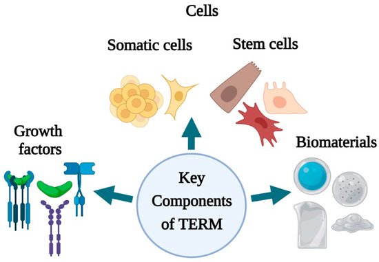
Skin tissue engineering has made remarkable progress in wound healing treatment with the advent of newer fabrication strategies using natural/synthetic polymers and stem cells. Currently, stem cells and biomaterials are popularly used in the skin tissue engineering approach in different wound healing treatments. In skin tissue engineering application, stem cell facilitates in the regeneration of disintegrated tissue. Whereas, biomaterials serve as a platform to improve the engraftment of implanted cells and facilitate the function of exogenous cells by mimicking the tissue microenvironment. Hence, the combination and synergistic effect of biomaterials and stem cells have the potential to broaden the application of skin tissue engineering in wound healing treatment therapies.  

wound healing tissue engineering skin regeneration

1. Introduction

The skin, an intricate structure composed of the epidermis, dermis, skin appendages, hair follicles, and sebaceous glands, is the body’s largest organ with direct exposure to the environment [1]. Healthy skin is essential in maintaining the human body’s physiological homeostasis as it protects the human body against infection, electrolyte loss, mechanical forces, and thermal imbalance. It also plays a pivotal role in dynamic processes such as hydration, initiation of vitamin D synthesis, and excretion [2]. Therefore, any disruption in skin integrity may lead to tissue disintegration, resulting in acute or chronic wounds. Acute wounds are traumatic injuries including burns or surgically created wounds that heal within an acceptable period of time, whereas chronic wounds including ulcers (diabetic, venous, pressure) or post-surgical wounds are those that fail to progress through the common healing process in a timely fashion, resulting in the lack of a significant recovery over a prolonged period of time [3].
The wound healing process comprises the coordination between the overlapping processes of inflammation, blood clotting, cellular proliferation, and extracellular matrix remodeling regulated by secretion of various growth factors, cytokines, and chemokines [1]. Malfunction in the following processes may lead to abnormal wound healing and failed regeneration leading to inconvenience in treatment, limiting wound repair, and tissue integrity restoration. Numerous conventional and regenerative studies have been catered towards achieving effective wound therapies to reduce health costs and ensure successful scar healing and long-term relief [4].
The emergence of skin tissue engineering has contributed to robust innovations in skin substitutes and replacement products for wound healing and tissue regeneration. The process includes various cells, biomaterials, biochemical, and physiochemical factors, and engineering technologies to improve or replace skin tissues [2]. Growth factors, stem cells, and scaffolds are collectively known as the tissue engineering triad, and scientists have been looking for the best combination to use these tools to develop safer and cost-effective approaches for wound healing and repair [3]. The combination of stem cells with a specifically designed novel 3D biomaterial has different impacts on engineered skin post-implantation [5]. An ideal biomaterial with multiple varieties of cultured cells and a collectively established broad knowledge of the healing process are the main criteria for the future development of skin substitutes [6].

2. Skin Tissue Engineering and Regenerative Medicine

Tissue engineering, an emerging interdisciplinary field in biomedical engineering, aims to regenerate new biological materials for replacing diseased or damaged tissues or organs [7]. A source of cells, biomaterials, and biomolecules is required along with an artificial extracellular matrix upon which cells can be supported and enriched for further survival. The engineered skin substitutes can be classified into three categories (i) based on materials: Biological, synthetic, and biosynthetic; based on covering time: Temporary and permanent; and (ii) based on layer: Epidermal, dermal, and bilayered skin substitutes, and can be used in combating acute and chronic skin wounds [8]. The first attempt in skin tissue engineering was taken by fabricating a cultured epidermal autograft (CEA) from the small piece of skin containing cultured human keratinocytes, which were later used clinically for burn treatment. However, these autologous cultured sheets exhibited certain demerits such as graft instability, prolonged culture time, formation of fragile skin after healing, and lack of dermal matrix support, limiting its application [2]. Since then, several innovative approaches have been adopted to create skin substitutes with essential properties including being easy to handle and apply to the wound site; enabling vital barrier function with appropriate water flux; being sterile, non-toxic, and non-antigenic; and evoking minimal inflammatory reactivity [4]. Besides, they should be incorporated into the host with minimal scarring or pain and facilitate angiogenesis while still being cost-effective [9]. Thus, a number of approaches based on the choices of cell types (keratinocytes, fibroblast, stem cells), their source (allogenic or autologous), and choice of biomaterials for matrix formation (synthetic, natural, ECM based) have been made to improve skin substitutes [4].
Other than tissue engineering, regenerative medicine has grown out of diverse disciplines such as surgery, organ transplantation, biomaterial science, developmental biology, and stem cell biology [10]. At present, the scope of regenerative medicine in wound treatment includes technologies and approaches that induce the body to redevelop missing tissue, regardless of their conformation, and engineered tissue or organs designed fully to replace the missing structures [7]. The merging of tissue engineering and regenerative medicine occurred with stem cell and therapeutic cloning research. This merging has been abetted by recognizing that various engineered skin constructs, some of which were originally designed to engraft and serve as replacement structures, stimulate endogenous processes that remodel the construct with the body’s tissue [10]. Another scope of regenerative medicine is to incorporate the relationship between tissues, organs, and systems, even the body as a whole, that enables us to combine several different molecular approaches into one course of treatment. The example includes reducing inflammation, stimulating tissue development pathways, recruiting endogenous stem cells, modulating immune function, and stimulating new blood vessel formation [4]. Hence, a deeper understanding of TERM, including the relationships between systems, will connect clinicians with scientific engineering skills to commercial teams and guide new technologies towards safe and effective treatment strategies in wound healing [10].

3. Techniques of Skin Tissue Engineering

In skin tissue engineering, new techniques such as bioprinting, bio-fabrication, and bio-inking coupled with advances in DNA microarray, proteomics, and stem cells have allowed the generation of skin replacements exploration [11]. The main elements of tissue engineering include biomaterials, cells, growth factors, other signaling molecules, and engineering components such as scaffolds, pumps, tubes, and bioreactors [2]. At present, 3-dimensional (3D) scaffold constructs made via bio-fabrication techniques exploit the field of skin tissue engineering as a key component in the wound healing process. Scaffolds play a unique role in the repair and restoration of disintegrated tissue by providing a suitable platform for various factors associated with cell survival, proliferation, and differentiation [6]. It can be constructed from natural and/or synthetic biomaterials, either materials that remain stable in a biological environment or materials that degrade in the human body [2]. Several techniques have been adopted for their constructions, but the four main approaches that are widely used include: (i) Sheets of cells secreting ECM [12]; pre-made porous scaffolds of synthetic, natural, and biodegradable biomaterial; (ii) decellularized ECM scaffolds, and (iii) cells entrapped in hydrogels [6]. The main objective of the scaffold is to represent the matrix as similar as possible to the native ECM as all cells are in close contact with ECM, which provides structural support to cells and tissues, stimulating migration proliferation, apoptosis, survival, and differentiation [2]. Hence, based on this, different parameters such as physio-chemical properties of the pristine materials, mechanical properties, shape, structure, pore sizes, and distribution need to be considered while fabricating scaffolds for wound healing and regeneration [13].
In practice, the techniques of fabrication of 3D scaffolds are subdivided into conventional and rapid prototyping (RP) methods, each producing different scaffolds with different characteristics [14]. Scaffold fabrication using conventional techniques include the construction of porous polymeric structures such as substrates for cell adhesion; however, it is challenging to obtain complex structures of microscale (containing an environment suitable for cell survival and function) and macroscale (permit the coordination of multicellular process, provide adequate transport of nutrients, and possess mechanical properties) using conventional methods [15]. On the other hand, the RP scaffold fabrication technique provides versatile opportunities for skin tissue engineering. It allows the independent control of macroscale and microscale features, facilitating the fabrication of multicellular structures needed for complex tissue functions [15]. Moreover, 3D vascular bed fabrication is possible using the RP technique, which allows the support of massive tissue formation. In addition, RP provides an opportunity to combine fabrication techniques with clinical imaging data, increasing the possibility of producing a bulk number of customized scaffolds in designated designs [16].
Apart from the conventional fabrication technique, 3D bioprinting involving the use of computer-controlled deposition of cells into precise 3D geometrical patterns has shown promising accuracy in cell delivery to replicate natural skin anisotropy [17]. Tarassoli et al. (2018) describe two main approaches to arranging cells in 3D patterns. The former is a top-down approach whereby cells are co-arranged with biomimetic scaffolds with tissue maturation in a bioreactor [18]. On the other hand, the latter involves a bottom-up fabrication technique in which temporary support instigates the secretion of the matrix by cells themselves. Despite the chosen 3D bioprinting technique, the functionality of a successfully engineered skin largely depends on the biomaterial and cells used. Some aspects that are considered during biomaterial selection include biocompatibility, biodegradability, bio inertness, strength, durability, and ductility [19][20]. For bioprinting purposes, biomaterials should be ‘printable’ depending on their rheology (divided into aspects such as shear thinning and viscosity) and cross-linking abilities (through a chemical, physical, stereo complex, or ionic mechanism) [17]. Cell selection is the second key component of bioprinting. So far, much of the research has focused on using keratinocytes, which can be propagated easily in cell culture, and fibroblasts, which have multilineage potential [21]. Besides, stem cells have been sought out as potential alternatives as they can both self-renew and differentiate into multiple cell types [17]. Although bioprinting technology offers promising outcomes in skin tissue engineering, a lack of understanding of bioink compatibility and biomaterial characteristics can be a major limitation in fully realizing this technology [22]. Hence, it is critical to innovate properties of bioinks that facilitate easy bioprinting processes, while preserving the viability and function of the printed tissue constructs

4. Components of Skin Tissue Engineering and Regenerative Medicine

Tissue engineering and regenerative medicine (TERM) can be considered a multidisciplinary and emerging field in technology used to regenerate damaged organs, produce complex tissues, and maintain normal cell homeostasis [7]. TERM aims to design new tissues and organ replacements that closely mimic a typical physiological environment for cells. It has caused a revolution in the present and future therapeutic possibilities for acute and chronic wound healing, improving restoration of biological function and rehabilitation [10]. Advanced multidisciplinary TERM approaches involving growth factors, stem cells, and biomaterials are being adopted to induce regeneration or indirectly change the wound environment and stimulate healing [2]. The key components of regenerative medicine and tissue engineering (growth factors, cells, stem cells, biomaterials) (Figure 1) have unveiled several perspectives for skin tissue engineering and regeneration that can be used to address different stages of wound healing.
Figure 1. Key components of skin tissue engineering and regenerative medicine (created with BioRender.com).
Growth factors (GFs) are defined as the biologically active polypeptides that control tissue repair via interacting with specific cell surface receptors. They play a prominent role in cell migration into the wound area, promote epithelialization, initiate angiogenesis, and stimulate matrix formation followed by remodeling the wounded area [11]. Epidermal growth factor (EGF) [23], basic fibroblast growth factor (bFGF) [21], transforming growth factor-beta (TGFβ3) [22], platelet-derived growth factor (PDGF) [24], and vascular endothelial growth factor (VEGF) [25] are some of the GFs that contribute to the wound-healing process. Several researchers have proven the role of each growth factor in wound healing by promoting angiogenesis, cell migration, re-epithelialization, and granulation tissue formation. Moreover, a handful of studies verified the potential of using growth factors in combination with carriers for effective delivery in maximizing wound healing.
Apart from growth factors, both cells and cellular skin substitutes (both differentiated and stem cells) have exhibited great potential by providing all the elements required for skin regeneration such as cells, mediators, and materials mimicking ECM [3]. The use of viable cells cultured in special conditions are used to produce cell sheet substitutes that contribute to wound repair. Among the available differentiated human cells, fibroblasts and keratinocytes are the primary sources for epidermal and dermal substitute production, respectively [26]. Taking advantage of the wound healing properties of fibroblasts and keratinocytes, specific cell composition constructs have been developed according to the treatment target for dermal and epidermal regeneration [2]. When applied to the wounded area, the cells supply signaling molecules, growth factors, and ECM proteins that aid healing [27]. Through a paracrine crosstalk mechanism, fibroblasts and keratinocytes communicate with each other, leading to cell recruitments and maintenance of skin homeostasis, which is desirable for complete wound healing. For this purpose, several double-layer dermal cellular skin substitutes have been synthesized commercially, incorporating both fibroblasts and keratinocytes for the repair and regeneration of chronic wounds [28]. EpiCel, Dermagraft, and Apligraf (Figure 2) are some of the instances of commercially available cellular skin substitute products that incorporate both keratinocytes and fibroblasts, respectively. These novel products are assembled according to their specific conformation and structure; in particular, pore sizes and their distribution are essential in providing a suitable matrix for effective cell migration and arrangement [29]. The novelty of these products represents a basis for revascularization, forming a proper microenvironment for cellular migration and proliferation [30]
Figure 2. Cellular skin substitutes made from fibroblast and keratinocyte (created with BioRender.com)
The use of stem cells (SC) has become a promising new approach in tissue engineering and regenerative medicine for skin injury treatment. Stem cells can be defined and characterized based on their capacity for self-renewal, asymmetric replication, and potency [31]. They are attributed with the ability to replenish lost cells throughout the lifespan of an organism via unlimited replication, providing a population of sister stem cells [32]. The main clinical focus of stem cell application in wound healing is to accelerate the healing process, prevent wound contracture and scar formation, and initiate earlier wound closure and regeneration of skin and its appendages. Besides, stem cells can secrete pro-regenerative cytokines, making them an attractive agent for treating chronic wounds [31]. Among the primary sources of cells that are in use for wound healing and the regeneration of injured skin are embryonic stem cells (ESCs), adult stem cells, and induced pluripotent stem cells (iPSCs) [33]
In tissue engineering, biomaterials play a prominent role in unlocking the regenerative potential innate to human tissues/organs, restoring deteriorated states, and re-establishing normal bodily function [34]. Biomaterial science and engineering have witnessed tremendous growth in the past five decades due to vast investment in developing new products [35]. In a broader sense, biomaterials can be defined as material devices or implants used to repair or replace native body tissues or as a provisional scaffolding material adopted to construct human-made tissues or organs [34]. Using biomaterial in tissue engineering aims to provide temporary mechanical support and mass transport to encourage cell adhesion, proliferation, migration, and differentiation and control the size and shape of the regenerated tissue [36]. Moreover, biomaterials known as temporary scaffolds act as an ECM template that is actively involved in delivering cues to the cells that form the regenerated tissues [4]. Numerous approaches are adopted for designing matrices, comprised of innovative biomaterials possessing two crucial traits: Biocompatibility (the materials must hold minimal toxicity and immunogenicity) and biodegradability (the materials must be easily removable upon completion of their function) [36]. Furthermore, they must also possess the ability to interact with a biological environment and particularly modulate cellular response [4]. Hence, biomaterials have become an active part of cellular function regulation and act as a support for tissue regeneration or a platform for drug delivery. 

5. Conclusion

In summary, the use of growth factors, cells, and biomaterials have received attention in skin tissue engineering and regenerative medicine due to their ability and capacity to improve wound healing. However, immune sensitivity, compromised survival, proliferation, and differentiation of cells and, unable to fabricate a suitable scaffold limit the application of cells and biomaterials in clinical trials as well as in vitro and in vivo settings. With the aid of appropriate technology, these barriers can be overcome. Natural and synthetic biomaterials can be rationally designed for wound healing treatment according to their biophysical and biochemical properties. The incorporation of cells (both differentiated and stem cells) into structured and modified biomaterials increases the competence of restoring and repairing dysfunctional skin tissue and promotes wound healing parameters such as improved epithelialization, granulation tissue formation, vascularization, and angiogenesis. The well-organized spatial properties of a biomaterial or scaffold, in turn, can provide a protective and sometimes inducible microenvironment for the cells, mimicking the natural ECM. In addition, biomaterials are also being used to regulate stem cell fate before and after delivery by providing mechanical and biochemical support. Despite the encouraging results in non-clinical studies, only a handful of biomaterials have been used for skin tissue engineering in patients. Thus, additional clinical trials that use biomaterial should be performed to elucidate the influence of materials’ biophysical and biochemical properties on wound healing, tissue repair, and regeneration of humans. Hence, future efforts are essential to improve the clinical outcome in designing and fabricating biomaterials using emerging techniques like 3D bioprinting, electrospinning, and nanotechnology to meet specific properties of the components that need to be delivered for wound healing and regeneration.

References

  1. Ho, J.; Walsh, C.; Yue, D.; Dardik, A.; Cheema, U. Current advancements and strategies in tissue engineering for wound healing: A comprehensive review. Adv. Wound Care (New Rochelle) 2017, 6, 191–209.
  2. Tottoli, E.M.; Dorati, R.; Genta, I.; Chiesa, E.; Pisani, S.; Conti, B. Skin wound healing process and new emerging technologies for skin wound care and regeneration. Pharmaceutics 2020, 12, 735.
  3. Rezaie, F.; Momeni-Moghaddam, M.; Naderi-Meshkin, H. Regeneration and repair of skin wounds: Various strategies for treatment. Int. J. Low. Extrem. Wounds 2019, 18, 247–261.
  4. Gurtner, G.C.; Chapman, M.A. Regenerative medicine: Charting a new course in wound healing. Adv. Wound Care 2016, 5, 314–328.
  5. Xu, Y.; Chen, C.; Hellwarth, P.B.; Bao, X. Biomaterials for stem cell engineering and biomanufacturing. Bioact. Mater. 2019, 4, 366–379.
  6. Chaudhari, A.A.; Vig, K.; Baganizi, D.R.; Sahu, R.; Dixit, S.; Dennis, V.; Singh, S.R.; Pillai, S.R. Future prospects for scaffolding methods and biomaterials in skin tissue engineering: A review. Int. J. Mol. Sci. 2016, 17, 1974.
  7. Boyce, S.T.; Lalley, A.L. Tissue engineering of skin and regenerative medicine for wound care. Burn. Trauma 2018, 6, 4.
  8. Dhasmana, A.; Singh, S.; Kadian, S.; Singh, L. Skin tissue engineering: Principles and advances. J. Dermatol. Skin 2018, 1, 3–6.
  9. Nicholas, M.N.; Jeschke, M.G.; Amini-Nik, S. Methodologies in creating skin substitutes. Cell. Mol. Life Sci. 2016, 73, 3453–3472.
  10. Gomes, M.E.; Rodrigues, M.T.; Domingues, R.M.A.; Reis, R.L. Tissue engineering and regenerative medicine: New trends and directions-A year in review. Tissue Eng. Part B Rev. 2017, 23, 211–224.
  11. Nourian Dehkordi, A.; Mirahmadi Babaheydari, F.; Chehelgerdi, M.; Raeisi Dehkordi, S. Skin tissue engineering: Wound healing based on stem-cell-based therapeutic strategies. Stem Cell Res. Ther. 2019, 10, 111.
  12. Nakayama, C.; Fujita, Y.; Matsumura, W.; Ujiie, I.; Takashima, S.; Shinkuma, S.; Nomura, T.; Abe, R.; Shimizu, H. The development of induced pluripotent stem cell-derived mesenchymal stem/stromal cells from normal human and RDEB epidermal keratinocytes. J. Dermatol. Sci. 2018, 91, 301–310.
  13. Blackstone, B.N.; Hahn, J.M.; McFarland, K.L.; DeBruler, D.M.; Supp, D.M.; Powell, H.M. Inflammatory response and biomechanical properties of coaxial scaffolds for engineered skin in vitro and post-grafting. Acta Biomater. 2018, 80, 247–257.
  14. Roseti, L.; Parisi, V.; Petretta, M.; Cavallo, C.; Desando, G.; Bartolotti, I.; Grigolo, B. Scaffolds for bone tissue engineering: State of the art and new perspectives. Mater. Sci. Eng. C 2017, 78, 1246–1262.
  15. Eltom, A.; Zhong, G.; Muhammad, A. Scaffold techniques and designs in tissue engineering functions and purposes: A Review. Adv. Mater. Sci. Eng. 2019, 3429527.
  16. Raeisdasteh Hokmabad, V.; Davaran, S.; Ramazani, A.; Salehi, R. Design and fabrication of porous biodegradable scaffolds: A strategy for tissue engineering. J. Biomater. Sci. Polym. Ed. 2017, 28, 1797–1825.
  17. Mondschein, R.J.; Kanitkar, A.; Williams, C.B.; Verbridge, S.S.; Long, T.E. Polymer structure-property requirements for stereolithographic 3D printing of soft tissue engineering scaffolds. Biomaterials 2017, 140, 170–188.
  18. Tarassoli, S.P.; Jessop, Z.M.; Al-Sabah, A.; Gao, N.; Whitaker, S.; Doak, S.; Whitaker, I.S. Skin tissue engineering using 3D bioprinting: An evolving research field. J. Plast. Reconstr. Aesthetic. Surg. 2018, 71, 615–623.
  19. Ramanathan, G.; Singaravelu, S.; Muthukumar, T.; Thyagarajan, S.; Perumal, P.T.; Sivagnanam, U.T. Design and characterization of 3D hybrid collagen matrixes as a dermal substitute in skin tissue engineering. Mater. Sci. Eng. C 2017, 72, 359–370.
  20. Park, Y.R.; Ju, H.W.; Lee, J.M.; Kim, D.-K.; Lee, O.J.; Moon, B.M.; Park, H.J.; Jeong, J.Y.; Yeon, Y.K.; Park, C.H. Three-dimensional electrospun silk-fibroin nanofiber for skin tissue engineering. Int. J. Biol. Macromol. 2016, 93, 1567–1574.
  21. Choi, S.M.; Lee, K.M.; Kim, H.J.; Park, I.K.; Kang, H.J.; Shin, H.C.; Baek, D.; Choi, Y.; Park, K.H.; Lee, J.W. Effects of structurally stabilized EGF and bFGF on wound healing in type I and type II diabetic mice. Acta Biomater. 2018, 66, 325–334.
  22. Li, M.; Qiu, L.; Hu, W.; Deng, X.; Xu, H.; Cao, Y.; Xiao, Z.; Peng, L.; Johnson, S.; Alexey, L.; et al. Genetically-modified bone mesenchymal stem cells with TGF-beta3 improve wound healing and reduce scar tissue formation in a rabbit model. Exp. Cell Res. 2018, 367, 24–29.
  23. Jeong, S.; Kim, B.; Park, M.; Ban, E.; Lee, S.H.; Kim, A. Improved diabetic wound healing by EGF encapsulation in gelatin-alginate coacervates. Pharmaceutics 2020, 12, 334.
  24. Xu, K.; An, N.; Zhang, H.; Zhang, Q.; Zhang, K.; Hu, X.; Wu, Y.; Wu, F.; Xiao, J.; Zhang, H.; et al. Sustained-release of PDGF from PLGA microsphere embedded thermo-sensitive hydrogel promoting wound healing by inhibiting autophagy. J. Drug. Deliv. Technol. 2020, 55, 101405.
  25. Chen, G.; Yu, Y.; Wu, X.; Wang, G.; Ren, J.; Zhao, Y. Bioinspired multifunctional hybrid hydrogel promotes wound healing. Adv. Funct. Mater. 2018, 28, 1801386.
  26. Kallis, P.J.; Friedman, A.J.; Lev-Tov, H. A guide to tissue-engineered skin substitutes. J. Drugs Dermatol. 2018, 17, 57–64.
  27. Ter Horst, B.; Chouhan, G.; Moiemen, N.S.; Grover, L.M. Advances in keratinocyte delivery in burn wound care. Adv. Drug Deliv. Rev. 2018, 123, 18–32.
  28. Nicholas, M.N.; Yeung, J. Current status and future of skin substitutes for chronic wound healing. J. Cutan. Med. Surg. 2017, 21, 23–30.
  29. Tavakoli, S.; Klar, A.S. Bioengineered skin substitutes: Advances and future trends. Appl. Sci. 2021, 11, 1493.
  30. Chang, D.K.; Louis, M.R.; Gimenez, A.; Reece, E.M. The basics of integra dermal regeneration template and its expanding clinical applications. Semin. Plast. Surg. 2019, 33, 185–189.
  31. Duscher, D.; Barrera, J.; Wong, V.W.; Maan, Z.N.; Whittam, A.J.; Januszyk, M.; Gurtner, G.C. Stem cells in wound healing: The future of regenerative medicine? A mini-review. Gerontology 2016, 62, 216–225.
  32. Kucharzewski, M.; Rojczyk, E.; Wilemska-Kucharzewska, K.; Wilk, R.; Hudecki, J.; Los, M.J. Novel trends in application of stem cells in skin wound healing. Eur. J. Pharmacol. 2019, 843, 307–315.
  33. Dash, B.C.; Xu, Z.; Lin, L.; Koo, A.; Ndon, S.; Berthiaume, F.; Dardik, A.; Hsia, H. Stem cells and engineered scaffolds for regenerative wound healing. Bioengineering 2018, 5, 23.
  34. Aramwit, P. Introduction to biomaterials for wound healing. In Wound Healing Biomaterials; Agren, M.S., Ed.; Woodhead Publishing: Cambridge, UK, 2016; pp. 3–38.
  35. Chen, F.M.; Liu, X. Advancing biomaterials of human origin for tissue engineering. Prog. Polym. Sci. 2016, 53, 86–168.
  36. Sheikholeslam, M.; Wright, M.E.E.; Jeschke, M.G.; Amini-Nik, S. Biomaterials for skin substitutes. Adv. Healthc. Mater. 2018, 7, 1700897.
More
Information
Subjects: Polymer Science
Contributors MDPI registered users' name will be linked to their SciProfiles pages. To register with us, please refer to https://encyclopedia.pub/register : ,
View Times: 2.1K
Revisions: 2 times (View History)
Update Date: 19 May 2021
1000/1000
Video Production Service