 +1 credit
| Version | Summary | Created by | Modification | Content Size | Created at | Operation |
|---|---|---|---|---|---|---|
| 1 | Miguel Angel Prieto Lage | + 6231 word(s) | 6231 | 2021-04-22 05:05:13 | | | |
| 2 | Camila Xu | Meta information modification | 6231 | 2021-04-23 12:15:06 | | |
Video Upload Options
Xanthophylls (fucoxanthin, astaxanthin, lutein, zeaxanthin, and β-cryptoxanthin) are a type of carotenoids with anti-tumor and anti-inflammatory activities, due to their chemical structure rich in double bonds that provides them with antioxidant properties.
1. Introduction
In recent years, consumer demand for naturally sourced products to promote health and reduce disease has grown steadily [1]. This demand has entailed an increased interest in new natural sources of food, pharmaceutical, and cosmetic products [2][3]. In this context, the marine environment has been considered a potential reservoir of natural compounds [4]. Among the organisms present in this environment, it is worth highlighting algae. Algae constitute a polyphyletic group of photosynthetic primary producers organisms, which represent an interesting source of chemical components with high-value biological activities. [5]. Although the total number of algal species is unknown, it is thought to vary between one and ten million [6].
The high value of algal extracts is due to their large number of molecules such as carbohydrates, proteins, peptides, lipids (including oils and polyunsaturated fatty acids, PUFAs), minerals, iodine, phenols (polyphenols, tocopherols), alkaloids, terpenes, and pigments (as chlorophylls, carotenoids, and phycobilins) [7][8]. Within these compounds, one of the groups with greater interest are pigments due to the concentrations in which they are present, these being higher than that of other compounds such as phenolic compounds. In fact, algae are considered pigment-producing organisms. They have a great variety of pigments, which can be classified into three large groups: chlorophylls, carotenoids, and phycobilins. Therefore, different carotenoids (CA) profiles can be used as a medium for algal classification [9]. In this way, a first classification of the algae allows us to make a division according to the size of the algae (microalgae or macroalgae) and the following divisions according to their tones, among other characteristics. As a result, the first group comprises greenish algae (Cyanophyceae), green algae (Chlorophyceae), diatoms (Bacillariophyceae), and golden algae (Chrysophyceae), among others. Meanwhile, the macroalgae family includes red (Rhodophyta), brown (Ochrophyta), and green algae (Chlorophyta) [10][11][12]. This diversity of species and, therefore, of its chemical compositions is interesting, since once compounds are properly isolated or extracted from algae, they may show a diverse range of biological activities, such as antioxidant, antimicrobial, anticancer, anti-allergic, antiviral, and anticoagulant activities, among others [7][8]. This diversity of biological activities implies that there is also a significant variety of potential applications in human health, agriculture, and in food and cosmetic industries [4], in which its application depends on its chemical composition.
On an industrial scale, the most interesting species are those that produce high percentages of CA. CA are usually located in chloroplasts or stored in vesicles and a cytoplasmic matrix of plants, algae, photosynthetic bacteria, and some fungi [9]. All CA are tetraterpenes, which are compounds that have a skeleton composed of 40 carbon atoms conjugated in polyene chains [9]. They are classified into two main groups: (i) compounds that have a hydrocarbon long chain known as carotenes and (ii) compounds that have an oxygen atom in its structure, known as xanthophylls. The first group includes α-carotene, β-carotene, lycopene, and phytoene, among others. The most representative molecules of the second group are fucoxanthin, astaxanthin, lutein, zeaxanthin, and β-cryptoxanthin. This difference in its structure makes xanthophylls more polar than carotenes due to the presence of oxygen in the form of methoxy, hydroxy, keto, carboxy, and epoxy positions. However, except for lutein, they are still non-polar compounds [13]. Its structure with alternating double bonds is responsible for many of its biological functions, being the main function in photosynthetic organisms to act as accessory pigments for the capture of light in photosynthesis, and to protect photosynthetic machinery against self-oxidation [14]. However, despite the wide diversity of molecules in the carotenoid family, with more than 700 compounds currently known, only about 30 CA have a significant role in photosynthesis [13]. In recent years, numerous studies have highlighted CA multiple effects on human health due to their antioxidant properties, preventing the damage caused by oxidative stress and therefore declining the risk of chronic diseases [14][15]. However, the biological properties of CA are not limited to their antioxidant properties. The scientific literature has shown CA actions as anti-tumor [16][17][18], anti-inflammatory [19][20], neuroprotective, antimicrobial, antidiabetic, and antiobesity [21][22]. Therefore, algae have several CA with market interest (β-carotene, fucoxanthin, astaxanthin, lutein, zeaxanthin, and violaxanthin), representing a natural and sustainable source of these compounds [9].
Among the xanthophylls of interest is fucoxanthin, which is one of the most abundant marine CA, accounting for approximately 10% of the total production of natural CA [23]. It is found in abundant concentrations in the chloroplasts of several brown seaweeds, such as Laminaria japonica, Undaria pinnatifida, Sargassum fusiformis, in several species belonging to the genera Sargassum (Sargassum horneri) and Fucus (Fucus serratus, Fucus vesiculosus) and in diatoms (Bacillariophyta) [9][24][25][26]. Another xanthophyll of interest is astaxanthin (AS), which is a red pigment. AS is considered a potent antioxidant as it has about ten times more antioxidant activity than other CA [27]. The main natural sources of this pigment are the microalgae Haematococcus pluvialis, Chlorella zofingiensis, and Chlorococcum sp. [28]. H. pluvialis is a single-celled green freshwater alga. It is the richest source of natural AS and is already produced on an industrial scale [26]. Procedures have been technologically advanced to grow Haematococcus containing 1.5–3.0% AS dry weight [27][29]. The richest source of β-carotene is the halotolerant green microalgae Dunaliella salina, accumulating up to 10% of it based on the dry weight of the microalgae [30][31]. When H. pluvialis and D. salina are cultivated in extreme conditions (such as high salinity, high luminosity, or lack of nutrients), AS and β-carotene, respectively, can reach more than 90% of the total carotenoids [7]. Lutein and zeaxanthin are pigments found in algal species such as Scenedesmus spp., Chlorella spp., Rhodophyta spp., or Spirulina spp. respectively [32]. Esteban et al., 2009 [33], reported that red algae (Rhodophyta) show a common carotenoid pattern of β-carotene and one to three xanthophylls: lutein, zeaxanthin, or anteraxanthin. Corallina elongata and Jania rubenseran were the only algae that contained anteraxanthin as the main xanthophyll. Spirulina platensis (strain pacifica) microalgae is a source of β-cryptoxantine, β-carotene, and zeaxanthin. β-cryptoxantine is a pigment that can also be found in plants [34]. The siphonaxanthin content in green algae such as Umbraulva japonica, Caulerpa lentillifera, and Codium fragile constitutes about 0.03%–0.1% of the dry weight [35]. The cyanobacteria Synechococcus sp. strain PCC7002 produces a monocyclic myxoxanthophyll, which is identified as Myxol-2 Fucoside (Myxoxanthophyll), in addition to producing other CA such as β-carotene, zeaxanthin, and sinecoxanthin [36]. The CA composition in cyanobacteria is very different from that of other algae, including for example β-carotene, zeaxanthin, myxol pentosides, and echineone [32].
Animals should get all these CA through the diet, as they are unable to synthesize them. CA are commonly incorporated as dietary supplements, feed additives, and food colorants in several sorts of food, such as dairy products and beverages, and also in the pharmaceutical and cosmetic industries [37]. As shown in Figure 1, CA have a high repertoire of commercial applications due to their multiple biological properties. Among the most notable applications are cosmetic, nutraceuticals, pharmaceutical purposes, and other human applications.
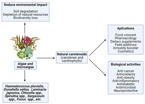
Figure 1. Positive effects on human health and industrial applications of carotenoids from natural sources.
Attributable to the various positive activities on human health and the multiple industrial applications of CA, global demand continues to increase. It is estimated that in 2026, the CA market will grow to USD 2.0 billion, registering an annual growth rate for CA of 4.2% [38]. The most relevant and important pigments on the market today are β-carotene and AS, followed by lutein, lycopene, and canthaxanthin [13][31]. So far, most commercial CA are artificially produced. However, the strong global interest in food of natural origin that is safe, healthy, and environmentally friendly has increased the demand for natural sources of CA [22]. Algae and algal extracts are a sustainable option for CA and have numerous benefits in comparation to alternative natural sources. For instance, its cultivation and production is cheap, easy, and ecological, its removal has higher yields and is simple, and raw materials are not scarce, nor are there seasonal limitations [32][39][40]. In order to obtain high concentrations of a certain compound, culture conditions and environmental stress can be modified to manipulate the biochemical composition of microalgae [39]. However, under optimal growth conditions, the concentration of CA pigments is often too low to produce microalgal-based pigments, making it economically unviable [13][40]. To improve its economic viability, it is vital to explore and understand how environmental factors and the integration of nutrients into the environment affect the production of compounds. Understanding how the metabolic pathways of species vary according to the culture conditions, the co-production and accumulation of multiple compounds in microalgae will be improved [41].
2. Mechanism of Action of Xanthophylls
2.1. Metabolism
The mechanism of action of xanthophylls is the specific binding through which the molecule produces its pharmacological effect. This effect will depend on the absorption, distribution, and metabolism of the compound, which are critical parameters of the pharmacokinetics of the xanthophylls. This can be seen in various studies that show the low presence of this type of compound in human tissues, which directly depends on their metabolism and intestinal absorption, and therefore, its bioavailability [42]. The metabolism of xanthophylls is poorly studied, especially for those that do not have provitamin A activity. Hence, more studies are needed to understand its metabolism and, therefore, be able to develop different applications according to the mechanism by which its biological activities occur.
In turn, this would allow the development of safe and effective applications in humans as well as increase its bioavailability [43]. For example, studies on FU metabolism revealed that this compounds itself is not present in plasma but rather its metabolites due to oxidative reactions that take place on FU in mammals. This reaction transforms both compounds into ketocarotenoids [44]. In addition, when FU is administered orally, it undergoes a process of hydrolysis at the intestinal level, giving rise to fucoxanthinol, while liver metabolization results in other active metabolites such as amarouciaxanthin A [45][46]. In fact, it was reported that dietary FU accumulated in the heart and liver as fucoxanthinol and in adipose tissue as amarouciaxanthin A, the latter being non-detectable by HPLC in human serum [47]. Therefore, the oral administration of this compound may only provide some bioactive metabolites, as it is completely metabolized. To release products that maintain its biological activities, it is necessary to develop alternatives that prolong its biological half-life [45], such as emulsions or encapsulations (Table 2).
Table 2. Delivery systems used to increase marine carotenoids’ bioavailability.
| Mol. | Delivery System | Assay | Benefits | Results | Use | Ref. |
|---|---|---|---|---|---|---|
| FU | Palm stearin solid lipid core | In vitro | Increase stability during storage | Release of FU of 22.92% during 2 h in SGF and 56.55% during 6 h SIF | Oral supplements | [48] |
| Nanoparticles of zein | ABTS DPPH | Increase antioxidant activity | More antioxidant than free FU | Foods and beverages | [49] | |
| Nanoemulsion | In vitro | Increase stability during storage; antiobesity | 95% of FU remains in the emulsion after 4 weeks | Food, beverages, nutraceutics | [50] | |
| Nanoemulsion (LCT) | In vitro digestion and bioability assays in rats | Increase stability | Increase FU level in serum blood (LCT > MCT) | Functional foods and nutraceutics | [51] | |
| Chitosan–glycolipid nanogels | In vitro | Significant increase in bioavailability | Lpx levels (nmol MDA/mL) higher in control (30.9) than in emulsions (17.0–12.15) | Foods and nutraceutics | [52] | |
| AS | Fish oil | In vitro | Useful for supplementation | Better antioxidant effect | Oral supplements | [53] |
| Encapsulation | TBARS Peroxide enzymes | Increase stability | Better antioxidant effect | Foods | [54] | |
| Pectin–chitosan multilayer | Stability Assays | Increase stability | Better stability than monolayer | Nutraceuticals, functional, medical foods | [55] | |
| l-lacic acid | Release and stability test | Increase stability | Enhance stability | Functional foods and nutraceutics | [56] | |
| Ascobyl palmitate emulsion | Stability assay | Sublingual delivery | Enhance sports performance, skin protection, cardioprotective | Dietetic supplementation in sports | [57] | |
| LU | β-CD | In vitro | Increase stability | More stable against oxidating agents | Foods | [58] |
| Glycyrrhizic acid, arabinogalactan | In vitro | Solubility enhancement | Prevention of H-aggregates formation, increase of photostability | Foods | [58] | |
| ZEA | Sea Buckthorn oil and water emulsion | Stability and digestive assays | Increase bioaccesibility | Increase 64.55% | Functional foods and nutraceutics | [59] |
| High-pressure treatment | Stability and digestive assays | Improve Nannochloropsis sp. ZEA disponibility | Foods | [60] | ||
| Glycyrrhizic acid, arabinogalactan | In vitro | Solubility enhancement | Prevention of H-aggregates formation, increase of photostability | Foods | [58] |
SGF: Simulated gastric fluid; SIF: Simulated intestinal fluid; LCT: Long-chain triglycerides; MCT: Medium-chain triglycerides.
A study carried out on rats reported that the pharmacokinetic parameters of AS only depend on the dose when it is administered intravenously due to the metabolism that takes place in the liver as a result of saturation of hepatic metabolism of AS [162]. As for AS metabolites described in humans, these are fundamentally 3-hydroxy-4-oxo-β-ionone and 3-hydroxy-4-oxo-7,8-dihydro-β-ionone [163]. The metabolization of AS after oral intake leads to 3-hydroxy-4-oxo-7,8-dihydro-β-ionol and 3-hydroxy-4-oxo-7,8-dihydro-β-ionone, being both compounds detected in plasma [164]. Several researchers hypothesize that the rate at which these reactions take place is determined by the structure of the ring, as well as by the length of the fatty acyl residue formed. Moreover, several enzymes, such as for example diacylglycerol acyltransferase 1, can catalyze the synthesis of AS esters in some strain. This is the case of the microalga Haematococcus pluvialis [165].
As for LU and its structural isomer, ZEA, studies carried out in humans have shown that both undergo an in vivo oxidation process that gives rise to several metabolites [61]. LU gives rise to a series of compounds (3′-epilutein, 3′-oxolutein) due to the presence of the enzyme that also mediated the conversion of fucoxanthinol to amarouciaxanthin A [62]. Other compounds such as 3-hydroxy-3′,4′-didehydro-β,γ-carotene and 3-hydroxy-2′,3′-didehydro-β,ε-carotene appear as result of acid hydrolysis in the stomach [63]. However, this compound is capable of remaining intact in its intact form in human ocular tissue due to the inability of the enzyme β-carotene-9′,10′-oxygenase to act on said organ. In this way, there is an extraordinary accumulation of these compounds in the ocular tissue, serving as a mechanism for the prevention of ocular diseases [64]. ZEA, being an isomer of LU, will undergo similar processes to LU. However, it is a much less studied molecule. In this way, ZEA will also be accumulated in the ocular tissue due to the inactivity of the enzymes responsible for the metabolism of ZEA in the organs of sight [65]. Therefore, to determine the bioavailability of LU it is necessary to quantify said metabolites, which also may have different bioactivities, with complementary studies.
2.2. Bioavailability and Bioaccessibility
Xanthophylls have been subjected to numerous studies due to its antioxidant activity and protective effect against several diseases [66]. In recent years, different studies have been carried out comparing the properties of synthetic CA with those of natural origin [67], noting that some of them can only be obtained from natural sources, where there is much more diversity. In addition, these CA obtained from algae can be co-extracted with other bioactive components such as polysaccharides or fatty acids. Therefore, the idea of incorporating CA in foods, nutraceuticals, or cosmetic products is of increasing interest due to their effective bioactive properties [68]. However, to develop and evaluate the viability of any food or cosmetic products that maintain these activities, it is necessary to know its bioactivity, bioavailability, and bioaccesibility [69]. These three parameters are influenced by several factors such as the food matrix; the type of cooking; the time of cooking; the CA involved; the presence of fats, fibers, proteins, and other nutrients in the diet; and the health or nutritional status in humans [70][71][72][73][74].
In humans, once CA are ingested, they are released from the food matrix through the action of gastric enzymes and must be emulsified with lipids in order to improve their absorption [75]. Moreover, its absorption mechanism will be determined by the concentration in which the compound is present. At low concentrations, absorption is mainly due to the action of type 1 class B scavenger receptor, which also captures high-density lipoproteins, platelet glycoprotein 4, and NPC1-like intracellular cholesterol transporter 1 [76]. At high concentrations, the main mechanism is passive diffusion through mucosa [77]. Enzymes released in the duodenum will also play an important role in the absorption, since in this part of the small intestine, pancreatic lipase is released. This enzyme assists the formation of mixed micelles of emulsified droplets with CA. This process depends on the concentration of bile acids among others [78]. Once the micelles are formed, they pass into the blood. Then, micelles are taken up by enterocytes, in which metabolization takes places due to the presence of the enzyme β-carotene oxygenase. The non-metabolized CA, such as LU and ZEA, are incorporated into chylomicrons or low-density lipoproteins (LDL) and are transported to the liver where they can be eliminated by the bile or metabolized and secreted in very low-density lipoprotein (VLDL) or high-density lipoproteins (HDL) to the peripheral tissues, as it can be seen in Figure 3 [75][79].
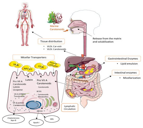
Figure 3. Uptake, transport, and secretion pathways of marine carotenoids in the human body.
All these absorption processes involve passing through membranes, which will be determined by the polarity of the membrane and the compounds. CA are frequently esterified with fatty acids, which decreases the polarity, so except for lutein, they are considered non-polar molecules. Among CA, xanthophylls have a bit higher polarity than carotenes. This is due to the small number of oxygen atoms in their structure (Figure 2). In addition, the polar groups of the molecules are at opposite ends of the molecule, so their forces cancel out. Therefore, the presence of hydroxyl groups makes them a bit more polar than carotenes, which do not contain oxygen but are still considered non-polar molecules [80]. CA polarity and flexibility seem to be correlate with bioaccessibility and uptake efficiency. This may be due to the fact that this type of CA presents a better affinity for lipid transporters and/or for plasma membranes, which would increase absorption [81]. Therefore, these compounds may be the CA with highest bioavailability. Different mechanisms have also been developed to increase the bioavailability of these compounds, of which the most common are the elaboration of emulsions or encapsulations.
3.2.1. Fucoxanthin
Different in vitro, in vivo, and clinical studies show that FU digestion and absorption gives rise to metabolites such as fucoxanthinol. In a study carried out with mice, FU was transformed into fucoxanthinol in the gastrointestinal mucosa by deacetylation due to the action of lipase and cholesterol esterase enzymes. Then, the fucoxanthinol that reached the liver was transformed to amarouciaxanthin by deoxidation. As a result, fucoxanthinol could be detected in the heart, spleen, liver, and lung, and amarouciaxanthin could be found in adipose tissue [44][47]. During all this process, pH is a limiting factor since, as it was observed in an in vitro simulated digestion study, enzymes can be inactivated due to low pH and, consequently, FU would remain intact [82]. A study of the colonic fermentation of FU reported that 50% of FU can be metabolized by action of the human microbiota, ensuring that the compound is bioaccessible [82]. However, the absorption of FU is lower than the rest of the CA despite achieving better accumulation [83]. This may be due to digestion of the compound. In fact, FU supplementation in adults correlated with fucoxanthinol increase in serum [84]. A human trial carried out with FU extracted from Undaria pinnatifida concluded that after the supplementation of an extract with 6.1 mg of FU, FU could not be detected in blood, and the metabolite fucoxanthinol was at very low concentration, which confirms the limited intestinal absorption of FU [85]. In order to improve its absorption, different mechanisms have been developed, of which the most common encapsulation is in micelles or liposomes [48]. The best results are obtained when long or medium-chain triglycerides are used to carry out the encapsulation [51]. Encapsulation can also be done with chitosan-glycolipid nanogels, which increase FU bioavailability by 68% according to in vitro studies [52]. Other options include encapsulation with proteins such as zein and caseinate, which provide better stability to FU and enhance its antioxidant and anti-tumor activity compared to free FU [49]. Yet, human studies are scarce and contradictory, since numerous factors that influence bioavailability are reported, such as the dietary fiber of the food matrix; the interaction with other nutrients such as lipids and proteins; the solubility of the molecule; or the affinity with intestinal transporters.
3.2.2. Astaxanthin
AS is considered the compound with the highest bioavailability among CA, followed by lutein, β-carotene, and lycopene [80]. However, its bioavailability depends on the type of matrix and on the stresses of this molecule in colonic Caco-2/TC7 cells [86]. A study carried out in an in vitro digestion model with human intestinal Caco-2 cells of three geometric isomers of AS conclude that the isomerization occurs at a gastrointestinal level, with the 13-cis-astaxanthin isomer showing the greater bioaccesibility and the higher concentrations in blood [87]. In human plasma, AS increases in a dose-dependent manner, achieving stimulation of the immune system, and decreasing oxidative stress and inflammation [88]. High doses (100 mg) present maximum levels of absorption at 11.5 h, while low doses (10 mg) reach them at 6.5 h [89]. Moreover, the bioavailability of said compound can be improved by emulsion with lipids, becoming between 1.7 and 3.7 times better compared to the reference formulation [90]. Other options include encapsulation with lipoprotein aggregates, maltodextrin, pectin, or chitosan [54]. Newer encapsulation methods have also been developed such as oleic acid–bovine serum albumin complexes nanoparticles [91], which are able to find products that, for example, use nanoemulsions with ascorbyl palmitate in sublingual application to favor the absorption and bioavailability of AS [57]. Nevertheless, as AS may be easily degraded by digestive acids, intake after digestion has shown increased levels of absorption [92]. Moreover, the consumption of AS in synergy with fish oil increased the lipid-lowering effects and increased phagocytic activity compared to the consumption of free AS [53]. On the contrary, sociological factors such as smoking habits also play an important role in bioavailability, since tobacco inhibits the bioavailability of AS [89]. AS has already been studied as dietary supplements in Europe, Japan, and the United States, demonstrating their safety in human clinical trials of up to 40 mg/day. Based on these data, the US Food and Drug Administration has approved AS from H. pluvialis for human consumption at 12 mg per day and up 24 mg per day for no more than 30 days [89].
3.2.3. β-Cryptoxanthin
The bioaccesibility of various xanthophylls has been demonstrated in numerous studies. In this regard, an in vitro gastric simulation study proved that all-trans-β-cryptoxanthin has 31.87% of bioaccesibility that could be improved by modifying the nature of the matrix [93]. Additional studies suggest a mechanism for the digestion and intestinal absorption of β-cryptoxanthin in its free and esterified forms. The study was made in a digestion model with Caco-2 cells and intestinal cells clone Caco-2 TC7, reporting that β-cryptoxanthin is more bioaccessible than β-carotene, but having worse uptake with Caco-2 TC7 cells [94]. At present, this lack of knowledge makes this compound subject to controversy, since there are studies with disparate results. For example, some of the sources that were consulted state that serum β-cryptoxanthin bioavailability is greater than β-carotene measured in humans after dietary intake [95].
3.2.4. Zeaxanthin
ZEA constitutes one of the three macular pigments, and it is characterized by having a preventive effect in age-related eye diseases [96]; consequently, its consumption is important, as humans are not able to synthesize it or store it at the ocular level [97]. In this sense, the bioavailability and bioavailability of this compound is essential to meet its beneficial effects on health [97]. However, in the case of the ZEA, temperature plays a fundamental role, since thermal processing promotes ZEA release and solubilization in the gastric environment [98]. In addition, its consumption associated with diets or foods rich in fat favors the formation of micelles. These micelles will increase the absorption of the compound at the intestinal level [99]. This is the reason why foods such as sea buckthorn, with a carotenoid-rich oil, possess high bioavailability of ZEA [59]. Thanks to this property, it is relatively easy to increase the bioaccesibility of ZEA, as shown by various studies. One of them endorses the use of coconut oil to increase 6% of ZEA bioaccesibility in goji berries [100]. However, despite the increase in the solubility of ZEA in lipid emulsions, it is necessary to subject the walls of the matrix to microstructural modifications, especially with microalgae, since they can influence the digestibility and bioaccesibility of CA [60]. Nevertheless, microalgae are useful as a source of ZEA in food formulations due to its good bioaccesibility and storage in studies carried out with mice [101]. Additionally, the relationship between ZEA content and bioavailability is another aspect to consider. For example, the bioaccesibility of ZEA in egg yolk is high [102], although the ZEA content is low.
3.3. Experimental Studies
The effects of CA on health have been long studied. As mentioned, some CA such as β-cryptoxanthin or β-carotene are precursors of retinol (vitamin A), while others such as fucoxanthin, lutein, or lycopene are not. As such, their intake relates to their role in retinol production, and to their antioxidant, anti-inflammatory, and anti-tumor activities [103]. In this regard, several in vitro as well as in vivo and observational or epidemiologic studies have been carried out in the last decades. Furthermore, the antioxidant role of CA has been long-known and evidenced for its use as antioxidant additive as well as antioxidant test assay [104]. The great majority of studies have assessed the intake of CA to test their effects, as it is the major ingress pathway of these molecules. As with other antioxidants of natural origin with observed health-promoting properties, it has been suggested that the potential chemopreventive effects of these molecules are derived from the synergy of their antioxidant and anti-inflammatory properties, besides their direct inhibition of certain factors involved in cell cycle and apoptosis [105]. This is due to the intimate relationship of oxidative stress as both a cause and result of inflammation and their relationship toward developing cancer [106][107]. Hence, the properties and effectiveness of CA have been tested and evaluated through various ways, both with molecular methods and relating their intake or serum levels with disease or mortality incidence. A summary of relevant findings will be addressed. Experimental designs and outcomes are shown in Table 3.
Table 3. Summary of studies and meta-analysis on the health-related properties and effects of carotenoids and observed results.
| Study | Model | Dose | Experimental Design | Observations | Ref. |
|---|---|---|---|---|---|
| Fucoxanthin | |||||
| Anti-inflammatory | In vitro. RAW 264.7 macrophages with LPS-induced inflammation | 15–60 μM | Expression of inflammatory mediators | D-d reduction of expression of IL6-IL-1, NO, and TNF-α | [108] |
| In vitro (Apo-9′). RAW 264.7 macrophages and zebrafish model | 25–100 μg/mL | Reduction of LPS-induced inflammation | D-d reduction of NO, ROS, TNF-α, and COX production | [109] | |
| In vitro and in vivo. RAW 264.7 and aqueous humor of rats | 10 mg/kg | Reduction of LPS-induced inflammation | D-d reduction of PGE2, NO, TNF-α by inhibiting iNOS and COX-2 | [110] | |
| Anti-cancer | Ex vivo. B16F10 cell culture implanted in mice | 200 μM | Growth inhibition of melanoma | D-d growth inhibition by inducing G0/G1 cell cycle arrest and apoptosis; inhibition production of retinoblastoma protein | [111] |
| In vitro. Human leukemic HL-60 cells | 15.2 μM | Inhibited the proliferation | DNA fragmentation | [112] | |
| Astaxanthin | |||||
| Anti-inflammatory | In vitro. RAW 264.7, splenocytes, and bone-narrow macrophages | 25 μM | Expression of inflammatory mediators in LPS-induced inflammation | D-d significant reduction of IL-6, IL-1β, and ROS production | [113] |
| In vivo. Mice with induced acute lung injury | 60 mg/kg/day for 14 days | Analysis of inflammation markers, tissue damage | Significant reduction of mortality, histological damage, inflammatory infiltration, and iNOS and NF-κβ levels | [114] | |
| Anti-cancer | In vitro. Human colon cancer lines HCT-116, SW480, WiDr, HT-29 and LS-174 | 5–25 µg/mL | Growth inhibition of with H. pluvialis astaxanthin-rich extract | D-d cell cycle arrest and apoptosis induction by lowering expression of Bcl-2, AKT and induced expression of apoptotic MAPK | [115] |
| In vivo. Chemically induced colitis and colon carcinogenesis mice | 200 ppm | Analysis of inflammatory biomarkers | D-d inhibition of NF-κβ, TNF-α, IL-1β, IL-6, and COX-2 expression; lower iNOS expression at high dosage | [116] | |
| Lutein | |||||
| Anti-inflammatory | Observational study. Early atherosclerosis patients (n = 65) | 20 mg/day for 3 months | Differences in serum cytokines, and metabolic biomarkers | Significant reduction in serum IL-6 MCP-1 and LDL-cholesterol after 3 months of supplementation | [117] |
| Observational study. Preterm infants (n = 203) | 30 mL/ kg/ day until 40 weeks post-menstrual age | Differences in inflammation biomarkers | Enhanced retinal development and reduced C-reactive protein levels | [118] | |
| Anti-cancer | In vivo. Rats | 3–30 g/L | Inhibition of N-methylnitrosourea-induced colon crypt foci formation | Significantly lowered formation of aberrant crypt foci | [119] |
| β-cryptoxanthin | |||||
| Anti-cancer | Prospective cohort study. Smokers and non-smokers from NHANES III (n = 10,382) | Dietary contribution | 20-year cohort | Higher serum levels of β-CRY were associated with lower death risk, but not for non-smokers | [120][121] |
| Ex vivo. Human gastric cell lines AGS and SGC-7901 implanted in mice | 0–40μM | Growth and proliferation inhibition | D-d growth and proliferation inhibitory activity by reducing cyclins, endothelial growth factor, PKA and increasing cleaved caspases expression | [122] | |
| In vivo. Mice | 10 mg/kg diet | Induced emphysema and lung tumorigenesis | D-d tumor mass reduction, decreased levels of IL-6 and AKT and restoration of silenced tumor-suppressor genes | [123] | |
| In vivo. Cigarette smoke-exposed ferrets | 7.5–37.5 μg/kg/day | Inflammation biomarkers and tissue damage analysis | D-d inhibition of NF-κβ, TNF-α, AP-1 expression as well as lung tissue squamous metaplasia and inflammation | [124] | |
| Siphonaxanthin | |||||
| Anti-cancer | In vitro. Human leukemia (HL-60) cells | 5–20 μM | Analysis on cell viability and apoptosis | D-d reduction of cell viability and induction of apoptosis by increasing levels of DR5, lower expression of Bcl-2 and increase in caspase-3 | [125] |
D-d: Dose-dependent; LPS: Lipoplysaccharide, ROS: Reactive oxygen species, IL: Interleukin, NRF2: Nuclear factor E2-related factor 2, PKA: Protein kinase A, AKT: Protein kinase B, ERK: Extracellular signal-regulated kinase, PAI-1: Plasminogen activator inhibitor-1, MMP: Metalloproteinases, Bcl-2: B-cell lymphoma 2, PG: Prostaglandin, RR: Relative risk, CI: Confidence interval.
3.3.1. Observation In Vitro
In vitro experiments testing properties of CA are of great value to analyze the role of specific molecules and discern potential participating molecules. Their apparent results have been reinforced in multiple animals and human studies, while in some cases, results have been mixed. In fact, most experiments with CA have been made in vitro. The in vitro studies analyzed in this article can be divided into two large groups. The first corresponds to those methods that quantify the antioxidant properties of xanthophylls. The second group includes those anti-inflammatory or anti-cancer tests in cell cultures. Inflammatory models usually comprise the use of human or murine macrophage cell cultures and measure differences in the expression or translation of pro-inflammatory mediators such as cytokines (tumor necrosis factor alpha (TNF-α), interleukins (IL)-1β and IL-6), nuclear factor (NF)-κβ (which mediates the expression of these cytokines), and the production of nitric oxide (NO) or enzymes related to the inflammatory process (cyclooxygenase (COX)-2, nitric oxide synthase (iNOS)) [105]. A study on RAW 264.7 murine macrophages, splenocytes, and bone marrow-derived mice macrophages obtained from mice fed with AS reported a significant reduction of IL-1β and IL-6 and generated ROS. Moreover, the authors described that AS inhibit nuclear translocation of NF-κβ and increase the expression of nuclear factor E2-related factor (NRF)-2, which subsequently involves a lower production of reactive oxygen species (ROS) and inflammatory response [113]. Experiments involving FU or some of its metabolites such as fucoxanthinol or apo-9′-fucoxanthinone in vitro have proven anti-inflammatory activities. On murine macrophages RAW 264.7 with a lipopolysaccharide (LPS)-induced inflammation model, FU and fucoxanthin isomers such as 9′-cis or 13′-fucoxanthin all displayed a significant dose-dependent inhibition of pro-inflammatory mediators IL6-IL-1, NO, and TNF-α [108]. Likewise, apo-9′-fucoxanthinone notably reduced levels of NO, ROS, TNF-α, and COX enzyme both in RAW 264.7 macrophages and zebrafish juveniles [109]. A study with different human colon and prostate cancer cell lines elucidated that besides the anti-inflammatory and antioxidant effect of β-carotene, it exerts a direct pro-apoptotic activity on cancerous cells by reducing the expression of caveolin-1 and inducing the activity of several caspases. This protein is heavily involved in cell cycle regulation, and its expression leads to increased protein kinase B levels, being both liable of cell proliferation. Conversely, caspases are signals for apoptosis. The authors were able to elucidate this significant pathway of cell growth inhibition, as this was observed in human colon and prostate cell lines that expressed caveolin-1 (HCT-116, PC-3), but not in those that do not produce it (Caco-2, LNCaP) [126].
3.3.2. Observation In Vivo
Although most of the articles studied dealt with in vitro studies, it is also possible to find various articles about in vivo studies of the activities of xanthophylls. Most of these in vivo studies have been carried out with model animals, including mice, rats, and ferrets. Regarding the results obtained, numerous studies reported that in both animals and humans, retinol levels decrease related to inflammatory responses [127]. For instance, β-cryptoxanthin displayed lower levels of TNF-α, as well as pro-inflammatory transcription factors such as NF-κβ and activator protein (AP)-1. Similarly, another study on the anticancer effect of β-cryptoxanthin on nicotin-induced lung carcinogenesis in mice reported significantly lower levels of IL-6 and AKT alongside the re-expression of tumor-suppressor genes that were silenced by nicotine administration [123]. This interaction between nicotine and β-cryptoxanthin was also analyzed in another in vivo study carried out in this case with ferrets. These ferrets were exposed to cigarette smoke for 3 months in order to induce pulmonary tissue inflammation and carcinogenesis, showing a dose-dependent reduction of both in the groups treated with β-cryptoxanthin [124]. On non-provitamin A CA, dextran sulfate sodium-induced colitis and colon carcinogenesis mice were treated with AS food supplementation. Tissue and gut mucose analysis displayed showed significantly lower NF-κβ, TNF-α, IL-1β, IL-6, iNOS, and COX-2 expression, relating these differences to the near nullification of the induced colitis and a lowered risk of colon carcinogenesis [116]. Regarding FU, which is one of the most promising xanthophylls, a study analyzed the anti-inflammatory activity of injected FU by inducing inflammation with LPS in mice and measuring pro-inflammatory mediators in their aqueous humor. FU exerted a significant reduction of prostaglandin (PG)E-2, NO, and TNF-α levels, also showing a lower infiltration of cells and proteins by the induced inflammation. The most relevant outcome of this study is that the effectiveness shown by FU was highly similar to prednisolone, which was used to establish a feasible comparison [110]. It is noteworthy that most carotenoids display anti-inflammatory and anticancer activities in a dose-dependent fashion, as in cell culture studies.
3.3.3. Observational and Epidemiological Studies
In the last decades, case-control and observational studies have also been carried out in humans to test the effectiveness of CA to extend life expectancy and other health-promoting effects such as reducing the risk of developing cancers, chronic inflammatory diseases, or cardiovascular diseases. Results on the possible chemopreventive effect of CA, especially of β-carotene, are mixed [128]. Nevertheless, this effectiveness has been reported in other studies. Various studies are available, for example, evaluating the potential health-promoting effects of LU. One of them analyzed the effect of LU supplementation in subjects from the Shanghai region with early symptoms of atherosclerosis. Albeit the study was carried out with a small sample (n = 65), it was observed that the levels of IL-6, MCP-1, and LDL-cholesterol were significantly lower [117]. In another study, food supplementation with β-carotene, lycopene, and lutein was provided to preterm infants. Although only C reactive was used as an inflammation marker, treated groups displayed significantly lower levels alongside improved retinal development in comparison with the control group [118]. The Alpha-Tocopherol, Beta-Carotene (ATBC) Cancer Prevention Study, which was carried out in 1994 with more than 25,000 (n = 29,133) median age male smokers, determined that intake of β-carotene and α-tocopherol supplements could increase the risk of lung cancer, after a ≤8 year follow-up [129]. Additionally, a 24-year follow-up of these subjects did not find a significant chemopreventive effect for supplementing β-carotene toward liver cancer incidence, but it did seem to exert a protective effect in diabetic subjects [130]. However, a recent prospective cohort study of a 30-year follow-up from these subjects determined a significant (p < 0.0001) correlation between CA serum levels and reduced all-cause mortality risk in the study quintiles that displayed higher CA in serum as a result of supplement intake, despite their advanced age and smoking habits [131]. These mixed results, also reported in other prospective cohort studies, show a general trend of a protective effect of CA toward cancer development and inflammation, of which research has focused extensively in β-carotene. However, the increased risks of lung cancer development observed in some studies could arguably be due to an excess of retinol in treated groups, as many studies used high-dosage CA supplements as treatment, while subjects may also intake these CA through diet [130]. Taking the case of the ATBC study, the β-carotene dose was of 20 mg, as much as three times the recommended dietary allowance of retinol [129]. Conversely, α-carotene, lycopene, and β-cryptoxanthin have been inversely correlated with developing lung cancer or at least showing a consistent chemopreventive effect [132]. Another study assessed serum CA levels from individuals from the US Third Nutrition and Health Examination Survey (NHANES III) [120], which evaluated health habits and analyzed the serum samples of the participants. In this prospective cohort study, α-carotene and β-cryptoxanthin also displayed effectiveness in lowering the risk of lung cancer development in smokers, but this effect was not apparent in non-smokers [121]. An extensive meta-analysis of human observational studies with a total sample size of more than 150,000 individuals (n = 174,067) assessed results from 13 studies, determining that provitamin A CA may exert a protective effect against cancer or cardiovascular mortality [133]. Yet, the authors noted that as mentioned, an excessive production of retinol because of supplementation may be responsible for the reported increased risks of lung cancer development in some case-control studies that considered these variables. It is noteworthy that the greatest meta-analysis up to date to our knowledge evaluated 34 observational studies with a total sample size of 592,479 participants and established correlations between intake or serum levels of α-carotene and lycopene but not β-carotene with lowered risk of developing prostate cancer [134]. These findings also noted that even if these carotenoids had an apparent chemopreventive activity, they were ineffective in preventing malignancy of prostate cancer once it was diagnosed. Altogether, albeit more extensive research with bigger sample sizes and the isolation of potential confusion factors is required, there is a great body of evidence suggesting that in controlled dose ranges, both provitamin A and non-provitamin carotenoids have chemopreventive effects on oxidative stress, inflammation, and cancer development through indirect and direct pathways.
References
- Plaza, M.; Santoyo, S.; Jaime, L.; García-Blairsy Reina, G.; Herrero, M.; Señoráns, F.J.; Ibáñez, E. Screening for Bioactive Compounds from Algae. J. Pharm. Biomed. Anal. 2010, 51, 450–455.
- Yamamoto, K.; Ishikawa, C.; Katano, H.; Yasumoto, T.; Mori, N. Fucoxanthin and Its Deacetylated Product, Fucoxanthinol, Induce Apoptosis of Primary Effusion Lymphomas. Cancer Lett. 2011.
- Kanda, H.; Kamo, Y.; Machmudah, S.; Wahyudiono; Goto, M. Extraction of Fucoxanthin from Raw Macroalgae Excluding Drying and Cell Wall Disruption by Liquefied Dimethyl Ether. Mar. Drugs 2014, 12, 2383–2396.
- Alves, C.; Pinteus, S.; Simões, T.; Horta, A.; Silva, J.; Tecelão, C.; Pedrosa, R. Bifurcaria Bifurcata: A Key Macro-Alga as a Source of Bioactive Compounds and Functional Ingredients. Int. J. Food Sci. Technol. 2016, 51, 1638–1646.
- AGRIOS, G.N. Plant diseases caused by parasitic higher plants, invasive climbing plants, and parasitic green algae. In Plant Pathology; Springer: San Diego, CA, USA, 2005; pp. 705–722.
- Ibañez, E.; Cifuentes, A. Benefits of Using Algae as Natural Sources of Functional Ingredients. J. Sci. Food Agric. 2013, 93, 703–709.
- Barkia, I.; Saari, N.; Manning, S.R. Microalgae for High-Value Products towards Human Health and Nutrition. Mar. Drugs 2019, 17, 304.
- Kosanić, M.; Ranković, B.; Stanojković, T. Biological Activities of Two Macroalgae from Adriatic Coast of Montenegro. Saudi J. Biol. Sci. 2015, 22, 390–397.
- Poojary, M.M.; Barba, F.J.; Aliakbarian, B.; Donsì, F.; Pataro, G.; Dias, D.A.; Juliano, P. Innovative Alternative Technologies to Extract Carotenoids from Microalgae and Seaweeds. Mar. Drugs 2016, 14, 1–34.
- El Gamal, A.A. Biological Importance of Marine Algae. Saudi Pharm. J. 2010, 18, 1–25.
- García, J.L.; de Vicente, M.; Galán, B. Microalgae, Old Sustainable Food and Fashion Nutraceuticals. Microb. Biotechnol. 2017, 10, 1017–1024.
- Andersen, R.A. Diversity of Eukaryotic Algae. Biodivers. Conserv. 1992, 1, 267–292.
- Gong, M.; Bassi, A. Carotenoids from Microalgae: A Review of Recent Developments. Biotechnol. Adv. 2016, 34, 1396–1412.
- Vílchez, C.; Forján, E.; Cuaresma, M.; Bédmar, F.; Garbayo, I.; Vega, J.M. Marine Carotenoids: Biological Functions and Commercial Applications. Mar. Drugs 2011, 9, 319–333.
- Beutner, S.; Bloedorn, B.; Frixel, S.; Blanco, I.H.; Hoffmann, T.; Martin, H.D.; Mayer, B.; Noack, P.; Ruck, C.; Schmidt, M.; et al. Quantitative Assessment of Antioxidant Properties of Natural Colorants and Phytochemicals: Carotenoids, Flavonoids, Phenols and Indigoids. The Role of β-Carotene in Antioxidant Functions. J. Sci. Food Agric. 2001, 81, 559–568.
- Saadaoui, I.; Rasheed, R.; Abdulrahman, N.; Bounnit, T.; Cherif, M.; Al Jabri, H.; Mraiche, F. Algae-Derived Bioactive Compounds with Anti-Lung Cancer Potential. Mar. Drugs 2020, 18, 197.
- Bolhassani, A. Cancer Chemoprevention by Natural Carotenoids as an Efficient Strategy. Anticancer. Agents Med. Chem. 2015, 15, 1026–1031.
- Garewal, H. Antioxidants in Oral Cancer Prevention. Am. J. Clin. Nutr. 1995, 62, 1410S–1416S.
- Kim, J.; Leite, J.; DeOgburn, R.; Smyth, J.; Clark, R.; Fernandez, M. A Lutein-Enriched Diet Prevents Cholesterol Accumulation and Decreases Oxidized LDL and Inflammatory Cytokines in the Aorta of Guinea Pigs. J. Nutr. 2011, 141, 1458–1463.
- Kim, K.N.; Heo, S.J.; Yoon, W.J.; Kang, S.M.; Ahn, G.; Yi, T.H.; Jeon, Y.J. Fucoxanthin Inhibits the Inflammatory Response by Suppressing the Activation of NF-ΚB and MAPKs in Lipopolysaccharide-Induced RAW 264.7 Macrophages. Eur. J. Pharmacol. 2010, 649, 369–375.
- Bhatt, T.; Patel, K. Carotenoids: Potent to Prevent Diseases Review. Nat. Products Bioprospect. 2020, 10, 109–117.
- Jain, A.; Sirisha, V.L. Algal Carotenoids: Understanding Their Structure, Distribution and Potential Applications in Human Health. Encycl. Mar. Biotechnol. 2020, 33–64.
- Pangestuti, R.; Kim, S.K. Biological Activities and Health Benefit Effects of Natural Pigments Derived from Marine Algae. J. Funct. Foods 2011, 3, 255–266.
- Wang, W.J.; Wang, G.C.; Zhang, M.; Tseng, C.K. Isolation of Fucoxanthin from the Rhizoid of Laminaria Japonica Aresch. J. Integr. Plant Biol. 2005, 47, 1009–1015.
- Peng, J.; Yuan, J.P.; Wu, C.F.; Wang, J.H. Fucoxanthin, a Marine Carotenoid Present in Brown Seaweeds and Diatoms: Metabolism and Bioactivities Relevant to Human Health. Mar. Drugs 2011, 9, 1806–1828.
- Ojulari, O.V.; Gi Lee, S.; Nam, J.O. Therapeutic Effect of Seaweed Derived Xanthophyl Carotenoid on Obesity Management; Overview of the Last Decade. Int. J. Mol. Sci. 2020, 21, 2502.
- Guerin, M.; Huntley, M.E.; Olaizola, M. Haematococcus Astaxanthin: Applications for Human Health and Nutrition. Trends Biotechnol. 2003, 21, 210–216.
- Camacho, F.; Macedo, A.; Malcata, F. Potential Industrial Applications and Commercialization of Microalgae in the Functional Food and Feed Industries: A Short Review. Mar. Drugs 2019, 17, 312.
- Lorenz, R.; Cysewski, G. Commercial Potential for Haematococcus Microalgae as a Natural Source of Astaxanthin. Trends Biotechnol. 2000, 18, 160–167.
- Murthy, K.N.C.; Vanitha, A.; Rajesha, J.; Swamy, M.M.; Sowmya, P.R.; Ravishankar, G.A. In Vivo Antioxidant Activity of Carotenoids from Dunaliella Salina - A Green Microalga. Life Sci. 2005, 76, 1381–1390.
- Silva, S.C.; Ferreira, I.C.F.R.; Dias, M.M.; Barreiro, M.F. Microalgae-Derived Pigments: A 10-Year Bibliometric Review and Industry and Market Trend Analysis. Molecules 2020, 25, 3406.
- Christaki, E.; Bonos, E.; Giannenasa, I.; Florou-Paneria, P. Functional Properties of Carotenoids Originating from Algae. J. Sci. Food Agric. 2013, 93, 5–11.
- Esteban, R.; Martínez, B.; Fernández-Marín, B.; Becerril, J.M.; García-Plazaola, J.I. Carotenoid Composition in Rhodophyta: Insights into Xanthophyll Regulation in Corallina Elongata. Eur. J. Phycol. 2009, 44, 221–230.
- Careri, M.; Furlattini, L.; Mangia, A.; Musci, M.; Anklam, E.; Theobald, A.; Von Holst, C. Supercritical Fluid Extraction for Liquid Chromatographic Determination of Carotenoids in Spirulina Pacifica Algae: A Chemometric Approach. J. Chromatogr. A 2001, 912, 61–71.
- Sugawara, T.; Ganesan, P.; Li, Z.; Manabe, Y.; Hirata, T. Siphonaxanthin, a Green Algal Carotenoid, as a Novel Functional Compound. Mar. Drugs 2014, 12, 3660–3668.
- Graham, J.E.; Bryant, D.A. The Biosynthetic Pathway for Myxol-2′ Fucoside (Myxoxanthophyll) in the Cyanobacterium Synechococcus Sp. Strain PCC 7002. J. Bacteriol. 2009, 191, 3292–3300.
- Michalak, I.; Chojnacka, K. Algae as Production Systems of Bioactive Compounds. Eng. Life Sci. 2015, 15, 160–176.
- Joel, J. Carotenoids Market by Type (Astaxanthin, Beta-Carotene, Lutein, Lycopene, Canthaxanthin, Zeaxanthin, and Others) for Feed, Food, Supplements, Cosmetics, and Pharmaceuticals-Global Industry Perspective, Comprehensive Analysis, Size, Share, Growth, Segmen. Available online: (accessed on 12 February 2021).
- da Silva Vaz, B.; Moreira, J.B.; de Morais, M.G.; Costa, J.A.V. Microalgae as a New Source of Bioactive Compounds in Food Supplements. Curr. Opin. Food Sci. 2016, 7, 73–77.
- Mulders, K.J.M.; Lamers, P.P.; Martens, D.E.; Wijffels, R.H. Phototrophic Pigment Production with Microalgae: Biological Constraints and Opportunities. J. Phycol. 2014, 50, 229–242.
- Ma, R.; Wang, B.; Chua, E.T.; Zhao, X.; Lu, K.; Ho, S.H.; Shi, X.; Liu, L.; Xie, Y.; Lu, Y.; et al. Comprehensive Utilization of Marine Microalgae for Enhanced Co-Production of Multiple Compounds. Mar. Drugs 2020, 18, 467.
- Rao, A.V.; Rao, L.G. Carotenoids and Human Health. Pharmacol. Res. 2007, 55, 207–216.
- Kotake-Nara, E.; Nagao, A. Absorption and Metabolism of Xanthophylls. Mar. Drugs 2011, 9, 1024–1037.
- Sugawara, T.; Baskaran, V.; Tsuzuki, W.; Nagao, A. Brown Algae Fucoxanthin Is Hydrolyzed to Fucoxanthinol during Absorption by Caco-2 Human Intestinal Cells and Mice. J. Nutr. 2002.
- Asai, A.; Sugawara, T.; Ono, H.; Nagao, A. Biotransformation of Fucoxanthinol into Amarouciaxanthin a in Mice and HepG2 Cells: Formation and Cytotoxicity of Fucoxanthin Metabolites. Drug Metab. Dispos. 2004.
- Yim, M.J.; Hosokawa, M.; Mizushina, Y.; Yoshida, H.; Saito, Y.; Miyashita, K. Suppressive Effects of Amarouciaxanthin A on 3T3-L1 Adipocyte Differentiation through down-Regulation of PPARγ and C/EBPα MRNA Expression. J. Agric. Food Chem. 2011, 59, 1646–1652.
- Hashimoto, T.; Ozaki, Y.; Taminato, M.; Das, S.K.; Mizuno, M.; Yoshimura, K.; Maoka, T.; Kanazawa, K. The Distribution and Accumulation of Fucoxanthin and Its Metabolites after Oral Administration in Mice. Br. J. Nutr. 2009, 102, 242–248.
- Wang, X.; Li, H.; Wang, F.; Xia, G.; Liu, H.; Cheng, X.; Kong, M.; Liu, Y.; Feng, C.; Chen, X.; et al. Isolation of Fucoxanthin from Sargassum Thunbergii and Preparation of Microcapsules Based on Palm Stearin Solid Lipid Core. Front. Mater. Sci. 2017, 11, 66–74.
- Li, H.; Xu, Y.; Sun, X.; Wang, S.; Wang, J.; Zhu, J.; Wang, D.; Zhao, L. Stability, Bioactivity, and Bioaccessibility of Fucoxanthin in Zein-Caseinate Composite Nanoparticles Fabricated at Neutral PH by Antisolvent Precipitation. Food Hydrocoll. 2018, 84, 379–388.
- Dai, J.; Kim, S.M.; Shin, I.S.; Kim, J.D.; Lee, H.Y.; Shin, W.C.; Kim, J.C. Preparation and Stability of Fucoxanthin-Loaded Microemulsions. J. Ind. Eng. Chem. 2014, 20, 2103–2110.
- Salvia-Trujillo, L.; Sun, Q.; Um, B.H.; Park, Y.; McClements, D.J. In Vitro and in Vivo Study of Fucoxanthin Bioavailability from Nanoemulsion-Based Delivery Systems: Impact of Lipid Carrier Type. J. Funct. Foods 2015.
- Ravi, H.; Baskaran, V. Chitosan-Glycolipid Nanocarriers Improve the Bioavailability of Fucoxanthin via up-Regulation of PPARγ and SRB1 and Antioxidant Activity in Rat Model. J. Funct. Foods 2017, 28, 215–226.
- Barros, M.P.; Marin, D.P.; Bolin, A.P.; De Cássia Santos Macedo, R.; Campoio, T.R.; Fineto, C.; Guerra, B.A.; Polotow, T.G.; Vardaris, C.; Mattei, R.; et al. Combined Astaxanthin and Fish Oil Supplementation Improves Glutathione-Based Redox Balance in Rat Plasma and Neutrophils. Chem. Biol. Interact. 2012, 197, 58–67.
- Burgos-Díaz, C.; Opazo-Navarrete, M.; Soto-Añual, M.; Leal-Calderón, F.; Bustamante, M. Food-Grade Pickering Emulsion as a Novel Astaxanthin Encapsulation System for Making Powder-Based Products: Evaluation of Astaxanthin Stability during Processing, Storage, and Its Bioaccessibility. Food Res. Int. 2020, 134, 109244.
- Liu, C.; Tan, Y.; Xu, Y.; McCleiments, D.J.; Wang, D. Formation, Characterization, and Application of Chitosan/Pectin-Stabilized Multilayer Emulsions as Astaxanthin Delivery Systems. Int. J. Biol. Macromol. 2019, 140, 985–997.
- Liu, G.; Hu, M.; Zhao, Z.; Lin, Q.; Wei, D.; Jiang, Y. Enhancing the Stability of Astaxanthin by Encapsulation in Poly (l-Lactic Acid) Microspheres Using a Supercritical Anti-Solvent Process. Particuology 2019, 44, 54–62.
- Fratter, A.; Biagi, D.; Cicero, A.F.G. Sublingual Delivery of Astaxanthin through a Novel Ascorbyl Palmitate-Based Nanoemulsion: Preliminary Data. Mar. Drugs 2019, 17, 508.
- Ligia Focsan, A.; Polyakov, N.E.; Kispert, L.D. Supramolecular Carotenoid Complexes of Enhanced Solubility and Stability — The Way of Bioavailability Improvement. Molecules 2019, 24, 3947.
- Tudor, C.; Bohn, T.; Iddir, M.; Dulf, F.V.; Focşan, M.; Rugină, D.O.; Pintea, A. Sea Buckthorn Oil as a Valuable Source of Bioaccessible Xanthophylls. Nutrients 2020, 12, 76.
- Bernaerts, T.M.M.; Verstreken, H.; Dejonghe, C.; Gheysen, L.; Foubert, I.; Grauwet, T.; Van Loey, A.M. Cell Disruption of Nannochloropsis Sp. Improves in Vitro Bioaccessibility of Carotenoids and Ω3-LC-PUFA. J. Funct. Foods 2020, 65, 103770.
- Khachik, F.; Steck, A.; Pfander, H. Bioavailability, Metabolism, and Possible Mechanism of Chemoprevention by Lutein and Lycopene in Humans. Food Factors Cancer Prev. 1997, 542–547.
- Arathi, B.P.; Sowmya, P.R.-R.; Vijay, K.; Baskaran, V.; Lakshminarayana, R. Biofunctionality of Carotenoid Metabolites: An Insight into Qualitative and Quantitative Analysis. In Metabolomics - Fundamentals and Applications; IntechOpen: London, UK, 2016; p. 19.
- Khachik, F.; Englert, G.; Beecher, G.R.; Cecil Smith, J. Isolation, Structural Elucidation, and Partial Synthesis of Lutein Dehydration Products in Extracts from Human Plasma. J. Chromatogr. B Biomed. Sci. Appl. 1995, 670, 219–233.
- Giordano, E.; Quadro, L. Lutein, Zeaxanthin and Mammalian Development: Metabolism, Functions and Implications for Health. Arch. Biochem. Biophys. 2018, 647, 33–40.
- Berg, J.; Lin, D. Lutein and Zeaxanthin: An Overview of Metabolism and Eye Health. J. Hum. Nutr. Food Sci. 2014, 2, 1039.
- Eggersdorfer, M.; Wyss, A. Carotenoids in Human Nutrition and Health. Arch. Biochem. Biophys. 2018, 652, 18–26.
- Maiani, G.; Castón, M.J.P.; Catasta, G.; Toti, E.; Cambrodón, I.G.; Bysted, A.; Granado-Lorencio, F.; Olmedilla-Alonso, B.; Knuthsen, P.; Valoti, M.; et al. Carotenoids: Actual Knowledge on Food Sources, Intakes, Stability and Bioavailability and Their Protective Role in Humans. Mol. Nutr. Food Res. 2009, 53, 194–218.
- Genç, Y.; Bardakci, H.; Yücel, Ç.; Karatoprak, G.Ş.; Akkol, E.K.; Barak, T.H.; Sobarzo-Sánchez, E. Oxidative Stress and Marine Carotenoids: Application by Using Nanoformulations. Mar. Drugs 2020, 18, 423.
- Fernández-García, E.; Carvajal-Lérida, I.; Pérez-Gálvez, A. In Vitro Bioaccessibility Assessment as a Prediction Tool of Nutritional Efficiency. Nutr. Res. 2009, 29, 751–760.
- Helena de Abreu-Martins, H.; Artiga-Artigas, M.; Hilsdorf Piccoli, R.; Martín-Belloso, O.; Salvia-Trujillo, L. The Lipid Type Affects the in Vitro Digestibility and β-Carotene Bioaccessibility of Liquid or Solid Lipid Nanoparticles. Food Chem. 2020, 311, 126024.
- Iddir, M.; Dingeo, G.; Porras Yaruro, J.F.; Hammaz, F.; Borel, P.; Schleeh, T.; Desmarchelier, C.; Larondelle, Y.; Bohn, T. Influence of Soy and Whey Protein, Gelatin and Sodium Caseinate on Carotenoid Bioaccessibility. Food Funct. 2020, 11, 5446–5459.
- Huo, T.; Ferruzzi, M.G.; Schwartz, S.J.; Failla, M.L. Impact of Fatty Acyl Composition and Quantity of Triglycerides on Bioaccessibility of Dietary Carotenoids. J. Agric. Food Chem. 2007, 55, 8950–8957.
- Bohn, T.; Mcdougall, G.J.; Alegría, A.; Alminger, M.; Arrigoni, E.; Aura, A.M.; Brito, C.; Cilla, A.; El, S.N.; Karakaya, S.; et al. Mind the Gap-Deficits in Our Knowledge of Aspects Impacting the Bioavailability of Phytochemicals and Their Metabolites-a Position Paper Focusing on Carotenoids and Polyphenols. Mol. Nutr. Food Res. 2015, 59, 1307–1323.
- Chitchumroonchokchai, C.; Failla, M.L. Bioaccessibility and Intestinal Cell Uptake of Astaxanthin from Salmon and Commercial Supplements. Food Res. Int. 2017, 99, 936–943.
- Tyssandier, V.; Lyan, B.; Borel, P. Main Factors Governing the Transfer of Carotenoids from Emulsion Lipid Droplets to Micelles. Biochim. Biophys. Acta - Mol. Cell Biol. Lipids 2001, 1533, 285–292.
- Borel, P.; Lietz, G.; Goncalves, A.; Szabo de Edelenyi, F.; Lecompte, S.; Curtis, P.; Goumidi, L.; Caslake, M.J.; Miles, E.A.; Packard, C.; et al. CD36 and Sr-Bi Are Involved in Cellular Uptake of Provitamin a Carotenoids by Caco-2 and Hek Cells, and Some of Their Genetic Variants Are Associated with Plasma Concentrations of These Micronutrients in Humans. J. Nutr. 2013, 143, 448–456.
- O’Connell, O.F.; Ryan, L.; O’Brien, N.M. Xanthophyll Carotenoids Are More Bioaccessible from Fruits than Dark Green Vegetables. Nutr. Res. 2007, 27, 258–264.
- Borel, P.; Grolier, P.; Armand, M.; Partier, A.; Lafont, H.; Lairon, D.; Azais-Braesco, V. Carotenoids in Biological Emulsions: Solubility, Surface-to-Core Distribution, and Release from Lipid Droplets. J. Lipid Res. 1996, 37, 250–261.
- Bohn, T.; Desmarchelier, C.; Dragsted, L.O.; Nielsen, C.S.; Stahl, W.; Rühl, R.; Keijer, J.; Borel, P. Host-Related Factors Explaining Interindividual Variability of Carotenoid Bioavailability and Tissue Concentrations in Humans. Mol. Nutr. Food Res. 2017, 61, 1–37.
- Sy, C.; Gleize, B.; Dangles, O.; Landrier, J.F.; Veyrat, C.C.; Borel, P. Effects of Physicochemical Properties of Carotenoids on Their Bioaccessibility, Intestinal Cell Uptake, and Blood and Tissue Concentrations. Mol. Nutr. Food Res. 2012, 56, 1385–1397.
- Reboul, E. Mechanisms of Carotenoid Intestinal Absorption: Where Do We Stand? Nutrients 2019, 11, 838.
- Guo, B.; Oliviero, T.; Fogliano, V.; Ma, Y.; Chen, F.; Capuano, E. Gastrointestinal Bioaccessibility and Colonic Fermentation of Fucoxanthin from the Extract of the Microalga Nitzschia Laevis. J. Agric. Food Chem. 2020, 68, 1844–1850.
- Sugawara, T.; Kushiro, M.; Zhang, H.; Nara, E.; Ono, H.; Nagao, A. Lysophosphatidylcholine Enhances Carotenoid Uptake from Mixed Micelles by Caco-2 Human Intestinal Cells. J. Nutr. 2001, 131, 2921–2927.
- Mikami, N.; Hosokawa, M.; Miyashita, K.; Sohma, H.; Ito, Y.M.; Kokai, Y. Reduction of HbA1c Levels by Fucoxanthin-Enriched Akamoku Oil Possibly Involves the Thrifty Allele of Uncoupling Protein 1 (UCP1): A Randomised Controlled Trial in Normal-Weight and Obese Japanese Adults. Sapporo Med. J. 2017, 86, 108–109.
- Asai, A.; Yonekura, L.; Nagao, A. Low Bioavailability of Dietary Epoxyxanthophylls in Humans. Br. J. Nutr. 2008, 100, 273–277.
- Mimoun-Benarroch, M.; Hogot, C.; Rhazi, L.; Niamba, C.N.; Depeint, F. The Bioavailability of Astaxanthin Is Dependent on Both the Source and the Isomeric Variants of the Molecule. Bull. Univ. Agric. Sci. Vet. Med. Cluj-Napoca. Food Sci. Technol. 2016, 73, 61.
- Yang, C.; Zhang, H.; Liu, R.; Zhu, H.; Zhang, L.; Tsao, R. Bioaccessibility, Cellular Uptake, and Transport of Astaxanthin Isomers and Their Antioxidative Effects in Human Intestinal Epithelial Caco-2 Cells. J. Agric. Food Chem. 2017, 65, 10223–10232.
- Park, J.S.; Chyun, J.H.; Kim, Y.K.; Line, L.L.; Chew, B.P. Astaxanthin Decreased Oxidative Stress and Inflammation and Enhanced Immune Response in Humans. Nutr. Metab. 2010, 7, 1–10.
- Vollmer, D.L.; West, V.A.; Lephart, E.D. Enhancing Skin Health: By Oral Administration of Natural Compounds and Minerals with Implications to the Dermal Microbiome. Int. J. Mol. Sci. 2018, 19, 3059.
- Odeberg, J.M.; Lignell, Å.; Pettersson, A.; Höglund, P. Oral Bioavailability of the Antioxidant Astaxanthin in Humans Is Enhanced by Incorporation of Lipid Based Formulations. Eur. J. Pharm. Sci. 2003, 19, 299–304.
- Liu, Y.; Huang, L.; Li, D.; Wang, Y.; Chen, Z.; Zou, C.; Liu, W.; Ma, Y.; Cao, M.J.; Liu, G.M. Re-Assembled Oleic Acid-Protein Complexes as Nano-Vehicles for Astaxanthin: Multispectral Analysis and Molecular Docking. Food Hydrocoll. 2020, 103, 105689.
- Olson, J.A. Absorption, Transport, and Metabolism of Carotenoids in Humans. Pure Appl. Chem. 1994, 66, 1011–1016.
- do Nascimento, T.C.; Pinheiro, P.N.; Fernandes, A.S.; Murador, D.C.; Neves, B.V.; de Menezes, C.R.; de Rosso, V.V.; Jacob-Lopes, E.; Zepka, L.Q. Bioaccessibility and Intestinal Uptake of Carotenoids from Microalgae Scenedesmus Obliquus. LWT 2021, 140, 110780.
- Dhuique-Mayer, C.; Borel, P.; Reboul, E.; Caporiccio, B.; Besancon, P.; Amiot, M.J. β-Cryptoxanthin from Citrus Juices: Assessment of Bioaccessibility Using an in Vitro Digestion/Caco-2 Cell Culture Model. Br. J. Nutr. 2007, 97, 883–890.
- Burri, B.J.; Chang, J.S.T.; Neidlinger, T.R. Βcryptoxanthin- and α-Carotene-Rich Foods Have Greater Apparent Bioavailability than Βcarotene-Rich Foods in Western Diets. Br. J. Nutr. 2011, 105, 212–219.
- Johnson, E.J. Role of Lutein and Zeaxanthin in Visual and Cognitive Function throughout the Lifespan. Nutr. Rev. 2014, 72, 605–612.
- Bernstein, P.S.; Li, B.; Vachali, P.P.; Gorusupudi, A.; Shyam, R.; Henriksen, B.S.; Nolan, J.M. Lutein, Zeaxanthin, and Meso-Zeaxanthin: The Basic and Clinical Science Underlying Carotenoid-Based Nutritional Interventions against Ocular Disease. Prog. Retin. Eye Res. 2016, 50, 34–66.
- Torregrosa-Crespo, J.; Montero, Z.; Fuentes, J.L.; García-Galbis, M.R.; Garbayo, I.; Vílchez, C.; Martínez-Espinosa, R.M. Exploring the Valuable Carotenoids for the Large-Scale Production by Marine Microorganisms. Mar. Drugs 2018, 16, 203.
- Fernández-García, E.; Carvajal-Lérida, I.; Jarén-Galán, M.; Garrido-Fernández, J.; Pérez-Gálvez, A.; Hornero-Méndez, D. Carotenoids Bioavailability from Foods: From Plant Pigments to Efficient Biological Activities. Food Res. Int. 2012, 46, 438–450.
- Hempel, J.; Schädle, C.N.; Sprenger, J.; Heller, A.; Carle, R.; Schweiggert, R.M. Ultrastructural Deposition Forms and Bioaccessibility of Carotenoids and Carotenoid Esters from Goji Berries (Lycium Barbarum L.). Food Chem. 2017, 218, 525–533.
- Gille, A.; Neumann, U.; Louis, S.; Bischoff, S.C.; Briviba, K. Microalgae as a Potential Source of Carotenoids: Comparative Results of an in Vitro Digestion Method and a Feeding Experiment with C57BL/6J Mice. J. Funct. Foods 2018, 49, 285–294.
- Rodrigues, D.B.; Chitchumroonchokchai, C.; Mariutti, L.R.B.; Mercadante, A.Z.; Failla, M.L. Comparison of Two Static in Vitro Digestion Methods for Screening the Bioaccessibility of Carotenoids in Fruits, Vegetables, and Animal Products. J. Agric. Food Chem. 2017, 65, 11220–11228.
- Niranjana, R.; Gayathri, R.; Nimish Mol, S.; Sugawara, T.; Hirata, T.; Miyashita, K.; Ganesan, P. Carotenoids Modulate the Hallmarks of Cancer Cells. J. Funct. Foods 2015, 18, 968–985.
- Marco, G.J. A Rapid Method for Evaluation of Antioxidants. J. Am. Oil Chem. Soc. 1968, 45, 594–598.
- Kaulmann, A.; Bohn, T. Carotenoids, Inflammation, and Oxidative Stress-Implications of Cellular Signaling Pathways and Relation to Chronic Disease Prevention. Nutr. Res. 2014, 34, 907–929.
- Moloney, J.N.; Cotter, T.G. ROS Signalling in the Biology of Cancer. Semin. Cell Dev. Biol. 2018, 80, 50–64.
- Crusz, S.M.; Balkwill, F.R. Inflammation and Cancer: Advances and New Agents. Nat. Rev. Clin. Oncol. 2015, 12, 584–596.
- Heo, S.J.; Yoon, W.J.; Kim, K.N.; Oh, C.; Choi, Y.U.; Yoon, K.T.; Kang, D.H.; Qian, Z.J.; Choi, I.W.; Jung, W.K. Anti-Inflammatory Effect of Fucoxanthin Derivatives Isolated from Sargassum Siliquastrum in Lipopolysaccharide-Stimulated RAW 264.7 Macrophage. Food Chem. Toxicol. 2012, 50, 3336–3342.
- Kim, E.A.; Kim, S.Y.; Ye, B.R.; Kim, J.; Ko, S.C.; Lee, W.W.; Kim, K.N.; Choi, I.W.; Jung, W.K.; Heo, S.J. Anti-Inflammatory Effect of Apo-9′-Fucoxanthinone via Inhibition of MAPKs and NF-KB Signaling Pathway in LPS-Stimulated RAW 264.7 Macrophages and Zebrafish Model. Int. Immunopharmacol. 2018, 59, 339–346.
- Shiratori, K.; Ohgami, K.; Ilieva, I.; Jin, X.H.; Koyama, Y.; Miyashita, K.; Yoshida, K.; Kase, S.; Ohno, S. Effects of Fucoxanthin on Lipopolysaccharide-Induced Inflammation in Vitro and in Vivo. Exp. Eye Res. 2005, 81, 422–428.
- Kim, K.; Ahn, G.; Heo, S.; Kang, S.; Kang, M.; Yang, H.; Kim, D.; Woon, S.; Kim, S.; Jeon, B.; et al. Inhibition of Tumor Growth in Vitro and in Vivo by Fucoxanthin against Melanoma B16F10 Cells. Environ. Toxicol. Pharmacol. 2012, 35, 39–46.
- Hosokawa, M.; Wanezaki, S.; Miyauchi, K.; Kurihara, H.; Kohno, H.; Kawabata, J.; Odashima, S.; Takahashi, K. Apoptosis-Inducing Effect of Fucoxanthin on Human Leukemia Cell Line HL-60. Food Sci. Technol. Res. 1999, 5, 243–246.
- Farruggia, C.; Kim, M.B.; Bae, M.; Lee, Y.; Pham, T.X.; Yang, Y.; Han, M.J.; Park, Y.K.; Lee, J.Y. Astaxanthin Exerts Anti-Inflammatory and Antioxidant Effects in Macrophages in NRF2-Dependent and Independent Manners. J. Nutr. Biochem. 2018, 62, 202–209.
- Bi, J.; Cui, R.; Li, Z.; Liu, C.; Zhang, J. Astaxanthin Alleviated Acute Lung Injury by Inhibiting Oxidative/Nitrative Stress and the Inflammatory Response in Mice. Biomed. Pharmacother. 2017, 95, 974–982.
- Palozza, P.; Torelli, C.; Boninsegna, A.; Simone, R.; Catalano, A.; Mele, M.C.; Picci, N. Growth-Inhibitory Effects of the Astaxanthin-Rich Alga Haematococcus Pluvialis in Human Colon Cancer Cells. Cancer Lett. 2009, 283, 108–117.
- Yasui, Y.; Hosokawa, M.; Mikami, N.; Miyashita, K.; Tanaka, T. Dietary Astaxanthin Inhibits Colitis and Colitis-Associated Colon Carcinogenesis in Mice via Modulation of the Inflammatory Cytokines. Chem. Biol. Interact. 2011, 193, 79–87.
- Xu, X.R.; Zou, Z.Y.; Xiao, X.; Huang, Y.M.; Wang, X.; Lin, X.M. Effects of Lutein Supplement on Serum Inflammatory Cytokines, ApoE and Lipid Profiles in Early Atherosclerosis Population. J. Atheroscler. Thromb. 2013, 20, 170–177.
- Rubin, L.P.; Chan, G.M.; Barrett-Reis, B.M.; Fulton, A.B.; Hansen, R.M.; Ashmeade, T.L.; Oliver, J.S.; MacKey, A.D.; Dimmit, R.A.; Hartmann, E.E.; et al. Effect of Carotenoid Supplementation on Plasma Carotenoids, Inflammation and Visual Development in Preterm Infants. J. Perinatol. 2012, 32, 418–424.
- Narisawa, T.; Fukaura, Y.; Hasebe, M.; Ito, M.; Aizawa, R.; Murakoshi, M.; Uemura, S.; Khachik, F.; Nishino, H. Inhibitory Effects of Natural Carotenoids, α-Carotene, β-Carotene, Lycopene and Lutein, on Colonic Aberrant Crypt Foci Formation in Rats. Cancer Lett. 1996, 107, 137–142.
- Altieri, M.; Nicholls, C.; Molina, M.G.D.; Ugas, R.; Midas, P.; Méndez, V.E. Plan and Operation of the Third National Health and Nutrition Examination Survey, 1988-94. Series 1: Programs and Collection Procedures. Vital Health Stat. 1. 1994, 7, 1–407.
- Min, K.B.; Min, J.Y. Serum Carotenoid Levels and Risk of Lung Cancer Death in US Adults. Cancer Sci. 2014, 105, 736–743.
- Gao, M.; Dang, F.; Deng, C. β-Cryptoxanthin Induced Anti-Proliferation and Apoptosis by G0/G1 Arrest and AMPK Signal Inactivation in Gastric Cancer. Eur. J. Pharmacol. 2019, 859, 172528.
- Iskandar, A.R.; Liu, C.; Smith, D.E.; Hu, K.Q.; Choi, S.W.; Ausman, L.M.; Wang, X.D. β-Cryptoxanthin Restores Nicotine-Reduced Lung SIRT1 to Normal Levels and Inhibits Nicotine-Promoted Lung Tumorigenesis and Emphysema in A/J Mice. Cancer Prev. Res. 2013, 6, 309–320.
- Liu, C.; Bronson, R.T.; Russell, R.M.; Wang, X.-D. β-Cryptoxanthin Supplementation Prevents Cigarette Smoke-Induced Lung Inflammation, Oxidative Damage, and Squamous Metaplasia in Ferrets. Cancer Prev. Res. 2011, 4, 1255–1266.
- Ganesan, P.; Noda, K.; Manabe, Y.; Ohkubo, T.; Tanaka, Y.; Maoka, T.; Sugawara, T.; Hirata, T. Siphonaxanthin, a Marine Carotenoid from Green Algae, Effectively Induces Apoptosis in Human Leukemia (HL-60) Cells. Biochim. Biophys. Acta - Gen. Subj. 2011, 1810, 497–503.
- Palozza, P.; Sestito, R.; Picci, N.; Lanza, P.; Monego, G.; Ranelletti, F.O. The Sensitivity to β-Carotene Growth-Inhibitory and Proapoptotic Effects Is Regulated by Caveolin-1 Expression in Human Colon and Prostate Cancer Cells. Carcinogenesis 2008, 29, 2153–2161.
- Rubin, L.P.; Ross, A.C.; Stephensen, C.B.; Bohn, T.; Tanumihardjo, S.A. Metabolic Effects of Inflammation on Vitamin A and Carotenoids in Humans and Animal Models. Adv. Nutr. An Int. Rev. J. 2017, 8, 197–212.
- Gallicchio, L.; Boyd, K.; Matanoski, G.; Tao, X.; Chen, L.; Lam, T.K.; Shiels, M.; Hammond, E.; Robinson, K.A.; Caulfield, L.E.; et al. Carotenoids and the Risk of Developing Lung Cancer: A Systematic Review. Am. J. Clin. Nutr. 2008, 88, 372–383.
- The ATBC Cancer Prevention Study Group. The Alpha-Tocopherol, Beta-Carotene Lung Cancer Prevention Study: Design, Methods, Participant Characteristics, and Compliance. Ann. Epidemiol. 1994, 4, 1–10.
- Lai, G.Y.; Weinstein, S.J.; Taylor, P.R.; McGlynn, K.A.; Virtamo, J.; Gail, M.H.; Albanes, D.; Freedman, N.D. Effects of α-Tocopherol and β-Carotene Supplementation on Liver Cancer Incidence and Chronic Liver Disease Mortality in the ATBC Study. Br. J. Cancer 2014, 111, 2220–2223.
- Huang, J.; Weinstein, S.J.; Yu, K.; Männistö, S.; Albanes, D. Serum Beta Carotene and Overall and Cause-Specific Mortality: A Prospective Cohort Study. Circ. Res. 2018, 123, 1339–1349.
- Erhardt, J.G.; Meisner, C.; Bode, J.C.; Bode, C. Lycopene, β-Carotene, and Colorectal Adenomas. Am. J. Clin. Nutr. 2003, 78, 1219–1224.
- Zhao, L.G.; Zhang, Q.L.; Zheng, J.L.; Li, H.L.; Zhang, W.; Tang, W.G.; Xiang, Y.B. Dietary, Circulating Beta-Carotene and Risk of All-Cause Mortality: A Meta-Analysis from Prospective Studies. Sci. Rep. 2016, 6, 1–10.
- Wang, Y.; Cui, R.; Xiao, Y.; Fang, J.; Xu, Q. Effect of Carotene and Lycopene on the Risk of Prostate Cancer: A Systematic Review and Dose-Response Meta-Analysis of Observational Studies. PLoS One 2015, 10, 1–20.

