Submitted Successfully!
To reward your contribution, here is a gift for you: A free trial for our video production service.
Thank you for your contribution! You can also upload a video entry or images related to this topic.
Version Summary Created by Modification Content Size Created at Operation
1 -- 2232 2024-03-18 10:42:48 |
2 Reference format revised. Meta information modification 2232 2024-03-19 02:55:39 |

Video Upload Options

Do you have a full video?

Confirm

Are you sure to Delete?
Cite
If you have any further questions, please contact Encyclopedia Editorial Office.
Malo, C.; Oliván, S.; Ochoa, I.; Shikanov, A. In Vitro Growth of Human Follicles. Encyclopedia. Available online: https://encyclopedia.pub/entry/56379 (accessed on 18 May 2024).
Malo C, Oliván S, Ochoa I, Shikanov A. In Vitro Growth of Human Follicles. Encyclopedia. Available at: https://encyclopedia.pub/entry/56379. Accessed May 18, 2024.
Malo, Clara, Sara Oliván, Ignacio Ochoa, Ariella Shikanov. "In Vitro Growth of Human Follicles" Encyclopedia, https://encyclopedia.pub/entry/56379 (accessed May 18, 2024).
Malo, C., Oliván, S., Ochoa, I., & Shikanov, A. (2024, March 18). In Vitro Growth of Human Follicles. In Encyclopedia. https://encyclopedia.pub/entry/56379
Malo, Clara, et al. "In Vitro Growth of Human Follicles." Encyclopedia. Web. 18 March, 2024.
In Vitro Growth of Human Follicles
Edit

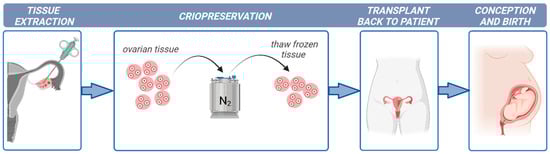
Ovarian tissue cryopreservation is gaining importance as a successful method to restore fertility to girls and young women at high risk of sterility. However, there are concerns regarding the safety of transplantation after ovarian tissue cryopreservation due to the high risk of reintroducing cancer cells and causing disease recurrence. In these cases, the development of culture systems that support oocyte development from the primordial follicle stage is required.

cancer cryopreservation fertility preservation

1. Introduction

Cancer is the leading cause of death in children around the world. Yearly, approximately 87,000 girls aged from 0 to 19 years old are diagnosed with cancer worldwide, according to the World Health Organization. Advances in childhood cancer research have increased the cure rate to over 80% in high-income countries [1]. However, the therapy side-effects on patient reproductive health have received little attention, with fertility failure being one of the most detrimental consequences. The risk of gonadotoxicity in oncologic patients after treatment is up to 80% depending on the type of anticancer treatment [2].
While fertility, in principle, can be preserved by freezing oocytes or embryos in post-pubertal women, the primary fertility preservation option that exists for pre-pubertal girls or oncologic patients undergoing immediate gonadotoxic cancer treatment is ovarian tissue cryopreservation (OTC) and its subsequent transplantation (OTT) [3][4] (illustrated in Figure 1). In addition, other non-oncological diseases such as autoimmune and hematological disorders are treated with chemotherapy and radiotherapy and require fertility preservation procedures [5].
Figure 1. Fertility preservation protocol in young women suffering from cancer. Created with BioRender.com.

2. Folliculogenesis in Human Ovaries

The normal development of the human ovary starts from day 26 of pregnancy when human primordial germ cells arrive from the yolk sac to the gonad (oogonia) [6]. Then, after many mitotic division cycles of the oogonia, meiotic division starts and the diplotene stage is achieved, when the oogonia become the larger oocyte. Follicle formation in humans begins in the fourth month, when a single layer enclosed in flat membrane cells called granulosa cells (GCs) surrounds the oocyte, forming the primordial follicles (30–50 µm in diameter) [7]. At birth, the female germline reserve of primordial follicles is located in the rigid cortex and contains oocytes arrested in the diplotene stage of the prophase of the first meiotic division. Primordial follicles are recruited throughout life to enter folliculogenesis and expand from the cortex towards the medulla, reaching antral follicles stages.
Follicle development involves a series of precisely regulated biological events (illustrated in Figure 2): activation of primordial follicles (35–40 μm) with GCs becoming cuboidal (called primary follicles; 50–60 μm diameter), growth of the primary follicles with an increase in the proliferation rate of GCs yielding a multilaminar granulosa layer (called secondary follicles; 115–125 μm diameter), creation of a theca layer that will produce steroid hormones through a complex interaction with the GCs, development of an antral cavity, and rupture of the ovulatory follicle releasing a cumulus–oocyte complex (COC) after antral follicles reach 20 mm in diameter [7][8][9].
Figure 2. In vivo folliculogenesis. Folliculogenesis can be separated into early folliculogenesis and late folliculogenesis. Early folliculogenesis is under paracrine control and is gonadotrophin-independent, whereas late folliculogenesis is under endocrine control and is gonadotrophin-dependent. Follicle and oocyte promedium sizes for human and mouse are mentioned, as well as time of appearance [9]. Created with BioRender.com.
Oocytes arrested in the meiotic stage of prophase I in early-stage follicles must acquire the developmental competence necessary to resume meiosis and complete maturation as well as support fertilization and embryonic growth. 
The ovarian tissue in all young females that is harvested for cryopreservation contains primordial follicles. At birth, the human ovary contains about 1–2 million primordial follicles. By the time a girl enters puberty, only about 25% (300,000 oocytes) remain. However, the majority of oocytes from these primordial follicles never ovulate and therefore never contribute to reproduction [10]

3. Culture Systems

3.1. Two-Dimensional Culture Systems

The first attempts for IVG in mammals were conducted using 2D culture systems [11][12]. Two-dimensional systems used for in vitro follicle culture include multi-wells [13], microdrops [14], or membranes coated with extracellular proteins [15][16]. Only in mice has the production of live young from cultured primordial follicles been successful [11][17]. This research group developed a two-stage culture system: primordial follicles were grown in tissue to secondary follicles, followed by isolation of the secondary follicles and culture to mature oocytes. However, only 59 live offspring (5.7% of embryos transferred) were obtained [17]. Achieving live birth from in vitro-cultured primordial follicles in mice is an important step in understanding some of the universal mammalian mechanisms of folliculogenesis, but this approach has not been translated to humans [18], mainly due to the longer duration of follicle development (10–12 days in mice compared to 2–3 months in humans) and the larger sizes reached by the follicles in humans (0.5–0.6 mm in mice compared to 6 mm in humans).
The major challenge of follicular IVG is to ensure the growth of the primary follicle, the development of granulosa and theca cells, and subsequently the development of an antrum [19]. Probably the most critical limitation is the failure to maintain the follicular spherical structure, disrupting the cellular interactions between the oocyte and GCs and compromising the further in vitro development. Several of the gap junctions and the intercellular communication between the oocyte and GCs are weakened during in vitro culture [20]. The conventional 2D culture of follicles impedes spherical growth and the preservation of the special arrangements between GCs and the oocyte tend to decrease, leaving the oocyte denuded and unable to complete the maturation process. In longer culture periods as when culturing primordial follicles, the importance of the special arrangements is even greater, leading to an interest in 3D culture to recapitulate the structure and function of the follicles [21].

3.2. Three-Dimensional Culture Systems

During the physiological reproductive cycle process, the ovarian microenvironment is in constant remodeling, providing clues to the potential role of these microenvironmental aspects in this process. Maintenance of the intricate 3D architecture and GCs and oocyte cell interaction may be critical for the successful in vitro maturation of follicles. Respecting its 3D structure is therefore crucial to maintain proper follicular physiology and obtain responses resembling the expected behavior of follicles in vivo. The 3D culture involves the use of the homogeneous hanging drop or hydrogel encapsulation to preserve the architecture. The early antral follicles culture is strongly influenced by the composition and architecture of its supporting tissue. This generates the need to develop extracellular matrixes and biomaterials that could imitate the ovarian physiologic milieu for optimal follicle development [22][23].
In addition to the spatial arrangement of the cells, the extracellular matrix (ECM) is a structural support network made up collagen, laminin, and fibrinogen, and is increasingly recognized as a master regulator in the communication between cells and in cell differentiation [24]. Matrices are needed to support follicle growth and maturation in 3D culture systems. Several biomaterials such as collagen, alginate, or Matrigel have been explored as an alternative to mimic the ECM within the ovary and to encapsulate and support human secondary and antral follicles. Although some attempts have been successful for culturing human follicles, collagen presents a few challenges such as a limited transparency for monitoring follicular development or the accelerated shrinkage of this material during prolonged culture. One commercially available ECM tested for follicular growth is Matrigel [25][26]. Matrigel is composed of collagen IV, laminin, fibronectin, entactin, and a variety of factors. 

3.3. Critical Cell–Cell and Cell–Matrix Interactions for Improved Oocyte Survival and Growth

Nonetheless, 3D culture still presents numerous challenges. For instance, the role of mechanical signaling has been mostly overlooked. There is increasing evidence that physical properties of the ECM play a critical role in follicle development. Indeed, mechanical stiffness may impact cellular proliferation and differentiation and even oocyte-specific gene expression levels in oocytes [27].
Combining different imaging modalities, Ouni et al. [28] studied some biophysical characteristics of the ovary microenvironment at different stages of a woman’s reproductive life, concluding that there is a correlation between rigidity and fertility. This link between ECM stiffness and antrum formation was also demonstrated in a study in rodents with the greatest antrum formation observed at 0.7% alginate hydrogel compared to 1.5 and 3% [29]. In contrast, an opposite link between rigidity and follicle growth was observed in primates, where follicle survival was higher in 0.5 vs. 0.25% calcium alginate [30]. The ovarian stroma of primates is more rigid than that of rodents, which may benefit from stiffer biomaterials. The physical attributes of the 3D matrix selected for IVG need to be tailored to meet species-specific requirements.
Within the ovary, there is an increase in vascularization as we move further away from the ovarian cortex up the medulla where the secondary follicles are, suggesting a strong need of oxygenation during the final follicular stages. The effect of oxygen tension on follicle and oocyte development has received little attention. Follicles have traditionally been grown in standard incubators with an atmospheric oxygen concentration. However, the oxygen pressure in the peritoneal cavity where the ovaries are located is approximately 5% O2 [31]. Primordial follicles exist in the relatively poorly vascularized cortex of the ovary and an abundance of blood vessels is found in the region of the ovary that contains secondary and antral follicles. It is possible that there is a dynamic oxygen transition from relative hypoxia in primordial follicles to a greater oxygen tension in preantral follicles. A lower oxygen tension during IVM improved blastocyst formation using mouse oocytes [32]

4. Organ-on-a-Chip Technology

Although the activation of growth of primordial follicles has been achieved, a limited number of follicles progress to secondary follicles [33]. Decent results were obtained in primates using expandable matrixes in 3D systems but the fertilization ability of the oocytes obtained was low and no blastocyst development was observed [34][35]. One of the reasons for the limited success of this technology can be found in the lack of more appropriate and physiological culture systems because it does not recapitulate the heterogeneous nature of the ECM in the ovary, with the medulla being much softer than the cortex. The ECM is believed to not only provide a 3D network to support the ovarian tissue architecture but also to regulate (together with hormones and nutrients) cell-ECM and cell–cell interactions that are important for follicle development. Choi et al. [36] revealed the crucial role of mechanical heterogeneity in the ovary in regulating follicle development by producing ovarian microtissues by encapsulating early secondary preantral follicles in microcapsules consisting of a softer, biodegradable collagen (0.5%) hydrogel core and a harder, slowly degradable alginate (2%) hydrogel shell. Folliculogenesis mainly depends upon hormones and nutrients, and their disturbance can cause abnormal follicle growth. Hence, a precise culture system that ensures the diffusion of nutrients and gases within the tissue, maximizing the retention of essential growth factors of oocyte maturation, is needed.

Considering the aforementioned aspects, a pioneering technology known as Organ-on-a-Chip (OOC) offers the means to replicate tissue architecture and emulate fluidic conditions in an in vitro setting. Broadly, these systems facilitate the miniaturization of experimental models, resulting in several advantages such as reduced working volumes, quicker reaction times, cost-effectiveness, and enhanced precision and control over experimental designs. This innovative approach holds significant promise in advancing research capabilities and improving the efficiency of experimental processes within the field.

OOC platforms, which are founded on microfluidic devices, facilitate three-dimensional organized cell culture by employing various types of ECMs. This methodology aims to emulate tissue architecture and replicate the cellular environment, mechanisms, and physiological responses of organs in an in vitro setting. The microenvironment is established through dynamic interactions among cells, fluids, and the ECM, influencing cellular processes and functions via biophysical and biochemical signals. A distinctive advantage of OOC lies in its ability to operate under flow conditions, enabling the continual replenishment of media, a process that mirrors in vivo blood supply or interstitial flow. Consequently, fluid flow ensures a consistent and adjustable supply of nutrients and oxygen, along with the supplementation of stage-specific growth factors. This dynamic environment sustains physical interactions while preventing cellular stress induced by the formation and accumulation of reactive oxygen substances [37][38].

Another pertinent aspect of this technology is the capability to tailor the devices based on design specifications including shaped microchannels, compartments, and reservoirs or the materials used to make the devices such as polydimethylsiloxane (PDMS; most prevalent), thermoplastics, or glass. Careful consideration is essential in material selection, as the inherent physicochemical properties of certain materials may impose limitations, including transparency, cost, flexibility or rigidity, and gas permeability, as well as considerations about time-consuming fabrication processes. The choice of material is contingent upon factors such as the intended application of the device, volume requirements, and production costs.

5. Conclusions

Ovarian follicular growth has great potential to restore fertility in young women suffering from cancer or adult women that need an imminent treatment and cannot undergo the cryopreservation of oocytes and embryos, avoiding the transplant and the risk of reintroducing malignant cells. However, the development of an optimal culture system to resemble in vivo folliculogenesis is necessary. The use of a dynamic culture system based on microfluidics, the definition of the mechanical characteristics of the matrix, and a stage-dependent modulation of this matrix composition should be addressed in order to obtain meiotically competent oocytes.
Advanced biomimetic devices such as microfluidic technology combined with ERC matrices may be valuable as a better in vitro culture system to preserve fertility, mimicking the dynamic supply of substances and gases in the ovary and recapitulating the 3D mechanical, physiological, and anatomical milieu in the ovary. This in vitro culture system is an ambitious pathway and is still maturing but may lead to a new assisted reproductive technique for clinical practice. OOC technology could revolutionize the field of reproductive biology.

References

  1. Howard, S.C.; Zaidi, A.; Cao, X.; Weil, O.; Bey, P.; Patte, C.; Samudio, A.; Haddad, L.; Lam, C.G.; Moreira, C.; et al. The My Child Matters programme: Effect of public-private partnerships on paediatric cancer care in low-income and middle-income countries. Lancet Oncol. 2018, 19, e252–e266.
  2. Salama, M.; Woodruff, T.K. Anticancer treatments and female fertility: Clinical concerns and role of oncologists in oncofertility practice. Expert. Rev. Anticancer Ther. 2017, 17, 687–692.
  3. Anderson, R.A.; Wallace, W.H.; Baird, D.T. Ovarian cryopreservation for fertility preservation: Indications and outcomes. Reproduction 2008, 136, 681–689.
  4. Rives, N.; Courbière, B.; Almont, T.; Kassab, D.; Berger, C.; Grynberg, M.; Papaxanthos, A.; Decanter, C.; Elefant, E.; Dhedin, N.; et al. What should be done in terms of fertility preservation for patients with cancer? The French 2021 guidelines. Eur. J. Cancer 2022, 173, 146–166.
  5. Filatov, M.A.; Khramova, Y.V.; Kiseleva, M.V.; Malinova, I.V.; Komarova, E.V.; Semenova, M.L. Female fertility preservation strategies: Cryopreservation and ovarian tissue in vitro culture, current state of the art and future perspectives. Zygote 2016, 24, 635–653.
  6. Van den Hurk, R.; Abir, R.; Telfer, E.E.; Bevers, M.M. Primate and bovine immature oocytes and follicles as sources of fertilizable oocytes. Hum. Reprod. Update 2000, 6, 457–474.
  7. Gougeon, A. Regulation of ovarian follicular development in primates: Facts and hypotheses. Endocr. Rev. 1996, 17, 121–155.
  8. Fortune, J.E. The early stages of follicular development: Activation of primordial follicles and growth of preantral follicles. Anim. Reprod. Sci. 2003, 78, 135–163.
  9. Jiang, Y.; He, Y.; Pan, X.; Wang, P.; Yuan, X.; Ma, B. Advances in Oocyte Maturation in Vivo and in Vitro in Mammals. Int. J. Mol. Sci. 2023, 24, 9059.
  10. Gougeon, A. Human ovarian follicular development: From activation of resting follicles to preovulatory maturation. Ann. Endocrinol. 2010, 71, 132–143.
  11. Eppig, J.J.; O’Brien, M.J. Development in vitro of mouse oocytes from primordial follicles. Biol. Reprod. 1996, 54, 197–207.
  12. Telfer, E.E.; McLaughlin, M.; Ding, C.; Thong, K.J. A two-step serum-free culture system supports development of human oocytes from primordial follicles in the presence of activin. Hum. Reprod. 2008, 23, 1151–1158.
  13. Nation, A.; Selwood, L. The production of mature oocytes from adult ovaries following primary follicle culture in a marsupial. Reproduction 2009, 138, 247–255.
  14. Cortvrindt, R.; Smitz, J.; Van Steirteghem, A.C. In-vitro maturation, fertilization and embryo development of immature oocytes from early preantral follicles from prepuberal mice in a simplified culture system. Hum. Reprod. 1996, 11, 2656–2666.
  15. Oktem, O.; Oktay, K. The role of extracellular matrix and activin-A in in vitro growth and survival of murine preantral follicles. Reprod. Sci. 2007, 14, 358–366.
  16. Figueiredo, J.R.; Hulshof, S.C.; Thiry, M.; Van den Hurk, R.; Bevers, M.M.; Nusgens, B.; Beckers, J.F. Extracellular matrix proteins and basement membrane: Their identification in bovine ovaries and significance for the attachment of cultured preantral follicles. Theriogenology 1995, 43, 845–858.
  17. O’Brien, M.J.; Pendola, J.K.; Eppig, J.J. A revised protocol for in vitro development of mouse oocytes from primordial follicles dramatically improves their developmental competence. Biol. Reprod. 2003, 68, 1682–1686.
  18. Picton, H.M.; Harris, S.E.; Muruvi, W.; Chambers, E.L. The in vitro growth and maturation of follicles. Reproduction 2008, 136, 703–715.
  19. Telfer, E.E. Future developments: In vitro growth (IVG) of human ovarian follicles. Acta Obstet. Gynecol. Scand. 2019, 98, 653–658.
  20. Eppig, J.J.; Pendola, F.L.; Wigglesworth, K.; Pendola, J.K. Mouse oocytes regulate metabolic cooperativity between granulosa cells and oocytes: Amino acid transport. Biol. Reprod. 2005, 73, 351–357.
  21. Gougeon, A.; Testart, J. Germinal vesicle breakdown in oocytes of human atretic follicles during the menstrual cycle. J. Reprod. Fertil. 1986, 78, 389–401.
  22. Green, L.J.; Shikanov, A. In vitro culture methods of preantral follicles. Theriogenology 2016, 86, 229–238.
  23. Jones, A.S.K.; Shikanov, A. Follicle development as an orchestrated signaling network in a 3D organoid. J. Biol. Eng. 2019, 13, 2.
  24. Griffith, L.G.; Swartz, M.A. Capturing complex 3D tissue physiology in vitro. Nat. Rev. 2006, 7, 211–224.
  25. Scott, J.E.; Carlsson, I.B.; Bavister, B.D.; Hovatta, O. Human ovarian tissue cultures: Extracellular matrix composition, coating density and tissue dimensions. Reprod. Biomed. Online 2004, 9, 287–293.
  26. Higuchi, C.M.; Maeda, Y.; Horiuchi, T.; Yamazaki, Y. A Simplified Method for Three-Dimensional (3-D) Ovarian Tissue Culture Yielding Oocytes Competent to Produce Full-Term Offspring in Mice. PLoS ONE 2015, 10, e0143114.
  27. Jiao, Z.X.; Woodruff, T.K. Follicle microenvironment-associated alterations in gene expression in the mouse oocyte and its polar body. Fertil. Steril. 2013, 99, 1453–1459.
  28. Ouni, E.; Peaucelle, A.; Haas, K.T.; Van Kerk, O.; Dolmans, M.-M.; Tuuri, T.; Otala, M.; Amorim, C.A. A blueprint of the topology and mechanics of the human ovary for next-generation bioengineering and diagnosis. Nat. Commun. 2021, 12, 5603.
  29. West, E.R.; Xu, M.; Woodruff, T.K.; Shea, L.D. Physical properties of alginate hydrogels and their effects on in vitro follicle development. Biomaterials 2007, 28, 4439–4448.
  30. Xu, M.; West-Farrell, E.R.; Stouffer, R.L.; Shea, L.D.; Woodruff, T.K.; Zelinski, M.B. Encapsulated three-dimensional culture supports development of nonhuman primate secondary follicles. Biol. Reprod. 2009, 81, 587–594.
  31. Tsai, A.G.; Friesenecker, B.; Mazzoni, M.C.; Kerger, H.; Buerk, D.G.; Johnson, P.C.; Intaglietta, M. Microvascular and tissue oxygen gradients in the rat mesentery. Proc. Natl. Acad. Sci. USA 1998, 95, 6590–6595.
  32. Adam, A.A.; Takahashi, Y.; Katagiri, S.; Nagano, M. Effects of oxygen tension in the gas atmosphere during in vitro maturation, in vitro fertilization and in vitro culture on the efficiency of in vitro production of mouse embryos. Jpn. J. Vet. Res. 2004, 52, 77–84.
  33. De Vos, M.; Smitz, J.; Woodruff, T.K. Fertility preservation in women with cancer. Lancet 2015, 385, 856.
  34. Xu, J.; Bernuci, M.P.; Lawson, M.S.; Yeoman, R.R.; Fisher, T.E.; Zelinski, M.B.; Stouffer, R.L. Survival, growth, and maturation of secondary follicles from prepubertal, young, and older adult rhesus monkeys during encapsulated three-dimensional culture: Effects of gonadotropins and insulin. Reproduction 2010, 140, 685–697.
  35. Xu, J.; Xu, M.; Bernuci, M.P.; Fisher, T.E.; Shea, L.D.; Woodruff, T.K.; Zelinski, M.B.; Stouffer, R.L. Primate follicular development and oocyte maturation in vitro. Adv. Exp. Med. Biol. 2013, 761, 43–67.
  36. Choi, J.K.; Agarwal, P.; Huang, H.; Zhao, S.; He, X. The crucial role of mechanical heterogeneity in regulating follicle development and ovulation with engineered ovarian microtissue. Biomaterials 2014, 35, 5122–5128.
  37. Heo, Y.S.; Lee, H.J.; Hassell, B.A.; Irimia, D.; Toth, T.L.; Elmoazzen, H.; Toner, M. Controlled loading of cryoprotectants (CPAs) to oocyte with linear and complex CPA profiles on a microfluidic platform. Lab A Chip 2011, 11, 3530–3537.
  38. Lee, S.; Chung, M.; Lee, S.R.; Jeon, N.L. 3D brain angiogenesis model to reconstitute functional human blood-brain barrier in vitro. Biotechnol. Bioeng. 2020, 117, 48–762.
More
Information
Contributors MDPI registered users' name will be linked to their SciProfiles pages. To register with us, please refer to https://encyclopedia.pub/register : , , ,
View Times: 145
Revisions: 2 times (View History)
Update Date: 19 Mar 2024
1000/1000