Submitted Successfully!
To reward your contribution, here is a gift for you: A free trial for our video production service.
Thank you for your contribution! You can also upload a video entry or images related to this topic.
Version Summary Created by Modification Content Size Created at Operation
1 -- 3248 2024-03-11 10:33:44 |
2 layout + 3 word(s) 3251 2024-03-12 03:44:58 | |
3 layout Meta information modification 3251 2024-03-12 03:46:20 |

Video Upload Options

Do you have a full video?

Confirm

Are you sure to Delete?
Cite
If you have any further questions, please contact Encyclopedia Editorial Office.
Zdrojewski, J.; Nowak, M.; Nijakowski, K.; Jankowski, J.; Scribante, A.; Gallo, S.; Pascadopoli, M.; Surdacka, A. Potential Immunohistochemical Biomarkers for Grading Oral Dysplasia. Encyclopedia. Available online: https://encyclopedia.pub/entry/56101 (accessed on 28 April 2024).
Zdrojewski J, Nowak M, Nijakowski K, Jankowski J, Scribante A, Gallo S, et al. Potential Immunohistochemical Biomarkers for Grading Oral Dysplasia. Encyclopedia. Available at: https://encyclopedia.pub/entry/56101. Accessed April 28, 2024.
Zdrojewski, Jakub, Monika Nowak, Kacper Nijakowski, Jakub Jankowski, Andrea Scribante, Simone Gallo, Maurizio Pascadopoli, Anna Surdacka. "Potential Immunohistochemical Biomarkers for Grading Oral Dysplasia" Encyclopedia, https://encyclopedia.pub/entry/56101 (accessed April 28, 2024).
Zdrojewski, J., Nowak, M., Nijakowski, K., Jankowski, J., Scribante, A., Gallo, S., Pascadopoli, M., & Surdacka, A. (2024, March 11). Potential Immunohistochemical Biomarkers for Grading Oral Dysplasia. In Encyclopedia. https://encyclopedia.pub/entry/56101
Zdrojewski, Jakub, et al. "Potential Immunohistochemical Biomarkers for Grading Oral Dysplasia." Encyclopedia. Web. 11 March, 2024.
Potential Immunohistochemical Biomarkers for Grading Oral Dysplasia
Edit

Oral cancer is becoming more and more frequent worldwide. Despite the widely available prevention, it is one of the most common cancers in the world, with 476,125 new cases and 225,900 deaths in 2020. Among the causes of carcinogenesis in the oral cavity, tobacco smoking or chewing, alcohol consumption, occupational exposure, risky sexual behaviour, genetic factors, and environmental pollution are widely mentioned. Smoking is the most prominent risk factor for oral cancer due to the carcinogenic chemicals in cigarette smoke, including nitrosamines, benzopyrenes, and aromatic amines. The risk of oral cancer is three times higher in smokers compared to non-smokers. In addition, the combination of cigarette smoking and frequent heavy alcohol consumption increases the risk of developing cancer by several times.

oral dysplasia oral epithelial dysplasia immunohistochemistry histological grading

1. Biomarkers Related to Cell Division and Proliferation

The cell cycle is regulated by the activity of various cyclins and cyclin-dependent kinases (Cdks). Cyclins form a complex with Cdks, and complex formation results in the activation of the Cdk active site. Cyclins without Cdk activation have no enzymatic activity but have binding sites for some substrates [1]. Cyclins are some of the most important cell cycle regulatory proteins and are linked to a specific phase of the cycle [2]. Both cyclins and their associated proteins are currently the subject of intense research, as perturbations of their expression and regulation can lead to tumorigenesis [3]. The majority of findings have reported on the overexpression of cyclins D and E in the development of many types of cancer [4].
In many studies, p63 and CD31 are the primarily examined markers. The p63 protein in normal cells is found in the basal layer of squamous epithelium [5]. Bavle et al. [6] found that p63 expression rose with increased severity of dysplasia and increased expression in suprabasal cells. The studies showed that p63 is required to maintain cell proliferation. It was observed that as the severity of dysplasia rose, the proliferation rate increased; however, cell differentiation was jeopardised [7]. As the disease progressed, the number of blood vessels increased and angiogenesis occurred. This is one of the factors that plays an important role in tumour growth and metastasis, providing nutrition to the developing tumour [8]. CD31 protein is a marker of angiogenesis, so it was used to detect vascular changes near the epithelium. The correlation of p63 with CD31 added value to the categorisation of leukoplakic lesions in the cases of low and moderate dysplasia [6].
Patel et al. [9] assessed p63 expression in different grades of dysplasia and Cyclin D1 expression. Cyclin D1 is classified as a proto-oncogene. P63 expression showed no statistically significant differences in different grades of dysplasia, and cyclin D1 showed only statistically significant differences between severe and mild grades of dysplasia. Gupta et al. [10] used VEGF and CD34 as dysplasia markers. The study evaluated the percentage of VEGF immunoreactivity, the intensity of VEGF staining, and CD34 immunostaining. The expression of VEGF and CD34 increased significantly during the transition from normal oral mucosa to severe oral epithelial dysplasia (OED).
CD44—cluster of differentiation 44—is a transmembrane glycoprotein [11]. Venkat Naga et al. [12] used a cluster of differentiation 44 (CD44) antibody to assess the correlation between this marker and oral dysplasia grading. The authors compared four groups: control tissue, mild epithelial dysplasia, moderate epithelial dysplasia, and severe epithelial dysplasia. A comparison of the groups showed statistically significant results. It suggested that CD44 may be a useful marker for diagnosing dysplastic lesions.
Interestingly, Aravind et al. [13] evaluated the osteopontin (OPN) expression in premalignant and malignant lesions. The authors observed a progressive increase in OPN expression, which was seen with increasing grades of dysplasia. Osteopontin seemed to be a promising biomarker in predicting the malignant potential of a premalignant lesion. Osteopontin, a phosphorylated sialoprotein, is a component of the mineralised extracellular matrices of bones and teeth [14] that has many functions in inflammation, immune responses, wound healing, cell adhesion, and cell migration through interactions with integrins and CD44 variants [15].
P53, also known as TP53, is a gene that encodes a protein that regulates the cell cycle and, therefore, acts as a tumour suppressor, regulating cell division by stopping cells from growing and proliferating too rapidly or in an uncontrolled manner [16]. As presented in Figure 1, p53 plays a critical role in the regulation of the DNA damage response. Under normal conditions, p53 is expressed at an extremely low level. The regulation of p53 activity is caused by the MDM2 protein, which contributes to the proteasomal degradation of this suppressor [17]. When DNA damage or energetic stress occurs in a cell, p53 expression is induced, causing the cell cycle to stop. This is a chance for the repairment processes, or the cells will develop apoptosis. The most important purpose of this protein is to eliminate cancer-prone cells from the replication pool [18]. When DNA damage, mitotic impairment, and oxidative stress are excessive, the p53 protein can be mutated to wild-type p53 protein (wtp53), which is inactivated under physiological conditions [19]. Mutations in the P53 gene and the functions of wtp53 expression have been linked to various human cancers [16].
Figure 1. The mechanism of p53 regulation in DNA damage response.
Researchers demonstrated p53’s role in differentiating grades of dysplasia. Pandya et al. [20] showed that the difference in expression was statistically significant between mild and severe dysplasia. The difference in TP53 expression between mild and severe dysplasia was statistically significant, according to Patil et al. [21]. The expression also increased with the increasing grades of epithelial dysplasia. Deregulation of this oncosuppressive protein may be important for the liability of the lesions to carcinogenesis. In the study by Sawada et al. [22], the higher the grade of dysplasia, the more frequently a TP53 mutation was observed. Imaizumi et al. [23] assessed p53 expression by immunofluorescence as a biomarker to differentiate between oral squamous epithelial lesions. The study consisted of 129 archival oral biopsy samples, including 18 benign squamous lesions, 37 low-grade dysplasias, 22 high-grade dysplasias, and 52 OSCCs. The authors found that the expression of p53 can be a valuable biomarker that helps to estimate the grade of oral epithelial dysplasia.
ΔNp63 is in the p53 family and is a p63 isoform, guiding the maturation of these stem cells through the regulation of their self-renewal and terminal differentiation. Yes-associated protein (YAP) is an oncoprotein in the cytoplasm in an inactive form [24]. YAP moves to the cell nucleus and activates the transcription of genes responsible for cell division and apoptosis [25]. Ono et al. [26] assessed the correlation between the expression of ΔNp63 and YAP and the grade of oral dysplasia. The authors found that in oral dysplasia, the expression of YAP and ΔNp63 was higher in high-grade than in low-grade disease. YAP and ΔNp63 expression correlated with grades of oral dysplasia.
The Ki-67 protein is widely used as a marker of human cancer cell proliferation [27]. Ki-67 plays a role in interphase and mitotic cells, and its distribution changes during the cell cycle. These localisations are associated with distinct functions [28]. Increased tumour cell proliferation is considered a significant natural factor in cancer detection. Ki-67 plays a significant role in cancer formation due to its positive association with tumour proliferation and invasion [29]. Ki-67 is the most suitable biological marker of mitotic activity due to its expression in the nucleus in a specific cell cycle period [30].
Mutations of P53 and high levels of Ki-67 protein are frequently observed in various types of human cancer. Ki-67 shows a stronger association with poor tumour differentiation and negatively affects patients’ survival in advanced stages [31]. Both P53 mutational status/type and high Ki-67 can also significantly impact overall survival [32]. The expression of p53 and Ki-67 increases as normal oral mucosa becomes dysplastic and undergoes malignant transformation [33]. Co-expression of p53 and Ki-67 is related to larger tumours and metastasis to lymph nodes; thus, this observation suggests that it can be used to identify high-risk lesions [34].
In their study, Kamala et al. [35] observed an increase in Ki-67 expression with the severity of dysplasia. The Ki-67 antigen can be used as a marker for histological evaluations of OED. According to Dash et al. [36], as the severity of OED increases, the number of cells showing positive Ki-67 expression also increases. This is also confirmed by Mondal et al. [37], who found that the differences in Ki-67 expression were statistically significant between normal mucosa and mild dysplasia, as well as between mild, moderate, and severe dysplasia. Ki-67 not only detects the hyperactive cells in OED, but its expression of Ki-67 can also be comparable to the clinical course or prognostication of a disease.
According to the study by Takkem et al. [38], Ki-67 expression was restricted to the basal layers of normal oral epithelium, while Ki-67-positive cells in OED were localised in the basal, suprabasal, and squamous layers; Ki-67 expression was increased in patients at high case risk. Ki-67-positive cells in well-differentiated OSCC were mainly located at the periphery of tumour nests; in moderately differentiated OSCC, they were located both at the periphery and in part of the centre of tumour nests, while they were scattered in the most poorly differentiated lesions. The study by Kamala et al. [35] aimed to determine the degree and pattern of expression of aberrant Ki67 in OSMF. The study confirmed a statistically significant correlation between the expression of Ki-67 with the clinical and histological grading of OSMF and the histological grading of OSCC.
Moreover, Swain et al. [39] examined Ki-67 with MCM2 expression in OED, OSCC, and normal mucosa. The study confirmed that the expression of these proteins increased progressively. The expression profile of MCM 2 and Ki-67 was increased with the increasing grades of epithelial dysplasia. In their studies, Gadbail et al. [40][41] used Ki-67, CD105, and α-SMA antigen to differentiate the OED grades. The expressions of Ki-67, CD105, and α-SMA markers complement the binary grading system of OED. Ki-67 showed significant increases from normal oral mucosa to low-grade and high-grade epithelial dysplasia.
Additionally, Suwasini et al. [42] found a statistically significant association between p53 and Ki-67. The results highlighted the potential use of the p53 protein and the Ki-67 antigen as significant molecular markers for early PMD detection and OSCC risk. This observation was also confirmed by Leung et al. [43]—Ki-67 and p53 were significantly increased with higher histological grades of OD. These observations showed the role of DNA-replicative stress in higher grades of dysplasia and transformation from OD to OSCC.
Monteiro et al. [44] analysed the immunoexpression of BubR1, Mad2, Bub3, Spindly, and Ki-67 proteins in 64 oral biopsies. Spindly is a protein that targets dynein/dynactin to kinetochores in mitosis. The authors observed that the expression of Spindly was significantly correlated with a high Ki-67 score and the grade of dysplasia. This observation confirmed that the expression of Ki-67 protein is associated with an increased risk for malignant transformation.
Stathmin is a member of a family of proteins that plays important roles in regulating the microtubule cytoskeleton [45]. This protein regulates microtubule dynamics by promoting the depolymerisation of microtubules and/or preventing the polymerisation of tubulin heterodimers [46]. Vadla et al. [47] evaluated the role of stathmin in OSCC and oral dysplasia and the correlation of stathmin expression with dysplasia grading. The study presented a statistically significant correlation between increased grades of oral dysplasia and expression levels of stathmin. This research confirmed the positive role of stathmin in disease progression and suggested that stathmin could be an early diagnostic biomarker for oral dysplasia.

2. Biomarkers Related to Epithelial–Mesenchymal Transition (EMT)

Epithelial stem cells maintain tissues throughout adult life and are controlled by epithelial–mesenchymal interactions to balance cell production and loss. A defining characteristic of an epithelium is the close contact that these cells have with the underlying mesenchyme [48]. Polarised epithelial cells normally interact with the basement membrane, causing several biochemical changes that enable them to adopt a mesenchymal cell phenotype, including enhanced migratory capacity, invasiveness, elevated resistance to apoptosis, and greatly increased production of ECM components. This biological process is called an epithelial–mesenchymal transition (EMT) [49]. This transformation can occur in physiological processes during embryogenesis, organ development, and tissue regeneration, as well as in tumorigenesis and cancer progression, including tumour cell invasion and metastasis [50].
Mesenchymal stem cells are stromal cells capable of self-renewal and multilineage differentiation. They show a greater ability to infiltrate the capillaries at the site of the primary tumour lesions [51][52]. This mechanism is a critical mechanism for the acquisition of the malignant phenotype in neoplastic epithelial processes. This subtype accompanies the formation of distant metastases, where, in secondary foci, cells change their phenotype through a reverse mesenchymal–epithelial transition (MET) [53][54].
The role of EMT in OSCC is to transform normal epithelial cells into malignant mesenchymal cells by losing intercellular adhesion, causing metastatic progression and infiltration [55]. In the epithelial stage, tumour cells are cubic and adherent to each other. Also, in this stage, tumour cells show positive E-cadherin expression and negative vimentin expression. In the mesenchymal stage, the tumour cells show higher vimentin expression, but the expression of E-cadherin is repressed. The tumour cells are fibroblast-like and lose their cell–cell junctions [56].

3. Biomarkers Related to Cell Death Regulation

Other altered proteins are the members of the Bcl-2 family. These proteins are considered as the principal players in the cascade of events that activate or inhibit apoptosis [57]. In this family, there are, for example, Bcl-XL, Bcl-2, and Bax. Bcl-2 acts as a checkpoint upstream of caspases and mitochondrial dysfunction [58]. Also, Bcl-2 can rescue maturation at several points of lymphocyte development. The Bcl-2 proto-oncogene was discovered at the chromosomal breakpoint of t (14;18) found in a human follicular lymphoma [59]. Pathak et al. [60] observed that the level of Bcl-2 increased with the grade of dysplasia. However, Bcl-2 expression was decreased in OSCC. Pallavi et al. [61] assessed the expression of Bcl-2 and c-Myc in OED and OSCC. Similarly, the authors noticed that Bcl-2 increased with grades of dysplasia. Bcl-2 proteins could positively affect lesion progression from premalignancy to malignancy.
Also, the PD-1/PD-L1 pathway can be a potential marker for oral dysplasia. Programmed Cell Death Protein 1 (PD-1) inhibits immune responses and modulates T-cell activity [62]. Kujan et al. [63] investigated the role of the PD-1/PD-L1 pathway in the development of dysplasia and OSCC. The study found that the PD-1/PD-L1 pathway can be associated with the development of OSCC and the grade of dysplasia. Programmed cell death 4 (PDCD4) functions as a tumour suppressor and an inhibitor of protein translation [64]. PDCD4 expression was observed in normal oral mucosa, OED, and OSCC. Desai and Kale [65] showed that the maximum expression was observed in normal oral mucosa, which reduced significantly in OED and OSCC.
Heat shock protein 27 (HSP27) belongs to the small-molecular-weight heat shock protein family and has a molecular weight of approximately 27 KDa [66]. This protein protects other proteins from damage due to environmental factors such as heat, toxins, free radicals, and ischaemia [67]. Karri et al. [68] found that a low expression of HSP27 could be an early molecular indicator of initial dysplastic changes in normal mucosa. Conversely, the overexpression of HSP27 could be a prognostic value of malignant transformation from oral dysplasia to oral squamous cell carcinoma. Cornulin (known as C1 Orf10, or squamous epithelial heat shock protein 53) is a member of the heat shock protein 70 (HSP70) family [69]. Cornulin plays an important role in the differentiation of the epidermis. The expression of cornulin causes cell cycle arrest at G1, and its downregulation plays a role in oral carcinogenesis [70]. Santosh et al. [71] found that cornulin expression decreased in oral dysplasia compared with normal oral mucosa and was absent in OSCC.

4. Biomarkers Related to Cellular Metabolism

A major component of the cellular response to oxygen deprivation is the transcription factor HIF-1 (hypoxia-inducible factor-1). HIF-1 consists of an HIF-1 beta unit and one of three units of HIF-1alpha, HIF-2alpha, or HIF-3alpha [72]. Patel et al. [73] assessed the expression of HIF-1alpha in OED and compared the expression between grades. The authors noticed that the expression of HIF-alpha statistically significantly increased as grades of oral dysplasia were higher. Also, HIF-alpha could be a marker of risk of malignant transformation.
Inducible nitric oxide synthase (iNOS) is an enzyme in oxygen and nitrogen metabolite metabolism [74]. Using immunohistochemical methods, Singh et al. [75] compared iNOS expression between oral leukoplakia and OSCC. The authors found that the expression of iNOS rose with the progressing clinical stages of oral leukoplakia and OSCC. Therefore, iNOS might be a diagnostic marker in oral leukoplakia and a prognostication marker of OSCC. Another enzyme, cyclooxygenase (COX or prostaglandin–endoperoxide synthase), is required to change arachidonic acid to prostaglandins [76]. Sharada et al. [77] examined the expression of COX-2 and type IV collagen in OED. The study found that its expression increased significantly as the grade of dysplasia was higher. This marker could be applied to assess the malignant potential.

5. Biomarkers Related to Extracellular Signalling Pathways

Paxillin is a 68 kDa, phosphotyrosine-containing protein that may play a role in several signalling pathways [78]. The study by Alam et al. [79] presented a statistically significant correlation between increased grades of oral dysplasia and expression of paxillin. Paxillin may play an important role in the pathogenesis of oral dysplasia and OSCC.
EGFR is a 170 kDa transmembrane glycoprotein receptor [80]. EGFR regulates cell growth, differentiation, and gene expression [81]. Fakurnejad et al. [82] demonstrated that an anti-EGFR agent could successfully discriminate high-grade dysplastic lesions from low-grade dysplasia. Melanoma inhibitory activity (MIA) and MIA2 are other receptors participating in tumour growth and invasion. Kawai et al. [83] evaluated MIA and MIA2 as expressed in the oral mucosa within early neoplastic lesions and suggested that MIA and MIA2 are useful novel immunohistochemical markers for discriminating between normal tissue and OED.
Laminins are another family of structural proteins. Laminins participate in organising the complex interactions of the basement membranes. Laminin-1 is in the Reichert membrane (extraembryonic basement membrane) [84]. A study by Vageli et al. [85] assessed laminin immunostaining in biopsies as a useful biomarker of actinic cheilitis and differential diagnosis between actinic cheilitis and lip cancer. This marker can differentiate between low- and high-grade dysplasia. This research can provide new insight into the mechanism of progression of actinic cheilitis into lip cancer. Also, Nguyen et al. [86] evaluated the immunoexpression of LAMC2. The expression of LAMC2 was significantly associated with the grade of dysplasia. LAMC2 may be a predictive marker for the malignant progression of leukoplakia.
In the study by Debta et al. [87], GLUT-1 also appeared as a marker for differentiating dysplasia severity. A statistically significant increasing level of GLUT-1 corresponded to more advanced grades of dysplasia and was consistent with the WHO system. GLUT-1 expression was significantly increased from normal to mild, moderate, and severe dysplasia. The expression of the GLUT-1 marker complemented the WHO grading system of OED. Also, Patlolla et al. [88] confirmed a significant correlation between the location of GLUT-1 within the cell and the grade of dysplasia.
Moreover, Udompatanakorn and Taebunpakul [89] assessed the pattern of expression of METTL3 in OED. METTL3 is an enzyme involved in the post-transcriptional methylation of internal adenosine residues [90]. The authors observed that the expression of METTL3 increased in oral dysplasia and OSCC. METTL3 expression might be a marker for the progression of oral dysplasia and transformation to OSCC.
Another marker is the minichromosome maintenance protein (MCM-2), which is a key component of the pre-replication complex. This protein may be involved in the formation of replication forks and in the migration of other proteins during DNA replication [91]. The study by Zakaria et al. [92] aimed to assess MCM-2 activity in oral epithelial dysplastic lesions. The MCM-2 immunostaining showed a statistically significant increase from mild to severe dysplasia, and the highest value was in invasive squamous cell carcinoma. MCM-2 activity is associated with the grade of dysplasia. This observation suggests that MCM-2 may be a potential biomarker for early squamous cell carcinoma.

References

  1. Wolgemuth, D.J. Function of Cyclins in Regulating the Mitotic and Meiotic Cell Cycles in Male Germ Cells. Cell Cycle Georget. 2008, 7, 3509–3513.
  2. Wang, Z. Cell Cycle Progression and Synchronization: An Overview. Methods Mol. Biol. 2022, 2579, 3–23.
  3. Loyer, P.; Trembley, J.H. Roles of CDK/Cyclin Complexes in Transcription and Pre-mRNA Splicing: Cyclins L and CDK11 at the Cross-Roads of Cell Cycle and Regulation of Gene Expression. Semin. Cell Dev. Biol. 2020, 107, 36–45.
  4. Zhang, W.; Liu, Y.; Jang, H.; Nussinov, R. Cell Cycle Progression Mechanisms: Slower Cyclin-D/CDK4 Activation and Faster Cyclin-E/CDK2. BioRxiv Prepr. Serv. Biol. 2023.
  5. Steurer, S.; Riemann, C.; Büscheck, F.; Luebke, A.M.; Kluth, M.; Hube-Magg, C.; Hinsch, A.; Höflmayer, D.; Weidemann, S.; Fraune, C.; et al. P63 Expression in Human Tumors and Normal Tissues: A Tissue Microarray Study on 10,200 Tumors. Biomark. Res. 2021, 9, 7.
  6. Bavle, R.M.; Paremala, K.; Venugopal, R.; Rudramuni, A.S.; Khan, N.; Hosthor, S.S. Grading of Oral Leukoplakia: Can It Be Improvised Using Immunohistochemical Markers P63 and CD31. Contemp. Clin. Dent. 2021, 12, 37–43.
  7. Truong, A.B.; Kretz, M.; Ridky, T.W.; Kimmel, R.; Khavari, P.A. P63 Regulates Proliferation and Differentiation of Developmentally Mature Keratinocytes. Genes Dev. 2006, 20, 3185–3197.
  8. Bergholz, J.; Xiao, Z.-X. Role of P63 in Development, Tumorigenesis and Cancer Progression. Cancer Microenviron. Off. J. Int. Cancer Microenviron. Soc. 2012, 5, 311–322.
  9. Patel, S.B.; Manjunatha, B.S.; Shah, V.; Soni, N.; Sutariya, R. Immunohistochemical Evaluation of P63 and Cyclin D1 in Oral Squamous Cell Carcinoma and Leukoplakia. J. Korean Assoc. Oral Maxillofac. Surg. 2017, 43, 324–330.
  10. Gupta, S.; Gupta, V.; Tyagi, N.; Vij, R.; Vij, H.; Sharma, E. Analysis of Role of Angiogenesis in Epithelial Dysplasia: An Immunohistochemical Study. J. Clin. Diagn. Res. 2017, 11, EC29–EC34.
  11. Tawara, M.; Suzuki, H.; Goto, N.; Tanaka, T.; Kaneko, M.K.; Kato, Y. A Novel Anti-CD44 Variant 9 Monoclonal Antibody C44Mab-1 Was Developed for Immunohistochemical Analyses against Colorectal Cancers. Curr. Issues Mol. Biol. 2023, 45, 3658–3673.
  12. Venkat Naga, S.K.S.; Shekar, P.C.; Kattappagari, K.K.; Prakash Chandra, K.L.; Reddy, G.S.; Ramana Reddy, B.V. Expression of Cluster Differentiation-44 Stem Cell Marker in Grades of Oral Epithelial Dysplasia: A Preliminary Study. J. Oral Maxillofac. Pathol. JOMFP 2019, 23, 203–207.
  13. Aravind, T.; Janardhanan, M.; Rakesh, S.; Savithri, V.; Unnikrishnan, U.G. Immunolocalization of Osteopontin in Dysplasias and Squamous Cell Carcinomas Arising from Oral Epithelium. J. Oral Maxillofac. Pathol. JOMFP 2017, 21, 18–23.
  14. Sodek, J.; Ganss, B.; McKee, M.D. Osteopontin. Crit. Rev. Oral Biol. Med. Off. Publ. Am. Assoc. Oral Biol. 2000, 11, 279–303.
  15. Mrochem, J.; Bartnik, W. Osteopontin—A New Marker in Neoplastic Diseases. Contemp. Oncol. Onkol. 2008, 12, 349–353.
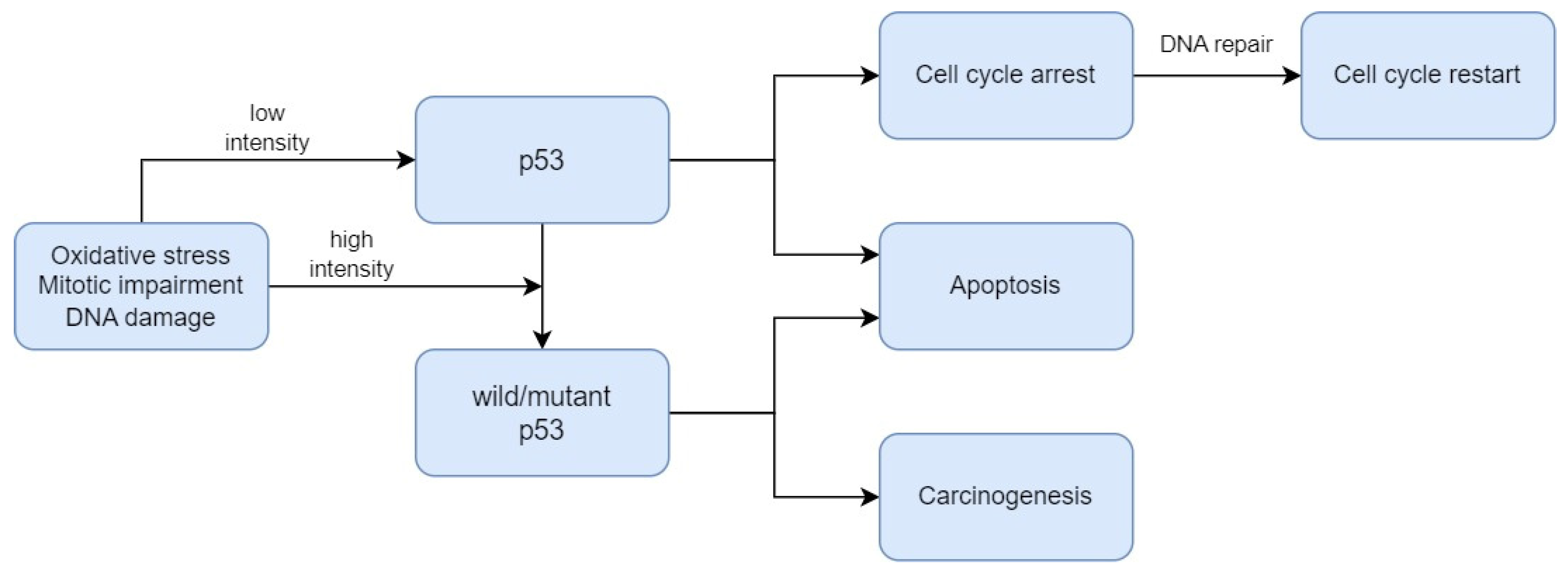
  16. Ozaki, T.; Nakagawara, A. Role of P53 in Cell Death and Human Cancers. Cancers 2011, 3, 994–1013.
  17. Williams, A.B.; Schumacher, B. P53 in the DNA-Damage-Repair Process. Cold Spring Harb. Perspect. Med. 2016, 6, a026070.
  18. Babamohamadi, M.; Babaei, E.; Ahmed Salih, B.; Babamohammadi, M.; Jalal Azeez, H.; Othman, G. Recent Findings on the Role of Wild-Type and Mutant P53 in Cancer Development and Therapy. Front. Mol. Biosci. 2022, 9, 903075.
  19. Borrero, L.J.H.; El-Deiry, W.S. Tumor Suppressor P53: Biology, Signaling Pathways, and Therapeutic Targeting. Biochim. Biophys. Acta Rev. Cancer 2021, 1876, 188556.
  20. Pandya, J.A.; Boaz, K.; Natarajan, S.; Manaktala, N.; Nandita, K.P.; Lewis, A.J. A Correlation of Immunohistochemical Expression of TP53 and CDKN1A in Oral Epithelial Dysplasia and Oral Squamous Cell Carcinoma. J. Cancer Res. Ther. 2018, 14, 666–670.
  21. Patil, S.; Gawande, M.; Chaudhari, M.; Sharma, P.; Hande, A.; Sonone, A. Prognostic Significance of P53 Expression in Various Grades of Epithelial Dysplasia. J. Datta Meghe Inst. Med. Sci. Univ. 2022, 17, 306–310.
  22. Sawada, K.; Momose, S.; Kawano, R.; Kohda, M.; Irié, T.; Mishima, K.; Kaneko, T.; Horie, N.; Okazaki, Y.; Higashi, M.; et al. Immunohistochemical Staining Patterns of P53 Predict the Mutational Status of TP53 in Oral Epithelial Dysplasia. Mod. Pathol. Off. J. US Can. Acad. Pathol. Inc. 2022, 35, 177–185.
  23. Imaizumi, T.; Matsuda, K.; Tanaka, K.; Kondo, H.; Ueki, N.; Kurohama, H.; Otsubo, C.; Matsuoka, Y.; Akazawa, Y.; Miura, S.; et al. Detection of Endogenous DNA Double-Strand Breaks in Oral Squamous Epithelial Lesions by P53-Binding Protein 1. Anticancer Res. 2021, 41, 4771–4779.
  24. Napoli, M.; Wu, S.J.; Gore, B.L.; Abbas, H.A.; Lee, K.; Checker, R.; Dhar, S.; Rajapakshe, K.; Tan, A.C.; Lee, M.G.; et al. ΔNp63 Regulates a Common Landscape of Enhancer Associated Genes in Non-Small Cell Lung Cancer. Nat. Commun. 2022, 13, 614.
  25. Abylkassov, R.; Xie, Y. Role of Yes-Associated Protein in Cancer: An Update. Oncol. Lett. 2016, 12, 2277–2282.
  26. Ono, S.; Nakano, K.; Takabatake, K.; Kawai, H.; Nagatsuka, H. Immunohistochemistry of YAP and dNp63 and Survival Analysis of Patients Bearing Precancerous Lesion and Oral Squamous Cell Carcinoma. Int. J. Med. Sci. 2019, 16, 766–773.
  27. Sun, X.; Kaufman, P.D. Ki-67: More than a Proliferation Marker. Chromosoma 2018, 127, 175–186.
  28. Booth, D.G.; Takagi, M.; Sanchez-Pulido, L.; Petfalski, E.; Vargiu, G.; Samejima, K.; Imamoto, N.; Ponting, C.P.; Tollervey, D.; Earnshaw, W.C.; et al. Ki-67 Is a PP1-Interacting Protein That Organises the Mitotic Chromosome Periphery. eLife 2014, 3, e01641.
  29. Liang, Y.; Ma, C.; Li, F.; Nie, G.; Zhang, H. The Role of Contactin 1 in Cancers: What We Know So Far. Front. Oncol. 2020, 10, 574208.
  30. Iqbal, A.; Tamgadge, S.; Tamgadge, A.; Pereira, T.; Kumar, S.; Acharya, S.; Jadhav, A. Evaluation of Ki-67 Expression in Oral Submucous Fibrosis and Its Correlation with Clinical and Histopathological Features. J. Microsc. Ultrastruct. 2019, 8, 20–24.
  31. Kim, C.-H.; Lee, H.S.; Park, J.-H.; Choi, J.-H.; Jang, S.-H.; Park, Y.-B.; Lee, M.G.; Hyun, I.G.; Kim, K.I.; Kim, H.S.; et al. Prognostic Role of P53 and Ki-67 Immunohistochemical Expression in Patients with Surgically Resected Lung Adenocarcinoma: A Retrospective Study. J. Thorac. Dis. 2015, 7, 822–833.
  32. Lalkota, B.P.; Srinivasa, B.J.; Swamy, M.V.; Hazarika, D.; Jeet, B.M.; Jyothi, K.; Ghosh, M.; Sayeed, S.M.; Nasiruddin, M.; Naik, R. The Role of P53 and Ki67 in Predicting Clinical Outcome in Breast Cancer Patients. J. Cancer Res. Ther. 2023, 19, 208.
  33. Humayun, S.; Prasad, V.R. Expression of P53 Protein and Ki-67 Antigen in Oral Premalignant Lesions and Oral Squamous Cell Carcinomas: An Immunohistochemical Study. Natl. J. Maxillofac. Surg. 2011, 2, 38–46.
  34. Kumar, P.; Kane, S.; Rathod, G.P. Coexpression of P53 and Ki 67 and Lack of C-erbB2 Expression in Oral Leukoplakias in India. Braz. Oral Res. 2012, 26, 228–234.
  35. Kamala, K.A.; Kanetkar, S.R.; Datkhile, K.D.; Sankethguddad, S. Expression of Ki67 Biomarker in Oral Submucous Fibrosis with Clinico-Pathological Correlations: A Prospective Study. Asian Pac. J. Cancer Prev. APJCP 2022, 23, 253–259.
  36. Dash, K.C.; Mahapatra, N.; Bhuyan, L.; Panda, A.; Behura, S.S.; Mishra, P. An Immunohistochemical Study Showing Ki-67 as an Analytical Marker in Oral Malignant and Premalignant Lesions. J. Pharm. Bioallied Sci. 2020, 12, S274–S278.
  37. Mondal, K.; Mandal, R.; Sarkar, B.C. Importance of Ki-67 Labeling in Oral Leukoplakia with Features of Dysplasia and Carcinomatous Transformation: An Observational Study over 4 Years. S. A. J. Cancer 2020, 9, 99–104.
  38. Takkem, A.; Barakat, C.; Zakaraia, S.; Zaid, K.; Najmeh, J.; Ayoub, M.; Seirawan, M.Y. Ki-67 Prognostic Value in Different Histological Grades of Oral Epithelial Dysplasia and Oral Squamous Cell Carcinoma. Asian Pac. J. Cancer Prev. APJCP 2018, 19, 3279–3286.
  39. Swain, S.; Nishat, R.; Ramachandran, S.; Raghuvanshi, M.; Behura, S.S.; Kumar, H. Comparative Evaluation of Immunohistochemical Expression of MCM2 and Ki67 in Oral Epithelial Dysplasia and Oral Squamous Cell Carcinoma. J. Cancer Res. Ther. 2022, 18, 997–1002.
  40. Gadbail, A.R.; Chaudhary, M.; Sarode, S.C.; Gondivkar, S.; Tekade, S.A.; Zade, P.; Hande, A.; Sarode, G.S.; Patil, S. Ki67, CD105, and α-SMA Expression Supports the Transformation Relevant Dysplastic Features in the Atrophic Epithelium of Oral Submucous Fibrosis. PLoS ONE 2018, 13, e0200171.
  41. Gadbail, A.R.; Chaudhary, M.S.; Sarode, S.C.; Gawande, M.; Korde, S.; Tekade, S.A.; Gondivkar, S.; Hande, A.; Maladhari, R. Ki67, CD105, and α-SMA Expressions Better Relate the Binary Oral Epithelial Dysplasia Grading System of World Health Organization. J. Oral Pathol. Med. Off. Publ. Int. Assoc. Oral Pathol. Am. Acad. Oral Pathol. 2017, 46, 921–927.
  42. Suwasini, S.; Chatterjee, K.; Purkait, S.K.; Samaddar, D.; Chatterjee, A.; Kumar, M. Expression of P53 Protein and Ki-67 Antigen in Oral Leukoplakia with Different Histopathological Grades of Epithelial Dysplasia. J. Int. Soc. Prev. Community Dent. 2018, 8, 513–522.
  43. Leung, E.Y.; McMahon, J.D.; McLellan, D.R.; Syyed, N.; McCarthy, C.E.; Nixon, C.; Orange, C.; Brock, C.; Hunter, K.D.; Adams, P.D. DNA Damage Marker Phosphorylated Histone H2AX Is a Potential Predictive Marker for Progression of Epithelial Dysplasia of the Oral Cavity. Histopathology 2017, 71, 522–528.
  44. Monteiro, L.; Silva, P.; Delgado, L.; Amaral, B.; Garcês, F.; Salazar, F.; Pacheco, J.-J.; Lopes, C.; Bousbaa, H.; Warnakulasuriya, S. Expression of Spindle Assembly Checkpoint Proteins BubR1 and Mad2 Expression as Potential Biomarkers of Malignant Transformation of Oral Leukoplakia: An Observational Cohort Study. Med. Oral Patol. Oral Cirugia Bucal 2021, 26, e719–e728.
  45. Rubin, C.I.; Atweh, G.F. The Role of Stathmin in the Regulation of the Cell Cycle. J. Cell. Biochem. 2004, 93, 242–250.
  46. Feng, S.; Song, Y.; Shen, M.; Xie, S.; Li, W.; Lu, Y.; Yang, Y.; Ou, G.; Zhou, J.; Wang, F.; et al. Microtubule-Binding Protein FOR20 Promotes Microtubule Depolymerization and Cell Migration. Cell Discov. 2017, 3, 17032.
  47. Vadla, P.; Deepthi, G.; Kumar, C.A.; Bashamalla, R.; Syeda, N.; Naramala, S. Immunohistochemical Expression of Stathmin in Oral Dysplasia: An Original Study with an Insight of Its Action on Microtubules. J. Oral Maxillofac. Pathol. JOMFP 2021, 25, 247–252.
  48. Blanpain, C.; Horsley, V.; Fuchs, E. Epithelial Stem Cells: Turning over New Leaves. Cell 2007, 128, 445–458.
  49. Kalluri, R.; Weinberg, R.A. The Basics of Epithelial-Mesenchymal Transition. J. Clin. Investig. 2009, 119, 1420–1428.
  50. Dongre, A.; Weinberg, R.A. New Insights into the Mechanisms of Epithelial-Mesenchymal Transition and Implications for Cancer. Nat. Rev. Mol. Cell Biol. 2019, 20, 69–84.
  51. Ding, D.-C.; Shyu, W.-C.; Lin, S.-Z. Mesenchymal Stem Cells. Cell Transplant. 2011, 20, 5–14.
  52. Huang, Y.; Hong, W.; Wei, X. The Molecular Mechanisms and Therapeutic Strategies of EMT in Tumor Progression and Metastasis. J. Hematol. Oncol. 2022, 15, 129.
  53. Chen, T.; You, Y.; Jiang, H.; Wang, Z.Z. Epithelial-Mesenchymal Transition (EMT): A Biological Process in the Development, Stem Cell Differentiation, and Tumorigenesis. J. Cell. Physiol. 2017, 232, 3261–3272.
  54. Toriumi, K.; Berto, S.; Koike, S.; Usui, N.; Dan, T.; Suzuki, K.; Miyashita, M.; Horiuchi, Y.; Yoshikawa, A.; Asakura, M.; et al. Combined Glyoxalase 1 Dysfunction and Vitamin B6 Deficiency in a Schizophrenia Model System Causes Mitochondrial Dysfunction in the Prefrontal Cortex. Redox Biol. 2021, 45, 102057.
  55. Krisanaprakornkit, S.; Iamaroon, A. Epithelial-Mesenchymal Transition in Oral Squamous Cell Carcinoma. ISRN Oncol. 2012, 2012, 681469.
  56. Ling, Z.; Cheng, B.; Tao, X. Epithelial-to-Mesenchymal Transition in Oral Squamous Cell Carcinoma: Challenges and Opportunities. Int. J. Cancer 2021, 148, 1548–1561.
  57. Hardwick, J.M.; Soane, L. Multiple Functions of BCL-2 Family Proteins. Cold Spring Harb. Perspect. Biol. 2013, 5, a008722.
  58. Chao, D.T.; Korsmeyer, S.J. BCL-2 Family: Regulators of Cell Death. Annu. Rev. Immunol. 1998, 16, 395–419.
  59. Hua, C.; Zorn, S.; Jensen, J.P.; Coupland, R.W.; Ko, H.S.; Wright, J.J.; Bakhshi, A. Consequences of the t(14;18) Chromosomal Translocation in Follicular Lymphoma: Deregulated Expression of a Chimeric and Mutated BCL-2 Gene. Oncogene Res. 1988, 2, 263–275.
  60. Pathak, A.; Shetty, D.C.; Dhanapal, R.; Kaur, G. To Analyse the Mitotic and Keratinisation Correlation with Bcl-2 Expression in Varying Grades of Oral Epithelial Dysplasia and Squamous Cell Carcinoma. J. Oral Maxillofac. Pathol. JOMFP 2022, 26, 316–321.
  61. Pallavi, N.; Nalabolu, G.R.K.; Hiremath, S.K.S. Bcl-2 and c-Myc Expression in Oral Dysplasia and Oral Squamous Cell Carcinoma: An Immunohistochemical Study to Assess Tumor Progression. J. Oral Maxillofac. Pathol. JOMFP 2018, 22, 325–331.
  62. Han, Y.; Liu, D.; Li, L. PD-1/PD-L1 Pathway: Current Researches in Cancer. Am. J. Cancer Res. 2020, 10, 727–742.
  63. Kujan, O.; Agag, M.; Smaga, M.; Vaishnaw, Y.; Idrees, M.; Shearston, K.; Farah, C.S. PD-1/PD-L1, Treg-Related Proteins, and Tumour-Infiltrating Lymphocytes Are Associated with the Development of Oral Squamous Cell Carcinoma. Pathology 2022, 54, 409–416.
  64. Wang, Q.; Yang, H.-S. The Role of Pdcd4 in Tumor Suppression and Protein Translation. Biol. Cell 2018, 110, 169–177.
  65. Desai, K.M.; Kale, A.D. Immunoexpression of Programmed Cell Death 4 Protein in Normal Oral Mucosa, Oral Epithelial Dysplasia and Oral Squamous Cell Carcinoma. J. Oral Maxillofac. Pathol. JOMFP 2017, 21, 462.
  66. Ferns, G.; Shams, S.; Shafi, S. Heat Shock Protein 27: Its Potential Role in Vascular Disease. Int. J. Exp. Pathol. 2006, 87, 253–274.
  67. Vidyasagar, A.; Wilson, N.A.; Djamali, A. Heat Shock Protein 27 (HSP27): Biomarker of Disease and Therapeutic Target. Fibrogenesis Tissue Repair 2012, 5, 7.
  68. Karri, R.L.; Subramanyam, R.V.; Venigella, A.; Babburi, S.; Pinisetti, S.; Rudraraju, A. Differential Expression of Heat Shock Protein 27 in Oral Epithelial Dysplasias and Squamous Cell Carcinoma. J. Microsc. Ultrastruct. 2020, 8, 62–68.
  69. Yagui-Beltran, A.; Craig, A.L.; Lawrie, L.; Thompson, D.; Pospisilova, S.; Johnston, D.; Kernohan, N.; Hopwood, D.; Dillon, J.F.; Hupp, T.R. The Human Oesophageal Squamous Epithelium Exhibits a Novel Type of Heat Shock Protein Response. Eur. J. Biochem. 2001, 268, 5343–5355.
  70. Chen, K.; Li, Y.; Dai, Y.; Li, J.; Qin, Y.; Zhu, Y.; Zeng, T.; Ban, X.; Fu, L.; Guan, X.-Y. Characterization of Tumor Suppressive Function of Cornulin in Esophageal Squamous Cell Carcinoma. PLoS ONE 2013, 8, e68838.
  71. Santosh, N.; McNamara, K.K.; Beck, F.M.; Kalmar, J.R. Expression of Cornulin in Oral Premalignant Lesions. Oral Surg. Oral Med. Oral Pathol. Oral Radiol. 2019, 127, 526–534.
  72. Lee, J.-W.; Bae, S.-H.; Jeong, J.-W.; Kim, S.-H.; Kim, K.-W. Hypoxia-Inducible Factor (HIF-1)Alpha: Its Protein Stability and Biological Functions. Exp. Mol. Med. 2004, 36, 1–12.
  73. Patel, N.R.; Jain, L.; Mahajan, A.M.; Hiray, P.V.; Shinde, S.S.; Patel, P.A. An Immunohistochemical Study of HIF-1 Alpha in Oral Epithelial Dysplasia and Oral Squamous Cell Carcinoma. Indian J. Otolaryngol. Head Neck Surg. Off. Publ. Assoc. Otolaryngol. India 2019, 71, 435–441.
  74. Kleinert, H.; Forstermann, U. Inducible Nitric Oxide Synthase. In xPharm: The Comprehensive Pharmacology Reference; Enna, S.J., Bylund, D.B., Eds.; Elsevier: New York, NY, USA, 2007; pp. 1–12. ISBN 978-0-08-055232-3.
  75. Singh, D.N.; Srivastava, K.C.; Potsangbam, A.D.; Shrivastava, D.; Nandini, D.B.; Singh, W.T.; Singh, K.S. A Case-Control Study Comparing and Correlating iNOS Expression among Various Clinicopathological Variants of Oral Leukoplakia and Oral Squamous Cell Carcinoma: A Immunohistochemistry Study. J. Pharm. Bioallied Sci. 2020, 12, S324–S331.
  76. Turini, M.E.; DuBois, R.N. Cyclooxygenase-2: A Therapeutic Target. Annu. Rev. Med. 2002, 53, 35–57.
  77. Sharada, P.; Swaminathan, U.; Nagamalini, B.; Vinod Kumar, K.; Ashwini, B. Histoscore and Discontinuity Score—A Novel Scoring System to Evaluate Immunohistochemical Expression of COX-2 and Type IV Collagen in Oral Potentially Malignant Disorders and Oral Squamous Cell Carcinoma. J. Orofac. Sci. 2021, 13, 96–104.
  78. Schaller, M.D. Paxillin: A Focal Adhesion-Associated Adaptor Protein. Oncogene 2001, 20, 6459–6472.
  79. Alam, S.; Astekar, M.S.; Sapra, G.; Agarwal, A.; Agarwal, A.M.; Vishnu Rao, S.G. Immunohistochemical Expression of Paxillin in Potentially Malignant Disorders and Squamous Cell Carcinoma Patients. J. Oral Maxillofac. Pathol. JOMFP 2022, 26, 322–329.
  80. Soonthornthum, T.; Arias-Pulido, H.; Joste, N.; Lomo, L.; Muller, C.; Rutledge, T.; Verschraegen, C. Epidermal Growth Factor Receptor as a Biomarker for Cervical Cancer. Ann. Oncol. 2011, 22, 2166–2178.
  81. Kim, J.W.; Kim, Y.T.; Kim, D.K.; Song, C.H.; Lee, J.W. Expression of Epidermal Growth Factor Receptor in Carcinoma of the Cervix. Gynecol. Oncol. 1996, 60, 283–287.
  82. Fakurnejad, S.; van Keulen, S.; Nishio, N.; Engelen, M.; van den Berg, N.S.; Lu, G.; Birkeland, A.; Baik, F.; Colevas, A.D.; Rosenthal, E.L.; et al. Fluorescence Molecular Imaging for Identification of High-Grade Dysplasia in Patients with Head and Neck Cancer. Oral Oncol. 2019, 97, 50–55.
  83. Kawai, R.; Sugita, Y.; Suzumura, T.; Hattori, T.; Yoshida, W.; Kubo, K.; Maeda, H. Melanoma Inhibitory Activity and Melanoma Inhibitory Activity 2 as Novel Immunohistochemical Markers of Oral Epithelial Dysplasia. J. Clin. Med. 2021, 10, 3661.
  84. Ekblom, P.; Lonai, P.; Talts, J.F. Expression and Biological Role of Laminin-1. Matrix Biol. J. Int. Soc. Matrix Biol. 2003, 22, 35–47.
  85. Vageli, D.; Doukas, P.G.; Zacharouli, K.; Kakanis, V.; Strataki, M.; Zioga, A.; Skoulakis, C.; Koukoulis, G.; Ioannou, M. Laminin Immunostaining in Biopsies as a Useful Biomarker of Early Invasion in Actinic Cheilitis and Differential Diagnosis Between Actinic Cheilitis and Lip Cancer: New Insights. Head Neck Pathol. 2022, 17, 331–338.
  86. Nguyen, C.T.K.; Okamura, T.; Morita, K.-I.; Yamaguchi, S.; Harada, H.; Miki, Y.; Izumo, T.; Kayamori, K.; Yamaguchi, A.; Sakamoto, K. LAMC2 Is a Predictive Marker for the Malignant Progression of Leukoplakia. J. Oral Pathol. Med. Off. Publ. Int. Assoc. Oral Pathol. Am. Acad. Oral Pathol. 2017, 46, 223–231.
  87. Debta, P.; Sarode, G.; Siddhartha, S.; Sarode, S.; Debta, F.M.; Swain, S.K.; Sahu, M.C.; Patro, S.; Patil, S. GLUT-1 Expression: An Aid in Complementing the WHO Oral Epithelial Dysplasia Grading System. J. Contemp. Dent. Pract. 2020, 21, 951–955.
  88. Patlolla, P.; Shyam, N.D.V.; Kumar, G.K.; Narayen, V.; Konda, P.; Mudududla, P. Evaluation of Glucose Transporter-1 Expression in Oral Epithelial Dysplasia and Oral Squamous Cell Carcinoma: An Immunohistochemical Study. J. Oral Maxillofac. Pathol. JOMFP 2020, 24, 578.
  89. Udompatanakorn, C.; Taebunpakul, P. The Expression of Methyltransferase-Like 3 in Oral Precancerous Lesions and Oral Squamous Cell Carcinoma. Eur. J. Dent. 2022, 17, 349–356.
  90. Singh, D.; Nishi, K.; Khambata, K.; Balasinor, N.H. Introduction to Epigenetics: Basic Concepts and Advancements in the Field. In Epigenetics and Reproductive Health; Tollefsbol, T., Ed.; Translational Epigenetics; Academic Press: Cambridge, MA, USA, 2020; Volume 21, pp. xxv–xliv.
  91. Takisawa, H.; Mimura, S.; Kubota, Y. Eukaryotic DNA Replication: From Pre-Replication Complex to Initiation Complex. Curr. Opin. Cell Biol. 2000, 12, 690–696.
  92. Zakaria, S.H.; Farag, H.A.; Khater, D.S. Immunohistochemical Expression of MCM-2 in Oral Epithelial Dysplasias. Appl. Immunohistochem. Mol. Morphol. AIMM 2018, 26, 509–513.
More
Information
Contributors MDPI registered users' name will be linked to their SciProfiles pages. To register with us, please refer to https://encyclopedia.pub/register : , , , , , , ,
View Times: 50
Revisions: 3 times (View History)
Update Date: 12 Mar 2024
1000/1000