Submitted Successfully!
To reward your contribution, here is a gift for you: A free trial for our video production service.
Thank you for your contribution! You can also upload a video entry or images related to this topic.
Version Summary Created by Modification Content Size Created at Operation
1 -- 5007 2024-01-23 12:50:10 |
2 Reference format revised. + 2 word(s) 5009 2024-01-24 01:38:31 |

Video Upload Options

Do you have a full video?

Confirm

Are you sure to Delete?
Cite
If you have any further questions, please contact Encyclopedia Editorial Office.
Cunha, A.; Silva, P.M.A.; Sarmento, B.; Queirós, O. Targeting Glucose Metabolism to Overcoming Drug Resistance. Encyclopedia. Available online: https://encyclopedia.pub/entry/54250 (accessed on 04 May 2024).
Cunha A, Silva PMA, Sarmento B, Queirós O. Targeting Glucose Metabolism to Overcoming Drug Resistance. Encyclopedia. Available at: https://encyclopedia.pub/entry/54250. Accessed May 04, 2024.
Cunha, Andrea, Patrícia M. A. Silva, Bruno Sarmento, Odília Queirós. "Targeting Glucose Metabolism to Overcoming Drug Resistance" Encyclopedia, https://encyclopedia.pub/entry/54250 (accessed May 04, 2024).
Cunha, A., Silva, P.M.A., Sarmento, B., & Queirós, O. (2024, January 23). Targeting Glucose Metabolism to Overcoming Drug Resistance. In Encyclopedia. https://encyclopedia.pub/entry/54250
Cunha, Andrea, et al. "Targeting Glucose Metabolism to Overcoming Drug Resistance." Encyclopedia. Web. 23 January, 2024.
Targeting Glucose Metabolism to Overcoming Drug Resistance
Edit

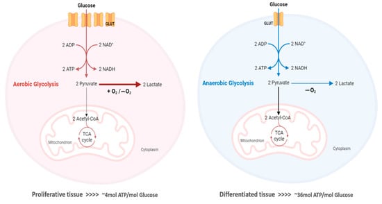
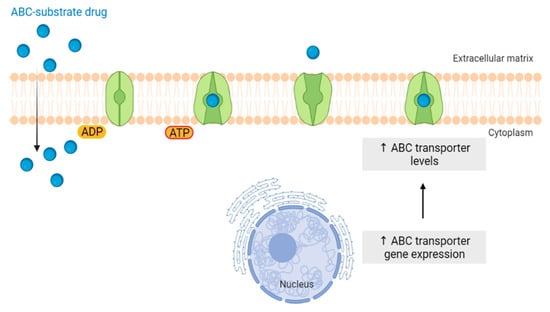
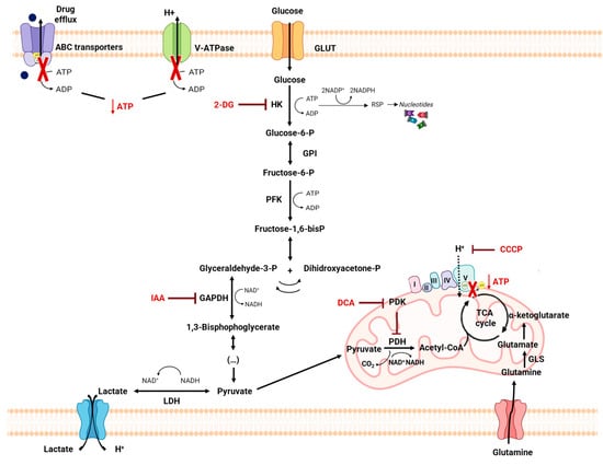
The “Warburg effect” consists of a metabolic shift in energy production from oxidative phosphorylation to glycolysis. The continuous activation of glycolysis in cancer cells causes rapid energy production and an increase in lactate, leading to the acidification of the tumour microenvironment, chemo- and radioresistance, as well as poor patient survival. Nevertheless, the mitochondrial metabolism can be also involved in aggressive cancer characteristics. The metabolic differences between cancer and normal tissues can be considered the Achilles heel of cancer, offering a strategy for new therapies. One of the main causes of treatment resistance consists of the increased expression of efflux pumps, and multidrug resistance (MDR) proteins, which are able to export chemotherapeutics out of the cell. Cells expressing MDR proteins require adenosine triphosphate (ATP) to mediate the efflux of their drug substrates. Thus, inhibition of the main energy-producing pathways in cancer cells, not only induces cancer cell death per se, but also overcomes multidrug resistance. 

tumor microenvironment tumor metabolism glycolysis Warburg effect resistance

1. Introduction

The conversion of normal cells or benign tissue into neoplastic precursors usually corresponds to malignant transformation. Additional alterations bestow these cells with unlimited proliferative potential, dissemination and metastasis, resulting in tumor progression [1]. In order to sustain the acquired features, metabolic reprogramming is essential. Changes in cellular metabolism promote the fast production of adenosine triphosphate (ATP) and an increase in the synthesis of biomolecules, including nucleotides, lipids and amino acids. Several mechanisms are known to modulate cancer metabolism, which affect essential pathways for both energy production and carbon metabolism, such as glycolysis and the tricarboxylic acid (TCA) cycle. As a consequence of these alterations, there is an increased consumption of glucose and also of glutamine in tumor cells in order to maintain their metabolic requirements [2]. Metabolic reprogramming is one of the emerging characteristics of tumor progression and is crucial to support the energy needs of cells during their continuous growth and proliferation. This metabolic reprogramming is also a key factor in the development of cancer resistance to treatment [2][3]. Often, during these treatments, cancer cells adapt, altering their metabolic pathways and becoming less susceptible to therapies. Targeting and exploiting such metabolic changes can be a promising approach to improve the chance of curing cancer. For this, the development of metabolism-targeting nanoparticles, carrying multiple therapeutic agents, are increasingly being exploited, aiming to overcome drug resistance and thus constituting an appellative tool in future cancer therapies.

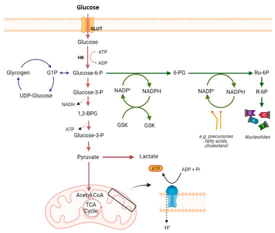
2. Glucose Metabolism

Most mammalian cells have glucose as their preferred metabolic substrate, which is used in the cytoplasm and/or mitochondria to provide energy for cell maintenance and proliferation [4] (Figure 1). Glycolysis, a metabolic pathway that does not require oxygen, partially oxidizes into two pyruvate molecules, producing two moles of ATP and two moles of nicotinamide adenine dinucleotide (NADH) per mole of consumed glucose [2][4]. In the presence of oxygen and active mitochondrial systems, healthy cells oxidize most of the pyruvate in the mitochondria, producing most of their ATP in this way (32 molecules of ATP from 1 single glucose molecule) [4][5]. When the anaerobic pathway is used, the pyruvate from glycolysis is reduced to lactate by the cytoplasmic enzyme lactate dehydrogenase (LDH), to regenerate the oxidized form NAD+ for glycolysis, producing 16 times less ATP per consumed glucose. The monocarboxylate transporters (MCTs) will then transport the excess lactate produced out of the cell through a proton-symport mechanism [4][5].
Figure 1. Glucose metabolism in mammalian cells. Illustrative scheme of glycolysis, TCA cycle, and the electron transport chain (red). Glucose from the blood stream is uptaken by the cells, converted into G6P by HK and posteriorly in pyruvate. In the absence of oxygen, pyruvate is converted into lactate, whereas in the presence of oxygen, the pyruvate is completely oxidized into Acetyl-CoA, which enters the mitochondrial TCA cycle. The generated NADH are then fed the OXPHOS-producing ATP (blue). The PPP (green) synthetizes the ribose-5-phosphate, which is needed for nucleic acid synthesis, and NADPH. The excess glucose is used to synthetize glycogen, via glycogenesis (purple). Created by the Authors with BioRender.com. ATP: adenosine triphosphate; G6P: glucose-6-phosphate; HK: hexokinase; NADH: nicotinamide adenine dinucleotide; NADPH: nicotinamide adenine dinucleotide phosphate; OXPHOS: oxidative phosphorylation; PPP: pentose phosphate pathway; TCA cycle: tricarboxylic acid cycle.
The first step in the glucose metabolism consists of its entrance into the cell. Glucose transporters (GLUTs) belong to the solute transporter (SLC2A) family of proteins and are present in many tissues/cells of the body, e.g., brain, erythrocytes, adipocytes, and liver, where they mediate glucose uptake [6]. The fourteen different isoforms of GLUTs are subdivided into three distinct protein classes, according to their sequence homology. Each GLUT isoform has a unique tissue distribution, substrate specificity, and a specific physiological function [7]. All GLUT proteins were originally assumed to catalyze the transport of hexoses into and out of cells. This is clearly the case for the class 1 GLUT proteins (GLUTs 1–4 and 14). However, class 2 (GLUTs 5, 7, 9 and 11) and class 3 (GLUTs 6, 8, 10, 12 and 13) GLUT proteins do not necessarily have a primary role in catalyzing glucose transport [8]. GLUT-1 is expressed in tissues with a high glycolytic rate, such as erythrocytes, which are responsible for glucose uptake in high-need cells [6][8]
Although there are hundreds of types of cancer, they share some specific characteristics, namely the reprogramming of the energy metabolism. Many cancer cells predominantly rely on glycolysis, instead of oxidative phosphorylation (OXPHOS), to produce energy from glucose, even in the presence of O2, with this metabolic shift being known as the “Warburg effect” or “aerobic glycolysis” [9]. Although OXPHOS is downregulated, cancer cells can still obtain the required ATP for cell survival and proliferation, increasing the glycolytic flux and metabolizing glucose at high rates, with lactate production [10]. This alteration in metabolism provides a selective advantage during tumor initiation and progression, sustaining the high proliferative rate of tumor cells and promoting resistance to cells. Nevertheless, in opposition to previous beliefs, this phenotype is not due to mitochondrial dysfunction and the whole ATP factory in cancer cells is important. In fact, not all tumor cell types completely restart glycolysis for the ATP supply, and some of them may equally or even predominantly use OXPHOS [11][12]
Many TCA cycle intermediates are used in biosynthetic processes; thus, a new carbon supply is required to maintain the activity of the TCA cycle. Glutaminolysis, where glutamine is used to fuel the TCA cycle, is one of the most important anaplerotic pathways in cancer [2]. In fact, glutamine deserves special attention, as it is the second most consumed metabolite by proliferating cells [2][4]. Glutamine has been shown to be essential for the synthesis of proteins, fatty acids, and nucleotides. Once inside the cell, glutaminase (GLS) converts glutamine into glutamate. Glutamate, in turn, can be converted into α-ketoglutarate, which is an intermediate of the TCA cycle. As tumor cells proliferate at higher rates, they are more glutamine-dependent than their non-tumoral counterparts [2][13]. However, a number of other metabolites have also been described to activate the TCA cycle in tumor cells [2].

3. The Warburg Effect

In 1920, Otto Warburg postulated that cancer cells are characterized by an increased glycolytic rate, with pyruvate mostly being converted to lactate, contrary to normal cells. This phenomenon became known as aerobic glycolysis or the “Warburg effect” [2][9][14]. This observation underlies the [18F]-fluorodeoxyglucose positron emission tomography (FDG-PET) of tumors, which is used in the diagnosis of cancer and in the detection of metastasis, due to the high consumption of the glucose analogue FDG by cancer cells [15].
Originally, Warburg postulated that the increased glycolytic activity observed in cancer cells should be due to impaired mitochondrial function. In fact, mutations in TCA cycle enzymes are present in several types of cancer, such as fumarate hydratase, succinate dehydrogenase, and isocitrate dehydrogenase [9][16][17]. However, even when mitochondrial function is normal, many cancer cells still prefer glycolysis, suggesting that glycolysis is associated with advantages to these cells [9]. As several glycolytic intermediates can be used in biosynthetic pathways, it is likely that the increase in the glycolytic rate supplies the biosynthetic needs of cancer cells [18]. In fact, the high consumption of glucose allows for the energy necessary for cell growth to be obtained and, under these conditions, the PPP pathway is also favored, generating NADPH and ribose-5-phosphate, which serve as a source for the formation of new nucleotides, lipids and proteins [10][11][19].
The overexpression of GLUTs is essential for cancer cells to meet their high demand for glucose, which is needed for their high glycolytic rates. In addition, cancer cells often present higher levels of MCTs, since they allow for the maintenance of intracellular pH and, consequently, the glycolytic way, as they are responsible for the export of lactate. Lactate secretion may help to create an acidic extracellular tumor microenvironment (TME) that favors tumor growth, promoting migration and invasion [2][5]. The low pH found in TME activates metalloproteinases released from the cancer cells, promoting the digestion of the surrounding matrix and leading to cells’ detachment from the solid substrate [20][21].
Thus, although ATP production through OXPHOS is more efficient, most cancer cells produce most of their ATP through glycolysis, even in the presence of oxygen [2][6][14] (Figure 2). In fact, 70–80% of human cancers present the Warburg phenotype, a metabolic alteration that results from the interaction between normoxic/hypoxic activation of the transcription factor HIF-1, oncogenes’ activation, loss of tumor suppressors, altered signaling pathways and interactions with components of the TME, as well as being associated with epigenetic mechanisms [16].
Figure 2. Schematic representation of the main differences between aerobic glycolysis (“Warburg effect”) in proliferative tissue and OXPHOS and anaerobic glycolysis in differentiated tissues. In the presence of O2, differentiated tissues (no proliferating) metabolize glucose to pyruvate via glycolysis and subsequently completely oxidize pyruvate to CO2 in the mitochondria (OXPHOS). At low levels of O2, pyruvate is partially oxidized by glycolysis, generating lactate (anaerobic glycolysis). The generation of lactate results in minimal ATP production when compared with OXPHOS. In contrast, cancer/proliferative cells predominantly produce energy through an increased rate of glycolysis, followed by a reduction of pyruvate into lactate in the cytosol, resulting in a high production of lactic acid. Created by the Authors with BioRender.com. ATP: adenosine triphosphate; OXPHOS: oxidative phosphorylation.
As glycolysis less efficient in energetic terms than OXPHOS, cancer cells increase their glycolytic flux by about 15 times, leading to a drastic increase in the rate of ATP production, in order to compensate the energy yield [5]. In addition, and as previously discussed, the “Warburg effect” contributes to counteracting apoptosis and promotes macromolecule biosynthesis. However, high rates of OXPHOS are displayed by some cancer cells. In fact, some cancer cells, even in a glycolytic cancer, switch their metabolism to OXPHOS, as this metabolic pathway is the predominant supplier of ATP in these cases [22][23][24]. There is a significant emphasis on enzymes like isocitrate dehydrogenase (IDH) 1 and IDH2, which catalyze the first oxidative reaction of the TCA cycle, resulting in the generation of NADH, and thus have particular importance in mitochondrial respiration [25].

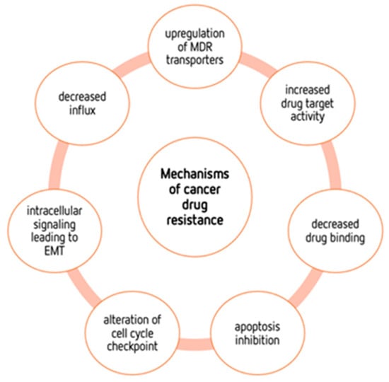
4. Mechanisms of Cancers’ Drug Resistance

In the last few decades, cancer treatment has made great, promising advances. Nevertheless, despite these advances, tumors seem to always find a way to resist practically all types of anticancer therapy, hindering their treatment potential [2][26]. Cancer patients who are resistant to therapy often develop more metastases, which are the main cause of cancer-related deaths in these cases [27][28]. Thus, it is important to develop new therapeutic approaches to overcome resistance to therapy [26][27]. The growing knowledge of the molecular mechanisms of cancer has allowed for the discovery and improvement of new therapeutic compounds with a better progression-free survival. Unfortunately, this does not always translate into overall survival benefits, as resistance is one of the main problems to overcome. This resistance may be due to intrinsic mechanisms or to acquired mechanisms, which arise after the exposure of cancer cells to chemotherapeutic drugs [26] (Figure 3).
Figure 3. Mechanisms of chemotherapeutic drug resistance in cancer cells. This resistance may be due to intrinsic mechanisms or due to acquired mechanisms, such as the ones listed in the figure.
This acquired resistance may result from several factors, namely the acquisition of mutations that cause a decrease in drug binding, an increase in drug target activity, or an upregulation of multidrug resistance (MDR) transporters [29]. For example, mutations of the TP53 gene, a tumor suppressor responsible for genome stability, are frequently observed in cancer cells and involved in cancer resistance to therapy [30]
EMT plays an important role in tumor progression, metastasis and therapy resistance and is often associated with metabolic alterations in cancer [27][31]. EMT is a highly conserved biological process that involves the transition of polarized, immobile epithelial cells into motile mesenchymal cells due to the loss of apicobasal polarity, loss of cell–cell contacts, reorganization of the actin cytoskeleton, and ability to invade the extracellular matrix as an individual cell [27]. Different studies using cancer cell lines demonstrated the responsibility of EMT in radio- or chemotherapy-driven resistance [27][32][33]. In fact, conventional anticancer drugs are mainly directed toward rapidly dividing cells, with EMT being associated with stem cell properties in cancer cells [34].
A large number of studies on metabolism-mediated drug resistance have focused on glycolysis and the TCA cycle, including the roles of glucose and glutamine in such phenotypes [2][35][36][37]. Nevertheless, fatty acids and BCAAs may also be associated with both energy production and tumorigenesis. Concerning amino acids, their metabolism may also constitute a target for treating drug-resistant tumors. Cancer cells may be dependent on specific amino acids, like serine, glycine, proline, aspartate, and arginine. In fact, amino acid metabolism has been extensively studied and recognized as an important factor in both drug resistance and energy production. 
Unfortunately, resistance to therapy not only includes resistance to conventional treatments, such as chemotherapy or radiation, but also immunological and targeted therapies [27], affecting the long-term therapeutic outcome of tumor patients [38]. Many scientific reports have shown that the MDR phenotype, which is characterized by a broad tumor’s resistance to multiple drugs and can differ either in its structure or in its mechanism of action, often correlates with the expression of active transport mechanisms responsible for the efflux of a wide variety of drugs, leading to a reduction in the effect of the drug, as there is a reduction in its intracellular levels [28][38][39]. These transporters, which are frequently highly expressed in resistant cancer cells, belong to the ATP-binding cassette (ABC) family, with P-glycoprotein (Pgp) being the first-identified and best-studied ABC transporter [38][39].

4.1. ABC Transporters

The ABC transporter family is composed of seven subfamilies (ABCA to ABCG), according to their genomic sequences and the core structure of transmembrane domains, but only a few of them transport drugs; therefore, they play an important role in their bioavailability [40][41][42]. In humans, the proteins of this ABC transporter superfamily comprise at least 48 genes with diverse functions [28][43]. Given their ability to extrude several conventional antitumor drugs, recent studies in cancer research focused on the members of this superfamily to understand the reasons for the failure of chemotherapy treatment (Figure 4) [28].
Figure 4. A simplified schematic diagram of ABC transporter overexpression leading to drug resistance in cancer cells. The ABC proteins (green) reduce intracellular drug concentration by actively transporting ABC substrate drugs (blue circles) out of the cancer cell, which leads to the MDR phenotype. Created by the Authors with BioRender.com. ABC: ATP-binding cassette; MDR: multidrug resistance.
Three major subfamilies of ABC transporters have been associated with the MDR phenotype and extensively studied: ABCB, comprising ABCB1 (Pgp/MDR1), ABCC, comprising ABCC1 (Multidrug-Resistance Protein 1 (MRP1)) and ABCG, comprising ABCG2 (Breast Cancer-Resistance Protein (BCRP)) in their respective members. These three proteins are major players in both primary and acquired resistance to chemotherapeutic drugs [28][44][45]. A key factor in the clarification of the mechanisms behind MDR was the discovery of the MDR1 and MRP1 transporters, which allowed for the identification of a variety of proteins with similar structures and transport capabilities. In addition to their role in transport of drugs, several members of the ABCB subfamily are also involved in intracellular peptides’ transport, including a key role in the presentation of major histocompatibility complex class I antigens [28]. MDR1, MRP1 and BCRP transporters can export an extensive range of chemotherapeutic compounds used in the treatment of cancer patients, making them attractive therapeutic targets [28]
In addition, cancer progression has been associated with the overexpression of some other ABC transporters, as in the case of melanoma, where a clinical correlation with ABCB5 expression was found [26][46]. To make the situation worse, several cancers overexpress more than one ABC transporter; this co-expression contributes to multiple-drug resistance [28][40]. Thus, to achieve a better clinical outcome, multi-carrier inhibitors are required [40]. For instance, the co-expression of MDR1, ABCB5 and ABCC2 was observed in a subpopulation of melanoma cells [26][46]. It has also been described that BCRP/MDR1 transporters are highly expressed in hematopoietic stem cells [26][47]. Furthermore, some studies demonstrated a possible relationship between ABC transporters and the in vivo formation of metastasis, although there is still no direct evidence of such an association [28][48].

4.2. Metabolic Alterations Involved in Drug Resistance in Cancer

Recently, it has been shown that the response to first-line chemotherapy treatment largely depends on the metabolism of cancer cells, which can be reprogrammed during the treatment [5]. The development of tumor-cell-associated resistance due to drug-induced selective pressures demonstrates specific resistant metabolic characteristics [36]. Several conventional chemotherapeutics activate apoptosis, killing cancer cells. However, if cancer cells find mechanisms to avoid chemotherapy’s cytotoxic effect, they will escape this programmed cell death and, as a consequence, the cancer will grow [37]. Several mechanisms are involved in the development of drug resistance in cancers, such as increased drug exportation, metabolic reprogramming and TME hypoxia [38][49]. The activation of different signaling pathways with the expression of signaling molecules is also involved in different mechanisms of drug resistance [38]. It is established that cells that express MDR proteins, such as Pgp or MRP, rely on ATP as their energy source to pump out drug substrates from within the cells. Consequently, the heightened expression of these proteins results in increased drug efflux due to the surplus production of cellular ATP, thereby inducing drug resistance [50]. Furthermore, as previously mentioned, TME plays an important role in the progression of cancers. Cancer cells have a greater need for nutrients to produce the necessary energy and sustain their anabolic needs. Thus, the availability of nutrients influences the proliferation rate of cancer cells. Despite this need, cancer cells have metabolic plasticity, which allows for them to adapt to conditions of reduced nutrient availability, and may, in turn, remodel the TME [51]. With changes in metabolism, the tumor microenvironment undergoes changes to ensure its survival, namely hypoxia, acidosis, and the formation of stroma cells. These changes, besides being particularly adverse to normal cells, are involved in the development of chemoresistance. Hypoxia can be caused by increased oxygen consumption, the rapid growth and proliferation of the tumor and also by the lack of a vascular system in certain tumor zones [10][51]. On the other hand, and as previously mentioned, hypoxia can lead to the greater use of glycolysis for the production of ATP in cancer cells, and this mechanism of obtaining energy leads to an accumulation of lactate in cells, facilitating the evasion of the immune system [52][53]. Lactate is transported to the outside of cells through the increased activity of pH regulators like ATPases, carbonic anhydrases and MCTs in order to maintain the intracellular acid-base balance.
At the mitochondrial level, mitochondrial DNA (mtDNA) depletion is related to tumor progression and metastasis, and may further act as a “progression signal” for chemoresistance [37][43]. Li et al. showed that mtDNA-depleted androgen-independent prostate carcinoma cells, despite growing slowly, are highly carcinogenic, revealing an overexpression of BCRP and extremely aggressive and radio- and chemoresistant characteristics [54]. In addition, the fact that these cancer cells present a slow growth may be an advantage in their resistance to chemotherapy treatments, since the cytotoxic agents used in conventional chemotherapy have a more direct impact on rapidly proliferative cells [37][45]. mtDNA depletion in hepatocarcinoma cells resulted in cisplatin, DOX, and SN-38 chemoresistance linked with the upregulation of the MDR1 gene and MRP1 and MRP2, which are particularly involved in MDR. In colon cancer cells that are mtDNA-depleted, the upregulation of MDR1 has also been observed [46][47].

4.3. Metabolic Modulation as an Approach to Overcome Drug Resistance

The metabolic reprogramming of cancer cells, besides its role in cancer proliferation and invasion, is also implicated in the acquisition of resistance to therapy in cancer patients. In this way, the recent increase in the knowledge of tumor cell metabolism and the subsequent exploration of metabolic alterations in these cells may offer an opportunity to discover new potential targets for therapeutic intervention and to overcome such resistance. This is particularly important in the different types of cancers that show resistance to drugs, to improve treatments and avoid adverse side effects. Disruption of the Warburg effect is the most often used means of sensitizing the cells to conventional antitumor drugs, exploiting cancer metabolic reprogramming [55]. Thus, glycolytic inhibitors can be used as a therapeutic strategy as they drastically decrease cellular ATP levels, which is necessary to maintain the activity of the drug efflux pumps [40] (Figure 5). This could be an effective strategy, as one of the best-described mechanisms of drug resistance is due to the increased level and/or activity of the efflux pumps that remove drugs from cells [49].
Figure 5. Metabolic modulation as an approach to overcome drug resistance. Glucose and glutamine metabolism, in tumor cells, supply vital components for the high requirements of both glycolysis and OXPHOS. The different compounds (IAA and 2DG) are glycolytic inhibitors. DCA inhibits PDK, reactivating PDH, and switching the metabolism from glycolysis towards OXPHOS. CCP is an uncoupler that inhibits ATP synthesis. The depletion of cancer cell energy probably leads to the inactivation of the pumps’ ABC transporters. Created by the Authors with BioRender.com. 2DG: 2-deoxyglucose; ABC: ATP-binding transporter; ATP: adenosine triphosphate; CCP: Carbonyl Cyanide m-chlorophenyl Hydrazone; DCA: dichloroacetate; IAA: iodoacetate; OXPHOS: oxidative phosphorylation; PDH: pyruvate dehydrogenase.
Amino acid metabolism can be also related to MDR phenotype, as it provides cancer cells with specific adaptive characteristics to neutralize the mechanism of action of the antitumor drugs to which they are exposed [56]. In fact, amino acids play an important role both in most biosynthetic pathways, which are upregulated in cancer cells, and in maintaining the redox homeostasis balance [42]. Among these, glutamine plays a crucial role in cancer metabolism and in drug resistance in cancer cells, since glutaminolysis supports the biosynthesis of many essential molecules [36][57] (Figure 5). The importance of glutamine is also due to the fact that it is the amino acid with the largest carbon source for the TCA cycle. In the context of tumor cells, glutamine metabolism can provide essential building blocks for the excessive demands of both glycolysis and OXPHOS [58]

4.4. Self-Delivery of Nanomedicine to Overcome Drug Resistance

Chemotherapy, radiation therapy and resection surgery remain the three “gold standard” anticancer therapies [59]. Whether radiotherapy and surgery can be indicated for localized cancers, chemotherapy is considered the most appropriate treatment for most patients with metastasis and advanced cancer, as chemotherapy drugs can be widely distributed in the organism through the bloodstream [60]. Nevertheless, the development of drug resistance and the low hydrosolubility of drugs are significant problems that restrict the clinical use of currently available chemotherapy drugs [60]. Major chemotherapeutic agents include compounds like platinum complexes, DOX, vinca alkaloids, and taxanes, and primarily affect nucleic acids and protein synthesis, interfering with cell cycle and triggering apoptosis [61][62]. However, most of the standard agents approved for clinical use do not have the capacity to differentiate normal cells from cancer cells. This leads to serious side effects, especially in rapidly growing cells, as these drugs generally compromise mitosis. These cells include hair follicles, bone marrow cells and the gastrointestinal system, leading to hair loss, immune system failure, and infections, respectively [63]. Thus, the decrease in the toxicity and side effects of the main chemotherapeutic agents is an urgent problem that needs to be overcome [61]. To overcome this problem, various compounds, such as 3BP, DCA and 2DG, that interfere with metabolism, have been tested and demonstrated their ability to decrease tumor cell metabolism [64]. However, there are disadvantages to a metabolism-based approach in cancer therapy, since the metabolic pathways required for cell survival are also present in normal cells. Thus, metabolism-based treatment can face the major hurdle of non-specific toxicity [5]. To decrease their toxic side effects and increase antitumor efficacy, a number of drug delivery systems have been developed, such as albumin-bound PTX (Abraxane®) or liposome-entrapped PTX and DOX, which have received clinical approval, as these formulations presented enhanced security but maintained their effectiveness [60][61][65].
Nanotechnology-based cancer therapies aim to find new therapeutic methodologies correlated with disease mechanisms. The use of nanoparticles to encapsulate the drugs may increase the specificity of delivery to cancer cells and decrease the interaction with other non-cancer cells involved in tumor growth and spreading [59]. Poly(lactic-co-glycolic acid) (PLGA), a synthetic thermoplastic aliphatic biodegradable and biocompatible polyester, is widely studied and is one of the most characterized polymers [66]. PLGA is degraded in non-toxic products (H2O and CO2) that are easily excreted [61][66]. Its polymeric NPs are degraded in vivo into lactate and glycolate. D-lactate is not metabolized prior to excretion and L-lactate is transformed into CO2, which is eliminated by pulmonary excretion, or converted to pyruvate, which fuels the TCA cycle. Glycolate can be directly excreted by the kidneys or can be oxidized to glyoxylate, which is, in turn, further metabolized producing glycine, serine, and pyruvate. Subsequently, pyruvate can re-enter the TCA cycle and follow the OXPHOS pathway [66]. The lactic acid (LA)/glycolic acid (GA) proportion is a good indicator not only when adjusting the degradation time, but also of the drug release rate [66][67]. Due to the absence of lateral methyl groups in GA, it has a higher hydrophilia, and thus, when higher amounts of GA are present, a higher degradation rate is observed [66][68]
Some PLGA polymers are FDA-approved materials and various PLGA NPs formulations have been clinically introduced, such as a formulation targeting advanced prostate cancer, ELIGARD® [63]. PLGA NPs were also shown to be effective in increasing the accumulation of docetaxel in gastric tumors, thus causing an increase in anticancer activity [69].
Ongoing research underscores the significance of the TME in driving tumor proliferation, invasion, metastasis, and resistance to therapeutic interventions. As mentioned, the TME provides protection for cancer cells, enabling them to evade conventional treatments like surgery, radiotherapy, and chemotherapy. Furthermore, the constituents of the TME play a pivotal role in fostering therapy resistance in solid tumors. Consequently, directing interventions toward the TME presents a promising avenue for advancing the field of cancer nanomedicine. The combination of antitumor drugs with drugs that interfere with resistance mechanisms has largely been made possible by advancements in nanotechnology [70]. Hence, directing efforts toward the TME presents an innovative approach to advancing the field of cancer nanomedicine [64][71]. Nanoparticles developed in response to TME cues, such as a low pH, redox conditions, and hypoxia, enhance the pharmacokinetics and therapeutic effectiveness of nanomedicine, but also have glycolytic inhibitors [64][72][73][74]. Although not directly associated with this, and as has been shown for DCA in a lung cancer cell model, the use of nanoparticles improves the delivery of the compound, which can be important in cases of resistance. 
Using NPs to direct therapy to energy metabolism and the TME could be a promising approach to sensitizing cells to conventional chemotherapy. Although the use of nanotechnology is still a recent field in cancer therapy, there is already enough evidence of its potential for successful treatment, allowing for a more accurate and specific delivery of antitumor drugs into cancer cells and avoiding many adverse side effects. Many barriers still need to be overcome regarding the success of NPs in clinical trials. Some of these barriers include the size and timing of certain NP therapies. The majority of experimental tests of NPs are cell-based and use animal models, which may not lead to convincing results in human testing. Furthermore, as the presence of metastases is a significant property of cancer, more studies should be carried out with models of cancer metastasis [75].

5. Conclusions

Although conventional chemotherapy is particularly toxic to tumor cells, it is often non-specific, and is responsible for the significant side effects associated with cancer treatment. However, there are differences between cancer cells and healthy cells that can be explored to increase treatment specificity against cancer. One of these differences consists of the “Warburg effect”, currently considered an emergent cancer hallmark, whereby the upregulation of the glycolytic rate in tumor cells is a key player in acid-resistant phenotypes through their adaptation to hypoxia and acidosis, as well as in tumor aggressiveness [2][9][76]. High glycolytic rates are widely reported to promote the chemoresistance of tumor cells to conventional therapy [2]. In fact, increased acidification of the extracellular space leads to lower drug stability and, consequently, lower drug efficacy. In parallel, the increased production of glycolytic intermediates promotes cell proliferation, since these are biosynthetic precursors, whereas ATP production sustains the activity of proteins involved in both drug efflux and cell division. Together, these effects underly multidrug resistance. Nevertheless, many cancer cells adapt to changes in TME, exhibiting metabolic plasticity and switching their metabolism from glycolysis to OXPHOS, and vice-versa. For example, OXPHOS could be the predominant metabolic pathway used by cancer stem cells, and is often involved in cancer resistance, metastasis, and tumor relapse [77]. Exploring specific characteristics of cancer cells, such as this change in metabolism, could be a promising strategy for the use of more effective and more specific drugs that primarily target cancer cells. In fact, metabolic changes in cancer cells can reveal specific vulnerabilities that could be targeted with precision therapies. However, the metabolic plasticity and interchange of glycolytic and oxidative cells, although occurring many times in the same cancer and being responsible for tumor heterogeneity, is not taken into account in cancer therapies. Thus, more integrated research is needed, investigating the main metabolic pathways used in different conditions and stages of each cancer type, and the influence of the TME characteristics (e.g., oxygen, pH, nutrients availability, immune components) on such metabolic adaptation and heterogeneity. An understanding of these metabolic switches, the identification of metabolic targets, and the use of combined therapies in a more targeted way through the use of nanoparticles could have a huge impact not only on the development of new drugs, but also on the ability to overcome drug resistance, one of the major problems that occurs during cancer treatment. 

References

  1. Porporato, P.E.; Filigheddu, N.; Pedro, J.M.B.; Kroemer, G.; Galluzzi, L. Mitochondrial metabolism and cancer. Cell Res. 2018, 28, 265–280.
  2. Zaal, E.A.; Berkers, C.R. The Influence of Metabolism on Drug Response in Cancer. Front. Oncol. 2018, 8, 500.
  3. Chen, X.; Chen, S.; Yu, D. Metabolic Reprogramming of Chemoresistant Cancer Cells and the Potential Significance of Metabolic Regulation in the Reversal of Cancer Chemoresistance. Metabolites 2020, 10, 289.
  4. Ortega, A.D.; Sanchez-Arago, M.; Giner-Sanchez, D.; Sanchez-Cenizo, L.; Willers, I.; Cuezva, J.M. Glucose avidity of carcinomas. Cancer Lett. 2009, 276, 125–135.
  5. Vanhove, K.; Graulus, G.J.; Mesotten, L.; Thomeer, M.; Derveaux, E.; Noben, J.P.; Guedens, W.; Adriaensens, P. The Metabolic Landscape of Lung Cancer: New Insights in a Disturbed Glucose Metabolism. Front. Oncol. 2019, 9, 1215.
  6. Cameron, M.E.; Yakovenko, A.; Trevino, J.G. Glucose and Lactate Transport in Pancreatic Cancer: Glycolytic Metabolism Revisited. J. Oncol. 2018, 2018, 6214838.
  7. Reckzeh, E.S.; Waldmann, H. Small-Molecule Inhibition of Glucose Transporters GLUT-1-4. Chembiochem 2020, 21, 45–52.
  8. Holman, G.D. Structure, function and regulation of mammalian glucose transporters of the SLC2 family. Pflug. Arch. 2020, 472, 1155–1175.
  9. Warburg, O. On the origin of cancer cells. Science 1956, 123, 309–314.
  10. Liberti, M.V.; Locasale, J.W. The Warburg Effect: How Does it Benefit Cancer Cells? Trends Biochem. Sci. 2016, 41, 211–218.
  11. Moreno-Sanchez, R.; Rodriguez-Enriquez, S.; Marin-Hernandez, A.; Saavedra, E. Energy metabolism in tumor cells. FEBS J. 2007, 274, 1393–1418.
  12. Gillies, R.J.; Robey, I.; Gatenby, R.A. Causes and consequences of increased glucose metabolism of cancers. J. Nucl. Med. 2008, 49 (Suppl. S2), 24S–42S.
  13. Yoo, H.C.; Yu, Y.C.; Sung, Y.; Han, J.M. Glutamine reliance in cell metabolism. Exp. Mol. Med. 2020, 52, 1496–1516.
  14. Sun, H.; Zhu, A.; Zhou, X.; Wang, F. Suppression of pyruvate dehydrogenase kinase-2 re-sensitizes paclitaxel-resistant human lung cancer cells to paclitaxel. Oncotarget 2017, 8, 52642–52650.
  15. Danhier, P.; Banski, P.; Payen, V.L.; Grasso, D.; Ippolito, L.; Sonveaux, P.; Porporato, P.E. Cancer metabolism in space and time: Beyond the Warburg effect. Biochim. Biophys. Acta Bioenerg. 2017, 1858, 556–572.
  16. Vaupel, P.; Multhoff, G. Revisiting the Warburg effect: Historical dogma versus current understanding. J. Physiol. 2021, 599, 1745–1757.
  17. Ghanbari Movahed, Z.; Rastegari-Pouyani, M.; Mohammadi, M.H.; Mansouri, K. Cancer cells change their glucose metabolism to overcome increased ROS: One step from cancer cell to cancer stem cell? Biomed. Pharmacother. 2019, 112, 108690.
  18. Nadzialek, S.; Vanparys, C.; Van der Heiden, E.; Michaux, C.; Brose, F.; Scippo, M.L.; De Coen, W.; Kestemont, P. Understanding the gap between the estrogenicity of an effluent and its real impact into the wild. Sci. Total Environ. 2010, 408, 812–821.
  19. Kroemer, G.; Pouyssegur, J. Tumor cell metabolism: Cancer’s Achilles’ heel. Cancer Cell 2008, 13, 472–482.
  20. Jesser, E.A.; Brady, N.J.; Huggins, D.N.; Witschen, P.M.; O’Connor, C.H.; Schwertfeger, K.L. STAT5 is activated in macrophages by breast cancer cell-derived factors and regulates macrophage function in the tumor microenvironment. Breast Cancer Res. 2021, 23, 104.
  21. Putney, L.K.; Barber, D.L. Expression profile of genes regulated by activity of the Na-H exchanger NHE1. BMC Genom. 2004, 5, 46.
  22. Zheng, J. Energy metabolism of cancer: Glycolysis versus oxidative phosphorylation (Review). Oncol. Lett. 2012, 4, 1151–1157.
  23. Fu, Y.; Liu, S.; Yin, S.; Niu, W.; Xiong, W.; Tan, M.; Li, G.; Zhou, M. The reverse Warburg effect is likely to be an Achilles’ heel of cancer that can be exploited for cancer therapy. Oncotarget 2017, 8, 57813–57825.
  24. Fan, T.W.; Kucia, M.; Jankowski, K.; Higashi, R.M.; Ratajczak, J.; Ratajczak, M.Z.; Lane, A.N. Rhabdomyosarcoma cells show an energy producing anabolic metabolic phenotype compared with primary myocytes. Mol. Cancer 2008, 7, 79.
  25. Minemura, H.; Takagi, K.; Sato, A.; Yamaguchi, M.; Hayashi, C.; Miki, Y.; Harada-Shoji, N.; Miyashita, M.; Sasano, H.; Suzuki, T. Isoforms of IDH in breast carcinoma: IDH2 as a potent prognostic factor associated with proliferation in estrogen-receptor positive cases. Breast Cancer 2021, 28, 915–926.
  26. Rezayatmand, H.; Razmkhah, M.; Razeghian-Jahromi, I. Drug resistance in cancer therapy: The Pandora’s Box of cancer stem cells. Stem Cell Res. Ther. 2022, 13, 181.
  27. De Las Rivas, J.; Brozovic, A.; Izraely, S.; Casas-Pais, A.; Witz, I.P.; Figueroa, A. Cancer drug resistance induced by EMT: Novel therapeutic strategies. Arch. Toxicol. 2021, 95, 2279–2297.
  28. Fletcher, J.I.; Haber, M.; Henderson, M.J.; Norris, M.D. ABC transporters in cancer: More than just drug efflux pumps. Nat. Rev. Cancer 2010, 10, 147–156.
  29. Vasan, N.; Baselga, J.; Hyman, D.M. A view on drug resistance in cancer. Nature 2019, 575, 299–309.
  30. Mantovani, F.; Collavin, L.; Del Sal, G. Mutant p53 as a guardian of the cancer cell. Cell Death Differ. 2019, 26, 199–212.
  31. Jia, D.; Park, J.H.; Kaur, H.; Jung, K.H.; Yang, S.; Tripathi, S.; Galbraith, M.; Deng, Y.; Jolly, M.K.; Kaipparettu, B.A.; et al. Towards decoding the coupled decision-making of metabolism and epithelial-to-mesenchymal transition in cancer. Br. J. Cancer 2021, 124, 1902–1911.
  32. Inoue, A.; Seidel, M.G.; Wu, W.; Kamizono, S.; Ferrando, A.A.; Bronson, R.T.; Iwasaki, H.; Akashi, K.; Morimoto, A.; Hitzler, J.K.; et al. Slug, a highly conserved zinc finger transcriptional repressor, protects hematopoietic progenitor cells from radiation-induced apoptosis in vivo. Cancer Cell 2002, 2, 279–288.
  33. Olmeda, D.; Moreno-Bueno, G.; Flores, J.M.; Fabra, A.; Portillo, F.; Cano, A. SNAI1 is required for tumor growth and lymph node metastasis of human breast carcinoma MDA-MB-231 cells. Cancer Res. 2007, 67, 11721–11731.
  34. Tavares-Valente, D.; Cannone, S.; Greco, M.R.; Carvalho, T.M.A.; Baltazar, F.; Queiros, O.; Agrimi, G.; Reshkin, S.J.; Cardone, R.A. Extracellular Matrix Collagen I Differentially Regulates the Metabolic Plasticity of Pancreatic Ductal Adenocarcinoma Parenchymal Cell and Cancer Stem Cell. Cancers 2023, 15, 3868.
  35. Yan, L.; Tu, B.; Yao, J.; Gong, J.; Carugo, A.; Bristow, C.A.; Wang, Q.; Zhu, C.; Dai, B.; Kang, Y.; et al. Targeting Glucose Metabolism Sensitizes Pancreatic Cancer to MEK Inhibition. Cancer Res. 2021, 81, 4054–4065.
  36. Guo, J.; Satoh, K.; Tabata, S.; Mori, M.; Tomita, M.; Soga, T. Reprogramming of glutamine metabolism via glutamine synthetase silencing induces cisplatin resistance in A2780 ovarian cancer cells. BMC Cancer 2021, 21, 174.
  37. Guo, J.; Yu, J.; Peng, F.; Li, J.; Tan, Z.; Chen, Y.; Rao, T.; Wang, Y.; Peng, J.; Zhou, H. In vitro and in vivo analysis of metabolites involved in the TCA cycle and glutamine metabolism associated with cisplatin resistance in human lung cancer. Expert Rev. Proteom. 2021, 18, 233–240.
  38. Lotz, C.; Kelleher, D.K.; Gassner, B.; Gekle, M.; Vaupel, P.; Thews, O. Role of the tumor microenvironment in the activity and expression of the p-glycoprotein in human colon carcinoma cells. Oncol. Rep. 2007, 17, 239–244.
  39. Skeberdyte, A.; Sarapiniene, I.; Aleksander-Krasko, J.; Stankevicius, V.; Suziedelis, K.; Jarmalaite, S. Dichloroacetate and Salinomycin Exert a Synergistic Cytotoxic Effect in Colorectal Cancer Cell Lines. Sci. Rep. 2018, 8, 17744.
  40. Xiao, H.; Zheng, Y.; Ma, L.; Tian, L.; Sun, Q. Clinically-Relevant ABC Transporter for Anti-Cancer Drug Resistance. Front. Pharmacol. 2021, 12, 648407.
  41. Robert, J.; Morvan, V.L.; Smith, D.; Pourquier, P.; Bonnet, J. Predicting drug response and toxicity based on gene polymorphisms. Crit. Rev. Oncol. Hematol. 2005, 54, 171–196.
  42. Aye, I.L.; Singh, A.T.; Keelan, J.A. Transport of lipids by ABC proteins: Interactions and implications for cellular toxicity, viability and function. Chem. Biol. Interact. 2009, 180, 327–339.
  43. Bugde, P.; Biswas, R.; Merien, F.; Lu, J.; Liu, D.X.; Chen, M.; Zhou, S.; Li, Y. The therapeutic potential of targeting ABC transporters to combat multi-drug resistance. Expert Opin. Ther. Targets 2017, 21, 511–530.
  44. Wang, S.A.; Young, M.J.; Wang, Y.C.; Chen, S.H.; Liu, C.Y.; Lo, Y.A.; Jen, H.H.; Hsu, K.C.; Hung, J.J. USP24 promotes drug resistance during cancer therapy. Cell Death Differ. 2021, 28, 2690–2707.
  45. Fletcher, J.I.; Williams, R.T.; Henderson, M.J.; Norris, M.D.; Haber, M. ABC transporters as mediators of drug resistance and contributors to cancer cell biology. Drug Resist. Updates 2016, 26, 1–9.
  46. Welte, Y.; Adjaye, J.; Lehrach, H.R.; Regenbrecht, C.R. Cancer stem cells in solid tumors: Elusive or illusive? Cell Commun. Signal. 2010, 8, 6.
  47. Scharenberg, C.W.; Harkey, M.A.; Torok-Storb, B. The ABCG2 transporter is an efficient Hoechst 33342 efflux pump and is preferentially expressed by immature human hematopoietic progenitors. Blood 2002, 99, 507–512.
  48. Zochbauer-Muller, S.; Filipits, M.; Rudas, M.; Brunner, R.; Krajnik, G.; Suchomel, R.; Schmid, K.; Pirker, R. P-glycoprotein and MRP1 expression in axillary lymph node metastases of breast cancer patients. Anticancer Res. 2001, 21, 119–124.
  49. Juan-Carlos, P.M.; Perla-Lidia, P.P.; Stephanie-Talia, M.M.; Monica-Griselda, A.M.; Luz-Maria, T.E. ABC transporter superfamily. An updated overview, relevance in cancer multidrug resistance and perspectives with personalized medicine. Mol. Biol. Rep. 2021, 48, 1883–1901.
  50. Maher, J.C.; Wangpaichitr, M.; Savaraj, N.; Kurtoglu, M.; Lampidis, T.J. Hypoxia-inducible factor-1 confers resistance to the glycolytic inhibitor 2-deoxy-D-glucose. Mol. Cancer Ther. 2007, 6, 732–741.
  51. Reina-Campos, M.; Moscat, J.; Diaz-Meco, M. Metabolism shapes the tumor microenvironment. Curr. Opin. Cell Biol. 2017, 48, 47–53.
  52. Liu, C.; Jin, Y.; Fan, Z. The Mechanism of Warburg Effect-Induced Chemoresistance in Cancer. Front. Oncol. 2021, 11, 698023.
  53. Tavares-Valente, D.; Sousa, B.; Schmitt, F.; Baltazar, F.; Queiros, O. Disruption of pH Dynamics Suppresses Proliferation and Potentiates Doxorubicin Cytotoxicity in Breast Cancer Cells. Pharmaceutics 2021, 13, 242.
  54. Li, X.; Zhong, Y.; Lu, J.; Axcrona, K.; Eide, L.; Syljuasen, R.G.; Peng, Q.; Wang, J.; Zhang, H.; Goscinski, M.A.; et al. MtDNA depleted PC3 cells exhibit Warburg effect and cancer stem cell features. Oncotarget 2016, 7, 40297–40313.
  55. Schiliro, C.; Firestein, B.L. Mechanisms of Metabolic Reprogramming in Cancer Cells Supporting Enhanced Growth and Proliferation. Cells 2021, 10, 1056.
  56. Pranzini, E.; Pardella, E.; Paoli, P.; Fendt, S.M.; Taddei, M.L. Metabolic Reprogramming in Anticancer Drug Resistance: A Focus on Amino Acids. Trends Cancer 2021, 7, 682–699.
  57. Li, T.; Copeland, C.; Le, A. Glutamine Metabolism in Cancer. Adv. Exp. Med. Biol. 2021, 1311, 17–38.
  58. Ma, W.W.; Jacene, H.; Song, D.; Vilardell, F.; Messersmith, W.A.; Laheru, D.; Wahl, R.; Endres, C.; Jimeno, A.; Pomper, M.G.; et al. fluorodeoxyglucose positron emission tomography correlates with Akt pathway activity but is not predictive of clinical outcome during mTOR inhibitor therapy. J. Clin. Oncol. 2009, 27, 2697–2704.
  59. Martins, J.P.; das Neves, J.; de la Fuente, M.; Celia, C.; Florindo, H.; Gunday-Tureli, N.; Popat, A.; Santos, J.L.; Sousa, F.; Schmid, R.; et al. The solid progress of nanomedicine. Drug Deliv. Transl. Res. 2020, 10, 726–729.
  60. Yang, Y.; Zheng, X.; Chen, L.; Gong, X.; Yang, H.; Duan, X.; Zhu, Y. Multifunctional Gold Nanoparticles in Cancer Diagnosis and Treatment. Int. J. Nanomed. 2022, 17, 2041–2067.
  61. Yao, W.; Yao, J.; Qian, F.; Que, Z.; Yu, P.; Luo, T.; Zheng, D.; Zhang, Z.; Tian, J. Paclitaxel-loaded and folic acid-modified PLGA nanomedicine with glutathione response for the treatment of lung cancer. Acta Biochim. Biophys. Sin. 2021, 53, 1027–1036.
  62. van den Boogaard, W.M.C.; Komninos, D.S.J.; Vermeij, W.P. Chemotherapy Side-Effects: Not All DNA Damage Is Equal. Cancers 2022, 14, 627.
  63. Sousa, A.R.; Oliveira, M.J.; Sarmento, B. Impact of CEA-targeting Nanoparticles for Drug Delivery in Colorectal Cancer. J. Pharmacol. Exp. Ther. 2019, 370, 657–670.
  64. Cunha, A.; Rocha, A.C.; Barbosa, F.; Baiao, A.; Silva, P.; Sarmento, B.; Queiros, O. Glycolytic Inhibitors Potentiated the Activity of Paclitaxel and Their Nanoencapsulation Increased Their Delivery in a Lung Cancer Model. Pharmaceutics 2022, 14, 2021.
  65. Barenholz, Y. Doxil(R)—The first FDA-approved nano-drug: Lessons learned. J. Control. Release 2012, 160, 117–134.
  66. Rezvantalab, S.; Drude, N.I.; Moraveji, M.K.; Guvener, N.; Koons, E.K.; Shi, Y.; Lammers, T.; Kiessling, F. PLGA-Based Nanoparticles in Cancer Treatment. Front. Pharmacol. 2018, 9, 1260.
  67. Lu, L.; Peter, S.J.; Lyman, M.D.; Lai, H.L.; Leite, S.M.; Tamada, J.A.; Uyama, S.; Vacanti, J.P.; Langer, R.; Mikos, A.G. In vitro and in vivo degradation of porous poly(DL-lactic-co-glycolic acid) foams. Biomaterials 2000, 21, 1837–1845.
  68. Luderer, F.; Lobler, M.; Rohm, H.W.; Gocke, C.; Kunna, K.; Kock, K.; Kroemer, H.K.; Weitschies, W.; Schmitz, K.P.; Sternberg, K. Biodegradable sirolimus-loaded poly(lactide) nanoparticles as drug delivery system for the prevention of in-stent restenosis in coronary stent application. J. Biomater. Appl. 2011, 25, 851–875.
  69. Cai, J.; Qian, K.; Zuo, X.; Yue, W.; Bian, Y.; Yang, J.; Wei, J.; Zhao, W.; Qian, H.; Liu, B. PLGA nanoparticle-based docetaxel/LY294002 drug delivery system enhances antitumor activities against gastric cancer. J. Biomater. Appl. 2019, 33, 1394–1406.
  70. Zhang, L.; Zhai, B.Z.; Wu, Y.J.; Wang, Y. Recent progress in the development of nanomaterials targeting multiple cancer metabolic pathways: A review of mechanistic approaches for cancer treatment. Drug Deliv. 2023, 30, 1–18.
  71. Ren, M.; Zheng, X.; Gao, H.; Jiang, A.; Yao, Y.; He, W. Nanomedicines Targeting Metabolism in the Tumor Microenvironment. Front. Bioeng. Biotechnol. 2022, 10, 943906.
  72. Sa, P.; Sahoo, S.K.; Dilnawaz, F. Responsive Role of Nanomedicine in the Tumor Microenvironment and Cancer Drug Resistance. Curr. Med. Chem. 2023, 30, 3335–3355.
  73. Sasaki, K.; Nishina, S.; Yamauchi, A.; Fukuda, K.; Hara, Y.; Yamamura, M.; Egashira, K.; Hino, K. Nanoparticle-Mediated Delivery of 2-Deoxy-D-Glucose Induces Antitumor Immunity and Cytotoxicity in Liver Tumors in Mice. Cell. Mol. Gastroenterol. Hepatol. 2021, 11, 739–762.
  74. Yang, B.; Chen, Y.; Shi, J. Tumor-Specific Chemotherapy by Nanomedicine-Enabled Differential Stress Sensitization. Angew. Chem. Int. Ed. Engl. 2020, 59, 9693–9701.
  75. Guimaraes, P.P.G.; Gaglione, S.; Sewastianik, T.; Carrasco, R.D.; Langer, R.; Mitchell, M.J. Nanoparticles for Immune Cytokine TRAIL-Based Cancer Therapy. ACS Nano 2018, 12, 912–931.
  76. Akins, N.S.; Nielson, T.C.; Le, H.V. Inhibition of Glycolysis and Glutaminolysis: An Emerging Drug Discovery Approach to Combat Cancer. Curr. Top. Med. Chem. 2018, 18, 494–504.
  77. Liu, G.; Luo, Q.; Li, H.; Liu, Q.; Ju, Y.; Song, G. Increased Oxidative Phosphorylation Is Required for Stemness Maintenance in Liver Cancer Stem Cells from Hepatocellular Carcinoma Cell Line HCCLM3 Cells. Int. J. Mol. Sci. 2020, 21, 5276.
More
Information
Subjects: Oncology
Contributors MDPI registered users' name will be linked to their SciProfiles pages. To register with us, please refer to https://encyclopedia.pub/register : , , ,
View Times: 135
Revisions: 2 times (View History)
Update Date: 24 Jan 2024
1000/1000