Submitted Successfully!
To reward your contribution, here is a gift for you: A free trial for our video production service.
Thank you for your contribution! You can also upload a video entry or images related to this topic.
Version Summary Created by Modification Content Size Created at Operation
1 -- 3215 2024-01-15 18:36:53 |
2 Reference format revised. Meta information modification 3215 2024-01-16 01:16:14 |

Video Upload Options

Do you have a full video?

Confirm

Are you sure to Delete?
Cite
If you have any further questions, please contact Encyclopedia Editorial Office.
D’avila, H.; Lima, C.N.R.; Rampinelli, P.G.; Mateus, L.C.O.; Sousa Silva, R.V.D.; Correa, J.R.; Almeida, P.E.D. Role of Extracellular Vesicles in SARS-CoV-2 Infection. Encyclopedia. Available online: https://encyclopedia.pub/entry/53851 (accessed on 07 July 2024).
D’avila H, Lima CNR, Rampinelli PG, Mateus LCO, Sousa Silva RVD, Correa JR, et al. Role of Extracellular Vesicles in SARS-CoV-2 Infection. Encyclopedia. Available at: https://encyclopedia.pub/entry/53851. Accessed July 07, 2024.
D’avila, Heloisa, Claudia Natércia Rocha Lima, Pollianne Garbero Rampinelli, Laiza Camila Oliveira Mateus, Renata Vieira De Sousa Silva, José Raimundo Correa, Patrícia Elaine De Almeida. "Role of Extracellular Vesicles in SARS-CoV-2 Infection" Encyclopedia, https://encyclopedia.pub/entry/53851 (accessed July 07, 2024).
D’avila, H., Lima, C.N.R., Rampinelli, P.G., Mateus, L.C.O., Sousa Silva, R.V.D., Correa, J.R., & Almeida, P.E.D. (2024, January 15). Role of Extracellular Vesicles in SARS-CoV-2 Infection. In Encyclopedia. https://encyclopedia.pub/entry/53851
D’avila, Heloisa, et al. "Role of Extracellular Vesicles in SARS-CoV-2 Infection." Encyclopedia. Web. 15 January, 2024.
Role of Extracellular Vesicles in SARS-CoV-2 Infection
Edit

Extracellular vesicles (EVs) have a significant impact on the pathophysiological processes associated with various diseases such as tumors, inflammation, and infection. They exhibit molecular, biochemical, and entry control characteristics similar to viral infections. Viruses, on the other hand, depend on host metabolic machineries to fulfill their biosynthetic requirements. Due to potential advantages such as biocompatibility, biodegradation, and efficient immune activation, EVs have emerged as potential therapeutic targets against the SARS-CoV-2 infection. 

extracellular vesicles lipid bodies COVID-19

1. Introduction

Respiratory viral diseases are a significant threat to human life and have been demonstrated to elevate pandemic risks, which is primarily due to their intricate transmission dynamics and viral evolutionary processes. In this context, the COVID-19 pandemic represented a significant global challenge. The etiological agent was identified as a novel coronavirus, named Severe Acute Respiratory Syndrome Coronavirus 2 (SARS-CoV-2), and the disease it causes was termed coronavirus disease 19 (COVID-19) [1]. It is now known that SARS-CoV-2 primarily spreads through respiratory droplets and aerosols generated by activities such as speaking, coughing, and sneezing [2][3][4][5].
Viruses and their hosts have undergone coevolution over an extended period, resulting in selective pressure on both the pathogen and the human immune system. On one hand, the immune system has adapted to counter viruses and infected cells, while viruses have developed intricate mechanisms to evade the immune response [6][7][8][9][10][11]. These pathogens manipulate host lipid metabolism to evade the immune system, promote their replication or trigger pathological responses [12][13]. Focusing on the interplay between SARS-CoV-2 and host lipid metabolism shows potential for the development of therapeutic strategies [12]. Despite the utilization of cellular organelles like lipid droplets (LDs), viral proteins, either independently or in concert with hijacked host proteins, swiftly alter the protein and lipid composition, as well as the structure, of LDs to achieve optimal conditions for replication [14]. Moreover, following replication and the assembly of new viral particles, (+) ssRNA viruses can commandeer membranes as carriers for non-lytic cellular exit, facilitating transmission to other susceptible hosts [14].
SARS-CoV-2 infection induces the formation of cytoplasmic membrane-bound vacuoles in respiratory epithelial cells, which are able to carry on virus-like particles. Both healthy and virally infected cells release EVs, which indeed serve as potential targets for the mediators of viral infection [15][16][17]. The action of EVs is mostly associated with their content, dependent on the state of homeostasis, as well as the cells of origin [18]. EVs are membrane-enclosed structures containing various biomolecules such as proteins, miRNAs, mRNAs, long non-coding RNAs (lncRNAs), DNA strands, lipids, and carbohydrates derived from their parent cells. Thus, they are capable of delivering these biomolecules to target cells, thereby reprogramming their metabolism, function, and morphology [19][20][21].
Increasing evidence underscores the crucial roles of secreted vesicles in the pathogenesis of several human diseases, notably cancer, neurodegenerative disorders and infections. The possible role of the EVs in incorporating pathogen-derived materials and becoming delivery vectors was described by Gould et al. in 2003 as the “Trojan exosome hypothesis” and was adapted in 2014 by Izquierdo-Useros N [22][23]. Over the past 40 years, scientists have explored the relationship between viral pathogenesis and exosomes [24][25]
A prominent characteristic of SARS-CoV-2 infection involves the utilization of host cell membranes to generate replication organelles, exhibiting a spectrum of morphologies encompassing double-membrane structures and vesicular compartments. These organelles fulfill the function of safeguarding viral RNA from the host innate immune system sensors, thereby contributing to viral evasion mechanisms [26][27][28] (Figure 1).
Figure 1. The SARS-CoV-2 infection cycle and the production of EVs. There are two distinct SARS-CoV-2 entry pathways. (1) CD9 works with TMPRSS2 to cleave viral fusion glycoproteins, facilitating coronavirus entrance. Alternatively, the virus binds to the ACE2 receptor in the lipid rafts region, where the virus–ACE2 complex is internalized via endocytosis. (2) The infection with a coronavirus causes the creation of new membrane structures of various sizes and forms in the perinuclear area, which are collectively referred to as replication organelles. Viral structural proteins and genomic RNA are produced at the replication site and subsequently translocated to the ER-Golgi intermediate compartment (ERGIC), where virus assembly and budding occur via an unknown mechanism. New viruses from the ERGIC lumen bud into the secretory route and reach the plasma membrane, where they are released into the extracellular environment after virus-containing vesicles fuse with the plasma membrane. In parallel, (3) SARS-CoV-2 arrives at lysosomes via the late endosome and (4) multivesicular bodies (MVBs), forming exosomes containing viral cargo (5), which are released into the extracellular environment. In parallel, EVs can be internalized by clathrin-mediated endocytosis (6). In detail, EVs carry proteins such as MHC, Tetraspanin, ACE2 and flotillin on their membrane while also containing viral and host cell components internally.

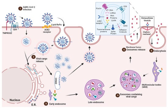
2. EVs Origins, Secretion and Communication

EVs are traditionally classified based on their size and mode of release from the cell. These vesicles are referred to by various names including exosomes, microvesicles (ectosomes), microparticles and apoptotic bodies (oncosomes). Exosomes are small EVs (40–100 nm) that bud from multivesicular bodies (MVBs) within cells and are released upon fusion of the MVBs with the cell plasma membrane, releasing their intraluminal vesicles in the extracellular environment in the form of exosomes [29][30][31][32].
EVs’ biogenesis is regulated by ceramide synthesis at the plasma membrane and endosomal levels [33][34]. The inhibition of neutral sphingomyelinase (nSMase) in both human and animal cells suppresses the release of exosomes while significantly boosting the release of microvesicles from the plasma membrane [34]. There are several proteins involved in exosome biogenesis such as endosomal sorting complex required for transport (ESCRT), vacuolar ATPase, and Vps4, which segregate and sort ubiquitylated proteins into intraluminal vesicles [19][35][36][37][38][39].
Microvesicles or ectosomes, on the other hand, are larger EVs (200–500 nm) believed to bud directly from the plasma membrane [32][40][41]. Similar to exosomes, the generation of microvesicles involves the active participation of several protein factors. These factors encompass Ca2+-dependent aminophospholipid translocases, namely flippases and floppases, sphingomyelinase 2 (nSMase2), scramblases, and calpain. They collectively orchestrate the rearrangement of phospholipids, membrane curvature, and actin cytoskeleton reorganization, ultimately resulting in the extracellular formation of microvesicles [42][43][44][45]. Apoptotic bodies or oncosomes are fragments of cells undergoing programmed cell death, representing the largest EVs with a diameter ranging from 1 to 5 μm [32][41][42][46].
EVs from eukaryotic cells play a vital role in both local and systemic cellular communication, serving as intracellular mediators upon release into the extracellular space [21]. Since EVs have the same membrane orientation as cells, they expose on their surface the extracellular domains of transmembrane proteins that can bind to nearby or long-distance targets. These EVs transfer their bioactive cargo to target cells, thereby influencing and altering their behaviors [47].
The uptake of EVs is influenced by several factors, encompassing their dimensions, surface composition (lipids, glycans, proteins), pH conditions, temperature, and oxidative/hypoxic environment [35]. The nature of both the donor and recipient cells can modulate this process [48]. EVs are secreted through exocytosis or in multivesicular endosomes (MVEs), and they interact with target cells through ligand/receptor signaling at the plasma membrane [21]
The cargo of EVs, which includes lipids, DNA, RNA and proteins, can alter the transcription and signaling activity of recipient cells, thereby regulating their phenotype and function [35]. The protein composition of various types of EVs largely reflects that of the parent cells, exhibiting a notable enrichment of specific molecules. These include adhesion molecules, membrane-trafficking proteins, cytoskeleton components, heat-shock proteins, cytoplasmic enzymes, signal transduction molecules, cytokines, chemokines, proteinases, and cell-specific antigens [49]. They originate from both immune and non-immune cells, which play a vital role in immune system regulation. EVs also possess the capacity to either facilitate immune stimulation or suppress it, thereby influencing the development of inflammatory, autoimmune, and infectious diseases.

3. EVs as Potential Role for Immune System Evasion during SARS-CoV-2 Infection

Viruses have developed intricate strategies to counteract the innate immune response, including interfering with antigen presentation, disrupting interferon signaling, and producing “decoy” sub-viral particles that bind to neutralizing antibodies (nAbs), thereby reducing their effective concentration available for neutralizing infectious virions [6][7][8][9][10][11].
The virions consist of a structural spike glycoprotein, an M-membrane protein (a type III transmembrane glycoprotein), an N-nucleocapsid protein (present within the phospholipid bilayer), and non-structural proteins [50][51]. Approximately one-third of the RNA sequence encodes four fundamental structural proteins: spike (S), envelope (E), membrane (M), and nucleocapsid (N) proteins. The remaining two-thirds of the viral genome consists of ORF1a and ORF1b, which encode non-structural replicase/transcriptase proteins [52].
SARS-CoV-2 enters the host through the respiratory tract and infects the cell mainly via the angiotensin-converting enzyme 2 (ACE2) receptor. ACE2 is a type I integral membrane protein involved in the renin–angiotensin system, which is found in the kidney, testis, intestine, lung, retina, cardiovascular system, adipose tissue, and central nervous system [53]. Once SARS-CoV-2 is internalized into the cell cytoplasm, its lipid bilayers are dismantled by lysosomal enzymes. Subsequently, SARS-CoV-2 utilizes the host cell’s RNA polymerase to replicate its viral single-stranded RNA, thereby increasing the viral load within the host cell [54] (Figure 1). 
The SARS-CoV-2 pathogenicity is highlighted by the entry of the virus through ACE2 receptors, cleavage of the complex, and activation of the S-protein by TMPRSS 2 [55]. According to structural analyses, the spike protein of SARS-CoV (SARS-S) contacts the apex of subunit I of the ACE2 catalytic domain. Once attached to ACE2 by SARS-CoV, the ectodomain of ACE2 is cleaved, which is accompanied by endocytosis of the transmembrane domain into the cell and sometimes internalized as an intact molecule [56]. The internalization and virus particle–host cell fusion is essential for virus entry [57] (Figure 1).
In the pathogenesis of COVID-19, cells that express ACE2 and CD9 can transfer these viral receptors to other cells via EVs, making recipient cells more susceptible for SARS-CoV-2 infection. Recent investigations have shed light not only on the presence of ACE2 within EVs but also on the capacity of the EVs to transfer ACE2 across various cell types [58]. In this context, SARS-CoV-2 gains access to target cells through binding to exosomal ACE2. However, recent studies have underscored the virus’s strong dependence on endocytic mechanisms with the ability to undergo swift endocytosis in cells that express high levels of ACE2 [59] (Figure 1).
Hence, an alternative mechanism for EV-mediated viral entry involves one of the most highly expressed surface proteins on EVs, tetraspanin CD9 [60]. Research has indicated that CD9 collaborates with TMPRSS2 in cleaving viral fusion glycoproteins, expediting the entry of coronaviruses [61], such as MERS-CoV [62], into lung cells. These findings suggest that CD9 and other tetraspanins on the exosomal surface may serve as mediators in SARS-CoV-2 infection [61] (Figure 1).
Many factors have been associated with both altered ACE2 expression and COVID-19 severity and progression, including age, sex, ethnicity, medication, and several comorbidities, such as cardiovascular disease and metabolic syndrome. Although ACE2 is widely distributed in various human tissues and many of its determinants have been well recognized, ACE2-expressing organs do not equally participate in COVID-19 pathophysiology, implying that other mechanisms are involved in orchestrating cellular infection, resulting in tissue damage [61].
Thus, one of the immune evasion strategies employed by SARS-CoV-2 involves the release of exosomes [63], which can carry viruses, viral proteins, and genetic material. Many studies have suggested the role of released SARS-CoV-2-loaded exosomes and other EVs as potential mechanisms for COVID-19 infection relapse [25][64]. The composition of plasma exosomes varies with the severity of the disease, with mild disease-associated vesicles modulating antigen-specific CD4 T cell responses and severe disease-associated vesicles linked to chronic inflammation [65]. Therefore, analyzing exosome components provides valuable insights into different disease states.
The virus co-opts the host’s lipid metabolism to optimize its replication dynamics. By targeting the host cells’ LDs, the principal reservoir of neutral lipids, the SARS-CoV-2 acquires energy substrates crucial for supporting its replication cycles [12] (Figure 2).
Figure 2. SARS-CoV-2 infection induces lipid droplet formation. (1) SARS-CoV-2 infection, via ACE2 receptor, can increase the activation of important transcription factors, such as SREBP1 and PPAR-gamma, leading to an increased induction of LDs biogenesis and the production of eicosanoids which modulate the host’s immune response. (2) EVs containing viral cargo are released into the extracellular environment. (3) Virus utilie the ER machinery for replication. (4) LDs interact with virus and support SARS-CoV-2 replication (box). Pharmacological inhibition and the genetic knockdown of SREBPs and DGAT1 inhibitor (A922500) can reduce SARS-CoV-2 replication and LD biogenesis. In detail, viral particles interact with LD, which is exhibiting structural proteins, such as PLIN-1, PLIN-2 and PLIN-3, and viral RNA. AA, arachidonic acid; COX-2, cyclooxygenase; DAG, diacylglycerol; PGE2, prostaglandin E2; TAG, triacylglycerol.

4. Impact of SARS-CoV-2 Infection in the Cellular Lipid Metabolism

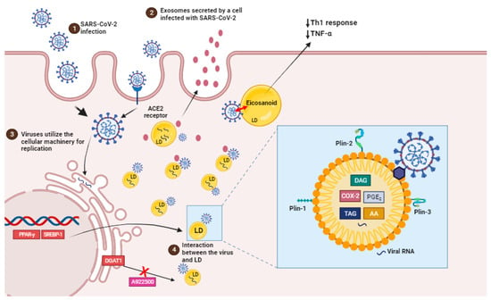
Recent data suggest that lipid domains, known as lipid droplets, play an important role in SARS-CoV-2 replication and the synthesis of inflammatory mediators during the disease as well as in other viral infections such as dengue HCV, DENV, and rotavirus.

The virus’s cellular tropism for lipid metabolism cell machinery suggests that the virus may exploit endogenous lipid materials of different forms, such as lipoproteins and exosomes, as “Trojan horses” to facilitate immune evasion in their systemic spreading [28]

LDs are intracellular structures that contain triglycerides, cholesterol esters, and enzymes involved in lipid synthesis and storage (Figure 2, box). The expression of proteins linked with lipid metabolism and de novo lipid synthesis is modified during SARS-CoV-2 infection, as well as the pathways involved in lipid uptake, such as CD36 and the primary transcriptional factors involved in lipogenesis, including (peroxisome proliferator-activated receptor) PPARγ and SREBP-1 (Figure 2) [12][66].

SREBP is a transcription factor family that regulates lipid homeostasis by directing the expression of a wide range of fatty acid (SREBP1) and cholesterol (SREBP2) metabolic enzymes [66]. SREBP isoforms have been shown to increase during SARS-CoV-2 infection. Their activation is linked to the immunological response via the increased assembly of the inflammasome complex, which results in the production of IL-1 [67].

Beyond their direct role in viral replication, LDs have also been associated with the establishment of a pro-inflammatory state in COVID-19. In human monocytes infected in vitro with SARS-CoV-2, the accumulation of LDs leads to the upregulation of lipid metabolism-related genes and the release of pro-inflammatory mediators such as leukotrienes (LTB4 and cysLT), chemokines (IL-8 and CXCL10), and inflammatory cytokines (IL-6, TNF, and IL-10). These inflammatory mediators amplify the immune response, leading to the recruitment and activation of immune cells at the site of infection and thereby contributing to COVID-19 development [68][69].
In fact, LDs are also sites of compartmentalized synthesis of eicosanoids during inflammatory conditions. Eicosanoids are inflammatory mediators that are derived from the breakdown of arachidonic acid (AA). Phospholipase A2 (PLA2) cleaves LDs and membrane phospholipids to produce AA. AA can be converted into prostaglandins (PGs), thromboxanes (TXs), or leukotrienes (LTs) by cyclooxygenases (COX) or lipoxygenases (LPX), which have regulatory roles in immunological homeostasis. PGE2, TXB2 and leukotriene B4 (LTB4) levels were higher in COVID-19 patients’ bronchoalveolar lavage fluid than in healthy controls. Also, eicosanoid levels were positively correlated with cytokines (IL-1, IL-6, TNF-, IL-12p70, IL-22, and IFN-2) and chemokines (CCL2, CCL11, CXCL9) [13]. PGE2, the most prevalent PG, is a key mediator in a variety of physiological processes, particularly the development and regulation of inflammation [70].
Leukotrienes are thought to be implicated in COVID-19 patients’ tissues because of increased neutrophil infiltration and neutrophil/lymphocyte ratios [71]. In addition, single-cell examination of COVID-19 patients’ bronchoalveolar immune cells and PBMCs reveals enhanced 5-lipoxygenase expression [72][73].
Furthermore, some viruses can employ LDs as a crucial site for viral component accumulation, assisting in the production of viral replication complexes [74][75][76][77]. It has been discovered that LDs can be used as platforms for the assembly and maturation of new virus particles just before release. SARS-CoV-2 proteins and ds-RNA are currently tightly connected with the LDs and, in some circumstances, colocalizing with LDs in an in vitro infection model, indicating a probable function for LDs in the SARS-CoV-2 replication cycle [12].
During COVID-19, the role of extracellular vesicles (EVs) in lipid metabolism remained limited. The transmission of viral components, including RNA, from infected to uninfected cells has been related to EVs. This could potentially facilitate the spread of the virus within the host, assisting viral replication and disease progression. 
In parallel, there is evidence that cells can release LDs into the extracellular space, such as LDs being secreted from milk duct cells into milk [78] and LDs moving between epithelial cells in vitro (KB HeLa cells) [79]. It was also recently proposed that LDs could be bundled within adipocyte-derived EVs, activating macrophages in surrounding adipose tissue [80]. There is a strong synergy between these cellular particles based on their similarities and the increasing role of LDs in intercellular communication. Some data suggest a previously unknown overlap and potential interaction between EV and LDs [81]

5. EVs as Therapeutic Target

EVs can act as a decoy for viruses [82] and bacterial toxins [83], suggesting a potential role as therapeutic agents. Before the COVID-19 pandemic, some research groups envisioned the use of exosomes as immunogenic components for the treatment of virus infections utilizing models of SARS coronavirus infection. Kuate et al. showed that EVs containing the SARS coronavirus spike S protein caused neutralizing antibody titers that were enhanced by priming with the SARS coronavirus spike vaccine [84]. Thus, coronavirus EVs may be effective for delivering therapeutic substances and inducing immune cell responses in the patient. Drugs or biological modulators that decrease viral propagation and replication in infected cells can be delivered into the EVs [84][85]. EVs have potential advantages, such as cell origin, safety, and consistency, which distinguish them from other delivery techniques such as liposomes [86].
Additionally, mRNAs, microRNAs, and DNA fragments carried by EVs can regulate gene expression in recipient cells [87]. The diverse range of EVs and their cargo underscores the potential of utilizing EVs for therapeutic purposes, as they possess a repertoire of bioactive molecules with a combinatorial capacity that would be challenging to replicate artificially [88]. As a result, miRNAs and lncRNAs extracted from exosomes could serve as biomarkers and potential drug carriers/delivery vehicles while acting as regulators of innate and acquired immunity through the stimulation of cytokine production, inflammatory responses, antigen presentation and lipid mediator synthesis.
The source of immunostimulatory exosomes for human antiviral vaccines is critical and must be further investigated [89].
EVs, on the other hand, show significant potential in the diagnosis of COVID-19 and the prediction of mild and severe disease progression. SARS-CoV-2 infection produces thrombosis, and disease severity is closely linked with thrombosis development. Some clinical investigations show a large rise in circulating EVs carrying tissue factor (TF)/CD142 levels in COVID-19 patients as well as a strong connection with thrombosis, malignant disease development, and hospitalization length [90].

6. Conclusions

The battle between viruses and their hosts has a long history. Whereas the immune system has evolved to protect from these pathogens, viruses have acquired clever evasion molecular mechanisms to avoid being detected and destroyed by the immune system. Given their extraordinarily high transmission rates and the possibility of recurrent outbreaks, there is an urgent need to find effective novel antiviral medicines for both treating present respiratory virus infections and preventing future outbreaks.
COVID-19 patients usually have dysregulated lipid profiles, which is associated with an adverse outcome of disease. As the virus is used to provide the building blocks for these vesicles and the viral envelope, they have developed sophisticated evasion strategies that explore lipid metabolism to replicate and avoid being recognized and destroyed by the immune system. Exosomes and respiratory viruses, such as coronavirus and other viruses, appear to make use of sorting complexes and cellular mechanisms that contribute to either maintain host homeostasis or allow viral replication, despite their distinct evolutionary origins. As a result, the development of exosome-based treatments, which include natural exosomes, nano-decoys, and antiviral-loaded exosomes, provide a range of treatment optimization choices that can successfully target, bind to, and limit the cellular absorption of various viruses.

References

  1. World Health Organization. Naming the Coronavirus Disease (COVID-19) and the Virus that Causes It. 2020. Available online: https://www.who.int/emergencies/diseases/novel-coronavirus-2019/technical-guidance/naming-the-coronavirus-disease-(covid-2019)-and-the-virus-that-causes-it (accessed on 10 October 2023).
  2. Kumar, D.; Manuel, O.; Natori, Y.; Egawa, H.; Grossi, P.; Han, S.H.; Fernández-Ruiz, M.; Humar, A. COVID-19: A Global Transplant Perspective on Successfully Navigating a Pandemic. Am. J. Transplant. 2020, 20, 1773–1779.
  3. Prather, B.K.A.; Wang, C.C.; Schooley, R.T. R 1422. Science 2020, 368, 1422–1424.
  4. van Doremalen, N.; Morris, D.H.; Holbrook, M.G.; Gamble, A.; Williamson, B.N.; Tamin, A.; Harcourt, J.L.; Thornburg, N.J.; Gerber, S.I.; Lloyd-Smith, J.O.; et al. Correspondance Aerosol and Surface Stability of SARS-CoV-2 as Compared with SARS-CoV-1. N. Engl. J. Med. 2020, 382, 1564–1567.
  5. Zou, L.; Ruan, F.; Huang, M.; Liang, L.; Huang, H.; Hong, Z.; Yu, J.; Kang, M.; Song, Y.; Xia, J.; et al. An Animal Model of Inhaled Vitamin E Acetate and EVALI-like Lung Injury. N. Engl. J. Med. 2020, 382, 1177–1179.
  6. Bailey, J.; Blankson, J.N.; Wind-Rotolo, M.; Siliciano, R.F. Mechanisms of HIV-1 Escape from Immune Responses and Antiretroviral Drugs. Curr. Opin. Immunol. 2004, 16, 470–476.
  7. Joyner, A.S.; Willis, J.R.; Crowe, J.E.; Aiken, C. Maturation-Induced Cloaking of Neutralization Epitopes on Hiv-1 Particles. PLoS Pathog. 2011, 7, e1002234.
  8. Lazarevic, I.; Banko, A.; Miljanovic, D.; Cupic, M. Immune-Escape Hepatitis B Virus Mutations Associated with Viral Reactivation upon Immunosuppression. Viruses 2019, 11, 778.
  9. Rydell, G.E.; Prakash, K.; Norder, H.; Lindh, M. Hepatitis B Surface Antigen on Subviral Particles Reduces the Neutralizing Effect of Anti-HBs Antibodies on Hepatitis B Viral Particles in Vitro. Virology 2017, 509, 67–70.
  10. Weber, F.; Haller, O. Viral Suppression of the Interferon System. Biochimie 2007, 89, 836–842.
  11. Yewdell, J.W.; Hill, A.B. Viral Interference with Antigen Presentation. Nat. Immunol. 2002, 3, 1019–1025.
  12. Dias da Silva Gomes, S.; Soares, V.C.; Ferreira, A.C.; Sacramento, C.Q.; Fintelman-Rodrigues, N.; Temerozo, J.R.; Teixeira, L.; da Silva, M.A.N.; Barreto, E.; Mattos, M.; et al. Lipid Droplets Fuel SARS-CoV-2 Replication and Production of Inflammatory Mediators. PLoS Pathog. 2020, 16, e1009127.
  13. Chen, P.; Wu, M.; He, Y.; Jiang, B.; He, M.L. Metabolic Alterations upon SARS-CoV-2 Infection and Potential Therapeutic Targets against Coronavirus Infection. Signal Transduct. Target. Ther. 2023, 8, 237.
  14. Altan-Bonnet, N. Lipid Tales on Viral Replication and Transmission Nihal. Trends Cell Biol. 2017, 27, 201–203.
  15. Akers, J.C.; Gonda, D.; Kim, R.; Carter, B.S.; Chen, C.C. Biogenesis of Extracellular Vesicles (EV): Exosomes, Microvesicles, Retrovirus-like Vesicles, and Apoptotic Bodies. J. Neurooncol. 2013, 1, 1–11.
  16. Hoen, E.N.; Cremer, T.; Gallo, R.C.; Margolis, L.B. Extracellular Vesicles and Viruses: Are They Close Relatives? Proc. Natl. Acad. Sci. USA 2016, 113, 9155–9161.
  17. Simons, M.; Raposo, G. Exosomes—Vesicular Carriers for Intercellular Communication. Curr. Opin. Cell Biol. 2009, 21, 575–581.
  18. Mueller, S.K.; Nocera, A.L.; Bleier, B.S. Exosome Function in Aerodigestive Mucosa. Nanomed. Nanotechnol. Biol. Med. 2018, 14, 269–277.
  19. Kowal, J.; Tkach, M.; Théry, C.; Kowal, J.; Tkach, M.; Biogenesis, C.T. Biogenesis and Secretion of Exosomes To Cite This Version: HAL Id: Inserm-02452742. Curr. Opin. Cell Biol. 2014, 29, 116–125.
  20. Statello, L.; Maugeri, M.; Garre, E.; Nawaz, M.; Wahlgren, J.; Papadimitriou, A.; Lundqvist, C.; Lindfors, L.; Collén, A.; Sunnerhagen, P.; et al. Identification of RNA-Binding Proteins in Exosomes Capable of Interacting with Different Types of RNA: RBP-Facilitated Transport of RNAs into Exosomes. PLoS ONE 2018, 13, e0195969.
  21. Mathieu, M.; Martin-jaular, L.; Lavieu, G.; Théry, C. Specificities of Secretion and Uptake of Exosomes and Other Extracellular Vesicles for Cell-to-Cell Communication. Nature Cell Biol. 2019, 21, 9–17.
  22. Gould, S.J.; Booth, A.M.; Hildreth, J.E.K. The Trojan Exosome Hypothesis. Proc. Natl. Acad. Sci. USA 2003, 100, 10592–10597.
  23. Izquierdo-Useros, N.; Lorizate, M.; McLaren, P.J.; Telenti, A.; Kräusslich, H.G.; Martinez-Picado, J. HIV-1 Capture and Transmission by Dendritic Cells: The Role of Viral Glycolipids and the Cellular Receptor Siglec-1. PLoS Pathog. 2014, 10, e1004146.
  24. Elrashdy, F.; Aljaddawi, A.A.; Redwan, E.M.; Uversky, V.N. On the Potential Role of Exosomes in the COVID-19 Reinfection/Reactivation Opportunity. J. Biomol. Struct. Dyn. 2020, 39, 5831–5842.
  25. Borowiec, B.M.; Volponi, A.A.; Mozdziak, P.; Kempisty, B.; Dyszkiewicz-Konwińska, M. Small Extracellular Vesicles and Covid19—Using the “Trojan Horse” to Tackle the Giant. Cells 2021, 10, 3383.
  26. Tsai, S.J.; Atai, N.A.; Cacciottolo, M.; Nice, J.; Salehi, A.; Guo, C.; Sedgwick, A.; Kanagavelu, S.; Gould, S.J. Exosome-Mediated MRNA Delivery in Vivo Is Safe and Can Be Used to Induce SARS-CoV-2 Immunity. J. Biol. Chem. 2021, 297, 101266.
  27. Troyer, Z.; Alhusaini, N.; Tabler, C.O.; Schlatzer, D.M.; Tilton, J.C.; Sweet, T.; Carias, L.; Inacio, K.; De Carvalho, L.; King, C.L.; et al. Extracellular Vesicles Carry SARS-CoV-2 Spike Protein and Serve as Decoys for Neutralizing Antibodies Zach. J. Extracell. Vesicles 2021, 10, e12112.
  28. Lam, S.M.; Huang, X.; Shui, G. Neurological Aspects of SARS-CoV-2 Infection: Lipoproteins and Exosomes as Trojan Horses. Trends Endocrinol. Metab. 2022, 33, 554–568.
  29. Johnstone, R.M.; Adam, M.; Hammond, J.R.; Orr, L.; Turbide, C. Vesicle Formation during Reticulocyte Maturation. Association of Plasma Membrane Activities with Released Vesicles (Exosomes). J. Biol. Chem. 1987, 262, 9412–9420.
  30. Bobrie, A.; Colombo, M.; Raposo, G.; Théry, C. Exosome Secretion: Molecular Mechanisms and Roles. Traffic 2011, 12, 1659–1668.
  31. Raposo, G.; Stoorvogel, W. Extracellular Vesicles: Exosomes, Microvesicles, and Friends. J. Cell Biol. 2013, 200, 373–383.
  32. Théry, C.; Witwer, K.W.; Aikawa, E.; Alcaraz, M.J.; Anderson, J.D.; Andriantsitohaina, R.; Antoniou, A.; Arab, T.; Archer, F.; Atkin-smith, G.K.; et al. Minimal Information for Studies of Extracellular Vesicles 2018 ( MISEV2018): A Position Statement of the International Society for Extracellular Vesicles and Update of the MISEV2014 Guidelines. J. Extracell. Vesicles 2018, 8, 1535750.
  33. Trajkovic, K.; Hsu, C.; Chiantia, S.; Rajendran, L.; Wenzel, D.; Wieland, F.; Schwille, P.; Brügger, B.; Simons, M. Ceramide Triggers Budding of Exosome Vesicles into Multivesicular Endosomes. Science 2008, 319, 1244–1247.
  34. Menck, K.; Sönmezer, C.; Worst, T.S.; Schulz, M.; Dihazi, G.H.; Streit, F.; Erdmann, G.; Kling, S.; Boutros, M.; Binder, C.; et al. Neutral Sphingomyelinases Control Extracellular Vesicles Budding from the Plasma Membrane. J. Extracell. Vesicles 2017, 6, 1378056.
  35. Liu, Y.J.; Wang, C. A Review of the Regulatory Mechanisms of Extracellular Vesicles-Mediated Intercellular Communication. Cell Commun. Signal. 2023, 21, 77.
  36. Jeppesen, D.K.; Fenix, A.M.; Franklin, J.L.; Rome, L.H.; Burnette, D.T.; Coffey, R.J.; Jeppesen, D.K.; Fenix, A.M.; Franklin, J.L.; Higginbotham, J.N.; et al. Reassessment of Exosome Composition Article Reassessment of Exosome Composition. Cell 2019, 177, 428–445.e18.
  37. Hurley, J.H. ESCRTs Are Everywhere. EMBO J. 2015, 34, 2398–2407.
  38. Zhang, J.; Kumar, S.; Jayachandran, M.; Hernandez, L.P.H.; Wang, S.; Wilson, E.M.; Lieske, J.C. Excretion of Urine Extracellular Vesicles Bearing Markers of Activated Immune Cells and Calcium/Phosphorus Physiology Differ between Calcium Kidney Stone Formers and Non-Stone Formers. BMC Nephrol. 2021, 22, 204.
  39. Jayachandran, M.; Yuzhakov, S.V.; Kumar, S.; Larson, N.B.; Enders, F.T.; Milliner, D.S.; Rule, A.D.; Lieske, J.C. Specific Populations of Urinary Extracellular Vesicles and Proteins Differentiate Type 1 Primary Hyperoxaluria Patients without and with Nephrocalcinosis or Kidney Stones. Orphanet J. Rare Dis. 2020, 15, 319.
  40. Heijnen, H.F.G.; Schiel, A.E.; Fijnheer, R.; Geuze, H.J.; Sixma, J.J. Activated Platelets Release Two Types of Membrane Vesicles: Microvesicles by Surface Shedding and Exosomes Derived from Exocytosis of Multivesicular Bodies and α-Granules. Blood 1999, 94, 3791–3799.
  41. Maas, S.L.N.; Breakefield, X.O.; Weaver, A.M.; Hospital, M.G. Extracellular Vesicles: Unique Intercellular Delivery Vehicles. Trends Cell Biol. 2017, 27, 172–188.
  42. Nabhan, J.F.; Hu, R.; Oh, R.S.; Cohen, S.N.; Lu, Q. Formation and Release of Arrestin Domain-Containing Protein 1-Mediated Microvesicles (ARMMs) at Plasma Membrane by Recruitment of TSG101 Protein. Proc. Natl. Acad. Sci. USA 2012, 109, 4146–4151.
  43. Wang, X.; Melino, G.; Shi, Y. Actively or Passively Deacidified Lysosomes Push β-Coronavirus Egress. Cell Death Dis. 2021, 12, 12–14.
  44. Li, B.; Antonyak, M.A.; Zhang, J.; Cerione, R.A. RhoA Triggers a Specific Signaling Pathway That Generates Transforming Microvesicles in Cancer Cells. Oncogene 2012, 31, 4740–4749.
  45. Yang, J.; Gould, S.J. The Cis -Acting Signals That Target Proteins to Exosomes and Microvesicles. Biochem. Soc. Trans 2013, 41, 277–282.
  46. Kerr, J.F.R.; Wyllie, A.H.; Currie, A.R. Apoptosis: A Basic Biological Phenomenon with Wide-Ranging Implications in Tissue kinetics. Br. J. Cancer 1972, 26, 239–257.
  47. Karnas, E.; Dudek, P.; Zuba-Surma, E.K. Stem Cell-Derived Extracellular Vesicles as New Tools in Regenerative Medicine—Immunomodulatory Role and Future Perspectives. Front. Immunol. 2023, 14, 1120175.
  48. Petroni, D.; Fabbri, C.; Babboni, S.; Menichetti, L.; Basta, G.; Del Turco, S. Extracellular Vesicles and Intercellular Communication: Challenges for In Vivo Molecular Imaging and Tracking. Pharmaceutics 2023, 15, 1639.
  49. Robbins, P.D.; Morelli, A.E. Regulation of Immune Responses by Extracellular Vesicles. Nat. Rev. Immunol. 2014, 14, 195–208.
  50. Yan, R.; Zhang, Y.; Li, Y.; Xia, L.; Guo, Y.; Zhou, Q. Structural Basis for the Recognition of SARS-CoV-2 by Full-Length Human ACE2. Science 2020, 367, 1444–1448.
  51. Zhang, H.; Lv, P.; Jiang, J.; Liu, Y.; Yan, R.; Shu, S.; Hu, B.; Xiao, H.; Cai, K.; Yuan, S.; et al. Advances in Developing ACE2 Derivatives against SARS-CoV-2. Lancet Microbe 2023, 4, e369–e378.
  52. Wan, Y.; Shang, J.; Graham, R.; Baric, R.S.; Li, F. Receptor Recognition by the Novel Coronavirus from Wuhan: An Analysis Based on Decade-Long Structural Studies of SARS Coronavirus. J. Virol. 2020, 94, e00127-20.
  53. Gheblawi, M.; Wang, K.; Viveiros, A.; Nguyen, Q.; Zhong, J.C.; Turner, A.J.; Raizada, M.K.; Grant, M.B.; Oudit, G.Y. Angiotensin-Converting Enzyme 2: SARS-CoV-2 Receptor and Regulator of the Renin-Angiotensin System: Celebrating the 20th Anniversary of the Discovery of ACE2. Circ. Res. 2020, 126, 1456–1474.
  54. Perrier, A.; Bonnin, A.; Desmarets, L.; Danneels, A.; Goffard, A.; Rouillé, Y.; Dubuisson, J.; Belouzard, X.S. Cro The C-Terminal Domain of the MERS Coronavirus M Protein Contains a Trans -Golgi Network Localization Signal. J. Biol. Chem. 2019, 294, 14406–14421.
  55. Senapati, S.; Banerjee, P.; Bhagavatula, S.; Kushwaha, P.P.; Kumar, S. Contributions of Human ACE2 and TMPRSS2 in Determining Host–Pathogen Interaction of COVID-19. J. Genet. 2021, 100, 12.
  56. Cabal, A.B.S.; Wu, T.Y. Recombinant Protein Technology in the Challenging Era of Coronaviruses. Processes 2022, 10, 946.
  57. Jackson, C.B.; Farzan, M.; Chen, B.; Choe, H. Mechanisms of SARS-CoV-2 Entry into Cells. Nat. Rev. Mol. Cell Biol. 2022, 23, 3–20.
  58. Wang, J.; Chen, S.; Bihl, J. Exosome-Mediated Transfer of ACE2 (Angiotensin-Converting Enzyme 2) from Endothelial Progenitor Cells Promotes Survival and Function of Endothelial Cell. Oxid. Med. Cell. Longev. 2020, 2020, 4213541.
  59. Bayati, A.; Kumar, R.; Francis, V.; McPherson, P.S. SARS-CoV-2 Infects Cells after Viral Entry via Clathrin-Mediated Endocytosis. J. Biol. Chem. 2021, 296, 100306.
  60. Fozouni, P.; Son, S.; Díaz de León Derby, M.; Knott, G.J.; Gray, C.N.; D’Ambrosio, M.V.; Zhao, C.; Switz, N.A.; Kumar, G.R.; Stephens, S.I.; et al. Amplification-Free Detection of SARS-CoV-2 with CRISPR-Cas13a and Mobile Phone Microscopy. Cell 2021, 184, 323–333.
  61. Xia, X.; Yuan, P.; Zheng, J.C. Emerging Roles of Extracellular Vesicles in COVID—Edged Sword? Immunology 2021, 163, 416–430.
  62. Earnest, J.T.; Hantak, M.P.; Li, K.; Mccray, P.B.; Perlman, S.; Gallagher, T. The Tetraspanin CD9 Facilitates MERS- Coronavirus Entry by Scaffolding Host Cell Receptors and Proteases. PLoS Pathog. 2017, 13, e1006546.
  63. Rubio-Casillas, A.; Redwan, E.M.; Uversky, V.N. SARS-CoV-2: A Master of Immune Evasion. Biomedicines 2022, 10, 1339.
  64. Horn, M.D.; MacLean, A.G. Extracellular Vesicles as a Means of Viral Immune Evasion, CNS Invasion, and Glia-Induced Neurodegeneration. Front. Cell. Neurosci. 2021, 15, 695899.
  65. Pesce, E.; Manfrini, N.; Cordiglieri, C.; Santi, S.; Bandera, A.; Gobbini, A.; Gruarin, P.; Favalli, A.; Bombaci, M.; Cuomo, A.; et al. Exosomes Recovered From the Plasma of COVID-19 Patients Expose SARS-CoV-2 Spike-Derived Fragments and Contribute to the Adaptive Immune Response. Front. Immunol. 2022, 12, 785941.
  66. Soares, V.C.; Dias, S.S.G.; Santos, J.C.; Azevedo-Quintanilha, I.G.; Moreira, I.B.G.; Sacramento, C.Q.; Fintelman-Rodrigues, N.; Temerozo, J.R.; da Silva, M.A.N.; Barreto-Vieira, D.F.; et al. Inhibition of the SREBP Pathway Prevents SARS-CoV-2 Replication and Inflammasome Activation. Life Sci. Alliance 2023, 6, e202302049.
  67. Li, Y.; Xu, S.; Jiang, B.; Cohen, R.A.; Zang, M. Activation of Sterol Regulatory Element Binding Protein and NLRP3 Inflammasome in Atherosclerotic Lesion Development in Diabetic Pigs. PLoS ONE 2013, 8, e67532.
  68. Fintelman-Rodrigues, N.; Sacramento, C.Q.; Lima, C.R.; da Silva, F.S.; Ferreira, A.; Mattos, M.; de Freitas, C.S.; Soares, V.C.; da Silva Gomes Dias, S.; Temerozo, J.R.; et al. Atazanavir Inhibits SARS-CoV-2 Replication and pro-Inflammatory Cytokine Production. bioRxiv 2020.
  69. Coperchini, F.; Chiovato, L.; Croce, L.; Magri, F.; Rotondi, M. The Cytokine Storm in COVID-19: An Overview of the Involvement of the Chemokine/Chemokine-Receptor System. Cytokine Growth Factor Rev. 2020, 53, 25–32.
  70. Ricciotti, E.; Fitzgerald, G.A. Prostaglandins and Inflammation. Arterioscler. Thromb. Vasc. Biol. 2011, 31, 986–1000.
  71. Kong, M.; Zhang, H.; Cao, X.; Mao, X.; Lu, Z. Higher Level of Neutrophil-to-Lymphocyte Is Associated with Severe COVID-19. Epidemiol. Infect. 2020, 148, e139.
  72. Wilk, A.J.; Rustagi, A.; Zhao, N.Q.; Roque, J.; Martínez-Colón, G.J.; McKechnie, J.L.; Ivison, G.T.; Ranganath, T.; Vergara, R.; Hollis, T.; et al. A Single-Cell Atlas of the Peripheral Immune Response in Patients with Severe COVID-19. Nat. Med. 2020, 26, 1070–1076.
  73. Liao, M.; Liu, Y.; Yuan, J.; Wen, Y.; Xu, G.; Zhao, J.; Cheng, L.; Li, J.; Wang, X.; Wang, F.; et al. Single-Cell Landscape of Bronchoalveolar Immune Cells in Patients with COVID-19. Nat. Med. 2020, 26, 842–844.
  74. Chatel-Chaix, L.; Bartenschlager, R. Dengue Virus- and Hepatitis C Virus-Induced Replication and Assembly Compartments: The Enemy Inside—Caught in the Web. J. Virol. 2014, 88, 5907–5911.
  75. Lee, J.Y.; Cortese, M.; Haselmann, U.; Tabata, K.; Romero-Brey, I.; Funaya, C.; Schieber, N.L.; Qiang, Y.; Bartenschlager, M.; Kallis, S.; et al. Spatiotemporal Coupling of the Hepatitis C Virus Replication Cycle by Creating a Lipid Droplet- Proximal Membranous Replication Compartment. Cell Rep. 2019, 27, 3602–3617.e5.
  76. Miyanari, Y.; Atsuzawa, K.; Usuda, N.; Watashi, K.; Hishiki, T.; Zayas, M.; Bartenschlager, R.; Wakita, T.; Hijikata, M.; Shimotohno, K. The Lipid Droplet Is an Important Organelle for Hepatitis C Virus Production. Nat. Cell Biol. 2007, 9, 1089–1097.
  77. Laufman, O.; Perrino, J.; Andino, R. Viral Generated Inter-Organelle Contacts Redirect Lipid Flux for Genome Replication. Cell 2019, 178, 275–289.
  78. Lu, Y.; Zhou, T.; Xu, C.; Wang, R.; Feng, D.; Li, J.; Wang, X.; Kong, Y.; Hu, G.; Kong, X.; et al. Occludin Is a Target of Src Kinase and Promotes Lipid Secretion by Binding to BTN1a1 and XOR. PLoS Biol. 2022, 20, e3001518.
  79. Collot, M.; Fam, T.K.; Ashokkumar, P.; Faklaris, O.; Galli, T.; Danglot, L.; Klymchenko, A.S. Ultrabright and Fluorogenic Probes for Multicolor Imaging and Tracking of Lipid Droplets in Cells and Tissues. J. Am. Chem. Soc. 2018, 140, 5401–5411.
  80. Flaherty, S.E., III; Grijalva, A.; Xu, X.; Ables, E.; Nomani, A.; Ferrante, A.W., Jr. A Lipase-Independent Pathway of Lipid Release and Immune Modulation by Adipocytes. Science 2019, 363, 989–993.
  81. Amarasinghe, I.; Phillips, W.; Hill, A.F.; Cheng, L.; Helbig, K.J.; Willms, E.; Monson, E.A. Cellular Communication through extracellular vesicles and lipid droplets. J. Extracell Biol. 2023, 2, e77.
  82. Pocsfalvi, G.; Mammadova, R.; Ramos Juarez, A.P.; Bokka, R.; Trepiccione, F.; Capasso, G. COVID-19 and Extracellular Vesicles: An Intriguing Interplay. Kidney Blood Press. Res. 2020, 45, 661–670.
  83. Keller, M.D.; Ching, K.L.; Liang, F.X.; Dhabaria, A.; Tam, K.; Ueberheide, B.M.; Unutmaz, D.; Torres, V.J.; Cadwell, K. Decoy Exosomes Provide Protection against Bacterial Toxins. Nature 2020, 579, 260–264.
  84. Kuate, S.; Cinatl, J.; Doerr, H.W.; Überla, K. Exosomal Vaccines Containing the S Protein of the SARS Coronavirus Induce High Levels of Neutralizing Antibodies. Virology 2007, 362, 26–37.
  85. Catalano, M.; O’Driscoll, L. Inhibiting Extracellular Vesicles Formation and Release: A Review of EV Inhibitors. J. Extracell. Vesicles 2020, 9, 1703244.
  86. Zhou, X.; Zhang, W.; Yao, Q.; Zhang, H.; Dong, G.; Zhang, M.; Liu, Y.; Chen, J.K.; Dong, Z. Exosome Production and Its Regulation of EGFR during Wound Healing in Renal Tubular Cells. Am. J. Physiol. -Ren. Physiol. 2017, 312, F963–F970.
  87. Schneider, D.J.; Speth, J.M.; Penke, L.R.; Wettlaufer, S.H.; Swanson, J.A.; Peters-Golden, M. Mechanisms and Modulation of Microvesicle Uptake in a Model of Alveolar Cell Communication. J. Biol. Chem. 2017, 292, 20897–20910.
  88. Sánchez Berumen, G.; Bunn, K.E.; Pua, H.H.; Rafat, M. Extracellular Vesicles: Mediators of Intercellular Communication in Tissue Injury and Disease. Cell Commun. Signal. 2021, 19, 104.
  89. Zheng, Y.; Tu, C.; Zhang, J.; Wang, J. Inhibition of Multiple Myeloma-derived Exosomes Uptake Suppresses the Functional Response in Bone Marrow Stromal Cell. Int. J. Oncol. 2019, 54, 1061–1070.
  90. Rosell, A.; Havervall, S.; Von Meijenfeldt, F.; Hisada, Y.; Aguilera, K.; Grover, S.P.; Lisman, T.; MacKman, N.; Thålin, C. Patients With COVID-19 Have Elevated Levels of Circulating Extracellular Vesicle Tissue Factor Activity That Is Associated With Severity and Mortality—Brief Report. Arterioscler. Thromb. Vasc. Biol. 2021, 41, 878–882.
More
Information
Subjects: Immunology
Contributors MDPI registered users' name will be linked to their SciProfiles pages. To register with us, please refer to https://encyclopedia.pub/register : , , , , , ,
View Times: 95
Revisions: 2 times (View History)
Update Date: 16 Jan 2024
1000/1000
Video Production Service