Submitted Successfully!
To reward your contribution, here is a gift for you: A free trial for our video production service.
Thank you for your contribution! You can also upload a video entry or images related to this topic.
Version Summary Created by Modification Content Size Created at Operation
1 -- 2570 2023-12-26 08:38:21 |
2 format correct + 2 word(s) 2572 2023-12-27 08:29:05 | |
3 format correct Meta information modification 2572 2023-12-27 08:30:10 |

Video Upload Options

Do you have a full video?

Confirm

Are you sure to Delete?
Cite
If you have any further questions, please contact Encyclopedia Editorial Office.
Gallenga, C.E.; Maritati, M.; Del Boccio, M.; D’aloisio, R.; Conti, P.; Mura, M.; Contini, C.; Gallenga, P.E. SAFE—S Strategy for Trachoma. Encyclopedia. Available online: https://encyclopedia.pub/entry/53125 (accessed on 08 July 2024).
Gallenga CE, Maritati M, Del Boccio M, D’aloisio R, Conti P, Mura M, et al. SAFE—S Strategy for Trachoma. Encyclopedia. Available at: https://encyclopedia.pub/entry/53125. Accessed July 08, 2024.
Gallenga, Carla Enrica, Martina Maritati, Marco Del Boccio, Rossella D’aloisio, Pio Conti, Marco Mura, Carlo Contini, Pier Enrico Gallenga. "SAFE—S Strategy for Trachoma" Encyclopedia, https://encyclopedia.pub/entry/53125 (accessed July 08, 2024).
Gallenga, C.E., Maritati, M., Del Boccio, M., D’aloisio, R., Conti, P., Mura, M., Contini, C., & Gallenga, P.E. (2023, December 26). SAFE—S Strategy for Trachoma. In Encyclopedia. https://encyclopedia.pub/entry/53125
Gallenga, Carla Enrica, et al. "SAFE—S Strategy for Trachoma." Encyclopedia. Web. 26 December, 2023.
SAFE—S Strategy for Trachoma
Edit

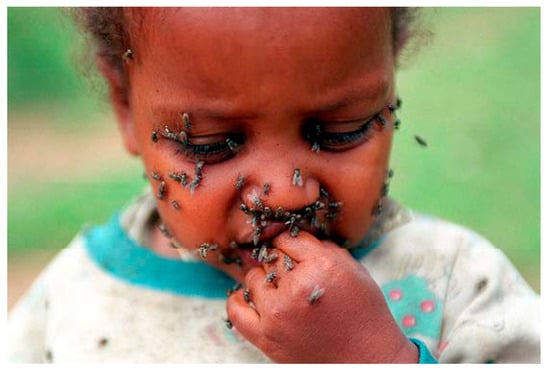
The biological history of Chlamydia trachomatis is intertwined with the evolution of the man. Infecting Elemental Bodies (EBs), having penetrated mucosal epithelial cells, wrap themselves in a cloak (ĸλαμις) of glycogen that ensures their obligatory intracellular survival and protects this differentiation into Reticulate Bodies (RBs) that feed on cellular ATP. Multiple chemokines and cytokines are involved under the direction of IL-6 in the florid phase and IL-17A in the scar phase. The WHO has successfully identified the SAFE strategy against trachoma (Surgery, Antibiotics, Facial cleansing, Environment) as the blueprint to eliminate the disease by 2020. Recently, interest has been increasingly focused on changing sexual attitudes in different areas of the world, leaving Musca sorbens, Scatophaga stercoraria, and stepsisters fairly blameless, but extolling the role of Chlamydia trachomatis in apparently “sterile” chronic prostatitis or conjunctivitis or, less frequently, in oropharyngitis and proctitis. The addition of an S (SAFE-S) standing for “sexual behavior” was then proposed to also attract the interest and attention not only of Ophthalmologists and Obstetricians/Gynecologists, Urologists/Andrologists, and the School Authorities for information on the prevention of sexually transmitted diseases, but also of Social Physicians and Pediatricians. This means that sexually transmitted infections should be screened in asymptomatic patients with risky sexual behavior or sexual contact with people diagnosed with a transmitted infection.

trachoma chlamydia WHO SAFE strategy RT-PCR conjunctivitis sexually transmitted diseases neglected transmitted diseases Musca sorbens vaccines

1. History and Italian Background

There are mentions of trachoma, known since antiquity, in Hammurabi’s Code of the 18th century B.C., also carved into the diorite of the 12th century B.C. in the Sousa stele, now in the Louvre, and in the 16th century B.C. Eber’s Papyrus, now in Leipzig (Figure 1).
Figure 1. Eber papyrus, approximately 1550 BC, Egypt, New Kingdom, XVIII dynasty. Medical papyrus on diseases and herbal knowledge, written in hieratic as a summa of older texts. Line 350. “nekha.t” = this can be interpreted as ‘trachome’, conjunctivitic granulosis; ripple of water = “n” as a phoneme; clumps of papyrus = “kh” as a phoneme, sounds like “ch” in German; vulture bird = “a” as a phoneme (it is generally transliterated with sign “3” to indicate an open “A”); bread loaf = “t” as a phoneme; pustule is a determinative for blister, wound or phlogosis; three vertical lines = indicate a plural form (maybe a number of blisters or tearing). The word “nekha.t” is translated as “an eye disease” [belegt Med. eineAugenkrankheit] in Woerterbuch [the Dictionary]. (Cristiano Daglio. Le Malattie. S. Malgora: Ur Sunu, pp 65–81. Saviolo ed. Vercelli, Italy, 2008. Courtesy Dr. F. Buttiglione PhD and Serena Salmé).
Knowledge of the School of Alexandria is reported in the De re medica by Celsus, who was active in Rome in the first century AD. In the Epitome book III De Trachomate by Paulus Aegineta, in the seventh century A.D., the florid granular aspect of the conjunctiva is described resembling a fig and defined as “sycosis”, while “tylosis” is the chronic form (for treatment: hematite, wine, saffron, Viergespann), but it remains generally confused in the set of conjunctivitis-secreting pathologies [1]. Francis of Assisi suffered severely on his return from Egypt, where he met Sultan Malik al Kamil in the summer truce at the siege of Damietta in 1219, during the Fifth Crusade. Infected and back in Italy, he was treated with eyelid burns with red-hot iron, not complaining but praising God for the sufferings: he died at 44, almost blind.
Mackenzie [2] in 1854 wondered if the disease “est susceptible de se propager par infection, c’est à dire si les miasmes qui s’echappent des yeux de ceux qui en son affecés peuvent, en se repandant dans l’atmosphère, provoquer le développement de la meme affection dans d’autres yeux”, while citing spreads in communities, barracks, schools and families and the possible transmission with infected towels.
Patients were treated both as outpatient or inpatient in general surgeries or the medical hospital, together with syphilodermopathic patients, as in the Ophthalmic Hospital of Turin, Italy, founded by senator Prof. Casimiro Sperino, transforming in 1835, on behalf of Carlo Alberto King of Sardinia [3], the previous hospitals (the so called ‘lazzaretto’: pesthouse) laid for the cholera epidemic.
There, of the 12,114 patients hospitalized in the decade 1873–1883 in the Ophthalmology Clinic of the R. University of Turin headed by Prof. Carlo Reymond, Dr. Camillo Gallenga published 1.392 cases of corneal leucoma including granular conjunctivitis, phlyctenular and pustular affections of the conjunctiva, ulcers and corneal abscesses “of which often represent the final outcome[4], treating with Snellen’s procedure the entropion with trichiasis in severe forms of granular conjunctivitis [5].
Trachoma has been classified with etiological/microbiological identity in Ophthalmology treatises since the second World War, when the basophilic bodies of Halberstaedter (German dermatologist and radiologist) and von Prowazek (Austrian biologist/zoologist), identified in 1907 in Jakarta during a scientific expedition to study syphilis transmission [6], were definitively attributed to Chlamydia trachomatis (Ct). Halberstaedter and von Prowazek, in an expedition to Java, also infected orangutans with conjunctival scrapings from trachoma patients and such agents in conjunctival smears. They called them “chlamydozoa”.
Just two years later, in 1909, an early confirmation was published by Camillo Gallenga, actually Professor of Ophthalmology in the University of Parma [7], replicated the next year 1910 for the German literature in Klinische Monatsblatter Augenheilkunde [8], contrasting opposite skeptical opinions largely represented by Scientists and Ophthalmologists not only in Europe.
Axenfeld [9], in 1914, still attributed the spread of trachoma in Europe to Napoleon’s Egyptian campaign, linked to granulomatous conjunctivitis, whose diagnosis became certain when it was confirmed by “corpuscules trachomateux de Prowazek” stained with Giemsa, accordingly with the 1907 first description of the etiology, supported by the Italian literature [7][8].
The May–Grunwald Giemsa solution stains chromatin in red-purple (Romanowsky effect), nucleoli in deep blue and the cytoplasm of epithelial cells in light blue. The smallest inclusions, even two or four in number, are generally associated in small aggregates with an intensely basophilic matrix and with a homogeneous surface element that differs from the cytoplasm and colors in intense blue, like a cap. Although differentiated, these Reticulated Bodies (RBs) tend to adhere to the nucleus. The initial inclusions are replaced by Elementary Bodies (EBs), which color red-purple and remain well separated from each other [10]. The amorphous masses of the blue-colored inclusions envelop like a cloak (κλαμίς: the ancient Greek term for the short cloak worn by Greek military men, draped and secured with a brooch on the right shoulder) the purple-red granules of chromatin ‘carriers of the virus’. The Chlamydiae were named this way because the intracytoplasmic inclusions formed by this agent inside host cells cluster around (are ‘draped’ around) the nucleus of the cell [11]. Still, in 1925, Amilcare Bietti discusses the distinction or unity of trachoma and ophthalmo-blennorrhea by inclusion bodies in children [12]. He reports—disputing the hypothesis—that Lindner, on the contrary, was convinced that the two affections were identical, although the latter was unable to infect the genitals of a female baboon with human ocular trachomatous material. Riccardo Gallenga, while operating the plasmoma of the superior bulbar conjunctiva of an eighteen-year-old boy, trachomatous from infancy and now in the scarring phase (Trachoma register int. N° 113 18, 1931, Ophthalmology Clinic R. University of Turin, head Prof. Luigi Guglianetti) in the accurate description of the histological examination, reports “in the remarkably thickened epithelium... the presence of polynuclear leukocytes, lymphocytes and plasmacells”, but “I have not observed either epithelial cells or trachomatous granules[13] and describes, in the plasmome, the presence of “rare Russel bodies colored in pink with eosin and in yellow with van Gieson”: the reference documents the absence of Chlamydia in the bulbar conjunctiva of trachoma in the MacCallan cicatricial stage T4, and the precise knowledge of the results by Halberstäedter and von Prowazeck.
The public health response to the spread of trachoma throughout the Mediterranean basin in the 19th and first half of the 20th century involved anti-trachoma clinics and the establishment of dispensaries in all major cities and hospitals. In all of them, there was intense activity and attendance. So much so that in the years 1936–1942, before and during the Second World War (but before the RAF and USAF heavily bombed the city, port and hospital of Cagliari, Sardinia), the flow of patients to the anti-trachoma department of the Cagliari University Eye Clinic headed by Professor Riccardo Gallenga was so intense that it had to be regulated every morning by the traffic police. There, in 1938, by suturing the conjunctival biopsy from surgery for entropion by a T4 patient to the mucosa of his own conjunctival fornix, Riccardo Gallenga also demonstrated that cicatricial trachoma is no longer contagious (Lessons, The Ophthalmology Course, 5th year Medical School University of Turin, 1966; and OMC&O Turin conference, University Eye clinic, 1967) [14], accordingly with his previous results as Assistant Professor in Turin Eye Clinic [13].
In the second half of the 1950s in Sicily island, another highly involved area, the experiences gathered in the Catania University Eye Clinic, directed by Prof. Giuseppe Favaloro, allowed Giuseppe Scuderi to publish a textbook and atlas of trachoma histopathology [15], clarifying from a clinical and anatomopathological point of view many aspects related to trachomatous infection (Figure 2a–c).
Figure 2. (a) Tarsal conjunctiva. Florid trachoma. (TF, according to WHO classification). Papillary infiltration, forming nodules. Van Gieson ferric hematoxylin. 75×. (b) Tarsal conjunctiva. Scarring trachoma. From TI (T. Inflammation Intense: fibroblastic proliferation of the chorion, still site of histiolymphocytic infiltration) to TS (T. Scarring): conjunctival scars with fibrous white bands (arrow). Absence of infiltrate and Ct in the scar area. Giemsa 120× [16], (Courtesy Prof. Nicola Delle Noci). (c) Tarsal conjunctiva. (MacCallan T4); TS: Arlt’s line. (Reproduced with permission—reference number 230716-012078. From Weisenthal RW. External Disease and Cornea. Basic and Clinical Science Course, Section 8, American Academy of Ophthalmology, 2013–2014).

2. Contagion and Clinic, The SAFE-S Strategy

In the four clinical stages of trachoma (McCallan, 1908, cited by Frezzotti and Guerra [17]) from lymphoid hyperplasia to the florid highly contagious granulomatous stage 2—for which ancient treatments with massage, forceps, silver nitrate or copper sulfate brushing have found promoters and innovators over time—the evolution becomes complicated with the upper corneal “neovascular panniculus”. Leaving Herbert’s dimple in place of emptied limbal follicles, with large polynuclear macrophages and Moauro–Leber cells, the evolution of the disease follows the stages of xerophthalmia and tarsal scarring—Arlt’s line—inducing scar entropion, trichiasis and corneal ulcers with a likelihood of superinfection [4][7], causing blindness and needing surgery, but which is different from the past (Figure 3). However, the large burden of symptoms overlapping with other diseases or syndromes, and the priority identification as an exclusively ocular disease, delayed the identification of Ct by WHO as a sexually transmitted infection until 1976 [18].
Figure 3. Everting sutures for entropion. George Bartisch: Ophthalmodouleia, 1583. Courtesy Prof. Nicola Delle Noci [19].
Infection can be caused either directly or indirectly. In the indirect mechanism, flies, in particular, may be involved, which contribute to the spread of the disease by settling on the eyes of infected patients. Contact with eye or nasal secretions (including through cloths or clothing) of infected patients, on the other hand, constitutes a direct route of transmission of the disease.
The spread of the disease is inversely proportional to the socioeconomic development and sanitation conditions; in particular, risk factors can be identified in the “lack of clean hot and running water, lack of personal cleanliness, crowding, promiscuity, superstitions, and abundance of flies[17]. Infection does not confer permanent immunity, so there is a likelihood of re-infection if the environmental causes of infection are not changed. An effective protocol of cyclic therapy protracted for four months to counteract the Ct biological cycle (EB-RB-EB) was developed by Gallenga PE et al., in 2018, achieving a complete microbiological and clinical resolution, leading to a favorable evolution in some cases of male (caused by chronic prostatitis) and female infertility, thus overcoming the “cultural era” to move to the “molecular era” [20].
Antimicrobial resistance is a major concern with the potential to cause millions of deaths, even though few studies recorded the clinical resistance of neglected tropical diseases (NTD), namely human African trypanosomiasis, leishmaniasis, onchocerciasis, schistosomiasis, soil-transmitted helminths, and trachoma [21]. Researches are in the pipeline to find which of the more than 2000 Antimicrobial peptides (AMPs) with broad-spectrum antibacterial, antiviral and antiparasitic activity, that have been shown to be effective against a variety of NTDs including trachoma, could be developed and used as alternatives [22][23]. Pep-1, LL-37, and melittin showed outstanding abilities in inhibiting the growth of Ct. The same applies to gene therapy: IRF5 and IL-10RA are related to macrophage–chlamydia interactions. Their roles seem vital in order to cure Ct infection in the scenario of the increasing antibiotic resistance, also to azytromycin. However, multiple challenges for the development of these therapies remained [23].
From the pictures of dirty and tearful children (Figure 4) in environments with poor sanitation in the Afro-Arabo-Indo-Asian basin, the contagion by flies is evident, although this does not account for the primitive forms of the disease in adults and those outside the tropical–equatorial area [24]. The WHO [25][26][27] and the Ligue contre le tTrachome (LCT) have rightly and successfully identified the SAFE strategy against trachoma (Surgery, Antibiotics, Facial cleanliness, Environmental improvement) as the blueprint for eliminating the disease by 2020; however, the evidence of the sexually transmitted disease still makes Musca domestica, Musca sorbens [28], Scathophaga stercoraria [29][30] or Sarcophafaga carnaria, so beloved by fishermen, the culprits.
Figure 4. A Sri Lankan child with eyes infested with flies (M. sorbens). Courtesy Imperial College London. Institute of Global Health Innovation. The impact of Neglected Tropical Diseases on Universal eye health, 2016.

Having this scenario clear, the researchers proposed to complete the acronym SAFE with -S (SAFE-S), understood as ''strategy for controlling sexual well-being, i.e.: sexual behaviour'' [20]. Unprotected sex is guilty: the WHO recomendations for the next decade 2021-2030 must attract the interest and attention not only of Obstetricians/Gynecologists, Urologists/Andrologists and Pediatricians, but also High School Authorities that should be mandated to improve students' knowledge on sexually transmitted disease education and prophylaxis/protection.

3. Conclusions

The oculo-genital correlation and the need for adequate systemic coverage are confirmed by cases of chronic conjunctival disease resolved with ultrasound-guided infiltrative therapy of chronic prostatitis [31] or recurrent atypical chronic oropharyngeal inflammation resolved after chlamydial antibiotic therapy [32].

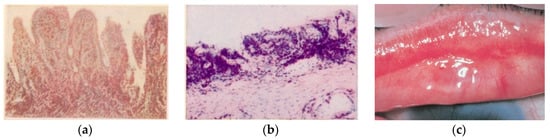
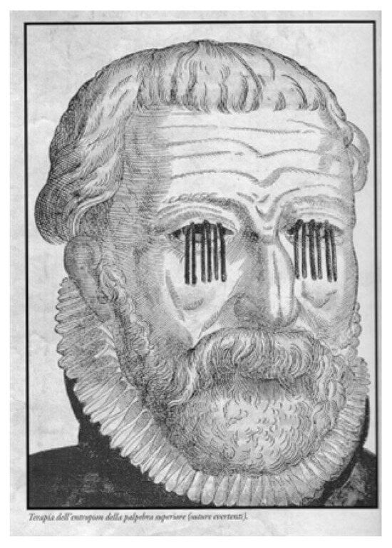
Therefore, the researchers consider the occasion of this edition useful to relaunch an alert also for the Practitioners and the Social Physicians to holistically evaluate the patient, and in front of multisite infection “check for the presence of typical signs (Arlt’s line [5]; Neri’s white line [33]) or consequences: entropion and pannus, infertility [34], spontaneous abortion, pelvic inflammatory disease” [1][35], cervical cancer in association with HPV, and proctitis especially in homosexual males [36]. Ct also appears to be involved in oculo-orbital lymphoma [37], previously associated only with Chlamydia psittaci. New research would be desirable to confirm or exclude the hypothesis of Chlamydia pneumoniae as a chronic inflammatory stimulus for neovascular Age-related Macular Degeneration (nAMD) [38], inducing the inflammatory state [39]. Thus, it seems appropriate to draw the attention to Chlamydia for an evaluation of florid or chronic conjunctivitis, as well as for oropharyngeal diseases, such as Neisseria gonorrhoeae and other sexually transmitted diseases [40] which could also trigger, sometimes, reactive arthritis [41][42]. The PAHO-WHO plans to control the worldwide trachoma infection by 2020, between 2018 and 2019 showed reduction of 14,4 million people that lived in areas with high trachoma prevalence, but still leaving a total burden of 2,5 million cases of trachomatous trichiasis in 2019. This urgently requires an increase in medical and surgical strategy with decisive support for research and development of an effective and durable vaccine against chlamydial and mycoplasmal infections [43][44], improving the prevention by considering SAFE-S strategy, rather than insect powder. 

References

  1. Oriel, J.D.; Ridgway, G.L. Genital Infection by Chlamydia Trachomatis; Elsevier Biomedical USA: Gainesville, FL, USA, 1982.
  2. Mackenzie, W. Traité Pratique des Maladies De l’Oeil; In Trad Franç, 4th ed.; Masson: Paris, France, 1856.
  3. Gallenga, P.E. Storia della cataratta nel secondo dopoguerra. In Evoluzione Della Chirurgia Della Cataratta in Italia; Storia, Buratto, L., Eds.; Fabiano: Moasca, Italy, 2019; pp. 1–40.
  4. Gallenga, C. Sul trattamento operativo delle cicatrici corneali. Gazz. Clin. 1885, 21, 1–9.
  5. Gallenga, C. Seconda osservazione di concrezione calcarea delle palpebre. Gazz. Clin. 1886, 3, 1–2.
  6. von Prowazek, S.; Halberstädter, L. Zur Aetiologie des Trachoms. Dtsch. Med. Wochenschr. 1907, 33, 1285–1287.
  7. Gallenga, C. Della Specificità Dei Corpuscoli Di Prowazeck E Halberstaedter Nella Congiuntivite Granulomatosa; Atti Società Italiana Patologia, VI riunione; Società Tipografica Modenese; Antica Tipografia Soliani: Modena, Italy, 1909.
  8. Gallenga, C. Zur Färbung der Prowazeck Halberstädterischen Trachom-Körperchen. Klin. Augenheilkd. 1910, 1, 195s.
  9. Axenfeld, T. Traité d’Ophtalmologie; Steinheil: Paris, France, 1914.
  10. Dark, A.J. Inclusion bodies in trachoma. Brit. J. Ophth. 1955, 39, 751.
  11. Gallenga, C.E.; Del Boccio, M.; Del Boccio, G.; Perri, P.; Contini, C. Who is afraid of a bug mantled like Zorro? EC Microbiol. 2019, 15, 511–513. Available online: www.researchgate.net/publication/334251061 (accessed on 20 January 2023).
  12. Bietti, A. Trattato di Oftalmoiatria; Istituto Editoriale Scientifico: Milan, Italy, 1925.
  13. Gallenga, R. Plasmoma Della Congiuntiva Bulbare Superiore; Atti Congr Soc It Oftalmol: Roma, Italy, 1931; L’Universale Tipografia Poliglotta: Roma, Italy, 1932.
  14. Gallenga, R. The Ophthalmology course, duplicated lecture notes (Gagna E. typographer. 5th year Medical School University of Turin). 1966; and OMC&O Turin conference, in the University Eye Clinic 1967.
  15. Scuderi, G. Istopatologia Del Trachoma; Minerva Medica: Turin, Italy, 1957.
  16. Contini, A. Un Sassarese in Un Tempio Della Scienza; EDES: Sassari, Italy; ISBN 9788860252081.
  17. Frezzotti, R.; Guerra, R. Oftalmologia Essenziale, 2nd ed.; CEA: Milano, Italy, 2006.
  18. Schachter, J.; Causse, G.; Tarizzo, M.L. Chlamydiae as agents of sexually transmitted disease. WHO Bull. 1976, 54, 245–254.
  19. Delle Noci, N. George Bartisch. L’Ophthalmodouleia e l’Oculistica Rinascimentale; Théa special ed; Centro Grafico srl: Foggia, Italy, 2019.
  20. Gallenga, P.E.; Del Boccio, M.; Gallenga, C.E.; Neri, G.; Pennelli, A.; Toniato, E.; Lobefalo, L.; Maritati, M.; Perri, P.; Contini, C. Diagnosis of a neonatal ophthalmic discharge, Ophtalmia Neonatorum, in the ‘Molecular age’: Investigation for a correct therapy. J. Biol. Regul. Homeost. Agents 2018, 32, 177–184.
  21. Akinsolu, F.T.; Nemieboka, P.O.; Njuguna, D.W.; Ahadji, M.N.; Dezso, D.; Varga, O. Emerging resistance of neglected tropical diseases: A scoping review of the literature. Int. J. Environ. Res. Public Health. 2019, 16, 1925.
  22. Lewies, A.; Wentzel, J.F.; Jacobs, G.; Du Plessis, L.H. The potential use of natural and structural Antimicrobial Peptides in the fight against neglected Tropical Diseases. Molecules 2015, 20, 15392–15433.
  23. Quin, J. Gene and peptide in the treatment of chlamydia trachomatis infections: Beginning of an era? Theoretical. Nat. Sci. 2023, 4, 18–25.
  24. Astrauskiene, D.; Griskevicius, A.; Luksiene, R.; Panaviene, V.; Venaliene, J. Chlamydia trachomatis, Ureaplasma urealyticum, and Mycoplasma hominis in sexually intact girls with arthritides. Scand. J. Rheumatol. 2012, 41, 275–279.
  25. Bailey, R.; Lietman, T. The SAFE strategy for the elimination of trachoma by 2020: Will work? Bull. World Health Organ. 2001, 79, 233–236.
  26. WHO. Alliance for the Global Elimination of Trachoma by 2020: Progress report, 2019. WER 2020, 30, 349–360.
  27. WHO. Ending the Neglect to Attain the Sustainable Development Goals: A Rationale for Continued Investment in Tackling Neglected Tropical Diseases 2021–2030; WHO: Geneva, Switzerland, 2022; ISBN 9789240052932.
  28. Robinson, A.; Bristow, J.; Holl, M.V.; Makalo, P.; Alemayehu, W.; Bailey, R.L.; Macleod, D.; Birkett, M.A.; Caulfield, J.C.; Sarah, V. Responses of the putative trachoma vector, Musca sorbens, to volatile semiochemicals from human faeces. PLoS Negl. Trop. Dis. 2020, 14, e0007719.
  29. Hirschberger, P.; Degro, H.N. Oviposition of the dung beetle Aphodius ater in relation to the abundance of yellow dung fly larvae (Scatophaga stercoraria). Ecol. Entomol. 1996, 21, 352–357.
  30. Blanckenhorn, W.U.; Pemberton, A.J.; Bussière, L.F.; Roembke, J.; Floate, K.D. A Review of the Natural History and Laboratory Culture Methods for the Yellow Dung Fly, Scathophaga stercoraria. J. Insect Sci. 2010, 10, 11.
  31. Gallenga, P.E.; Del Boccio, M.; Neri, G.; Tenaglia, R.; Martinotti, S. Conjunctival chronic pain by Chlamydia trachomatis solved after asymptomatic chronic prostatitis treatment with echoguided drugs infiltration: A case report. Int. J. Med. Clin. Res. 2011, 2, 93–97.
  32. Neri, G.; Del Boccio, M.; Pennelli, A.; Martinotti, S.; Tenaglia, R.; Pugliese, M.; Toniato, E.; Croce, A.; Gallenga, P.E. Jugulodigastric lymph node inflammation derived from chronic atypical oropharyngeal phlogosis recurring annually after flu virus vaccination: A holistic vision of a clinical case solved after chlamydicidal antibiotic therapy. Int. J. Immunopathol. Pharmacol. 2012, 25, 835–847.
  33. Neri, G.; Pugliese, M.; Castriotta, A.; Mastronardi, V.; Pasqualini, P.; Colasante, A.; Cazzato, F.; Talamonti, R.; Del Boccio, G. White-line: A new finding in laryngopharyngeal reflux objective evaluation. Med. Hypotheses 2013, 80, 769–772.
  34. Di Bonaventura, G.; Del Boccio, M.; Pennelli, A.; Rapinese, M.; Tenaglia, R.; Gallenga, P.E.; Neri, G.; Boccio, G.D. Unusual Oropharyngeal Asymptomatic Manifestations Caused by Atypical Pathogens Detected by PCR into Altered Ecosystems of an Infertile Couple. Microbiol. Res. J. Int. 2014, 5, 447–458.
  35. Neri, G.; Pennelli, A.; Del Boccio, M.; Neri, L.; Gallenga, C.E.; Toniato, E.; Tenaglia, R. Molecular Evidences of Chlamydia Trachomatis and Urogenital Mycoplasmas Infections from Birth Evolving into Multisite Infections. EC Microbiol. 2019, 15, 385–394. Available online: https://ecronicon.net/assets/ecmi/pdf/ECMI-15-00639.pdf (accessed on 20 January 2023).
  36. Mei, C. Confronto di Metodiche Analitiche, Immunocromatografiche e Biomolecolari per la Determinazione di Chlamydia Trachomatis in Campioni Biologici. Ph.D. Thesis, Università C. Bo, Urbino, Italy, 2017.
  37. Contini, C.; Seraceni, S.; Carradori, S.; Cultrera, R.; Perri, P.; Lanza, F. Identification of Chlamydia trachomatis in a patient with ocular lymphoma. Am. J. Hematol. 2009, 84, 597–599.
  38. Robman, L.; Mahdi, O.; McCarty, C.; Dimitrov, P.; Tikellis, G.; McNeil, J.; Byrne, G.; Taylor, H.; Guymer, R. Exposure to Chlamydia pneumoniae infection and progression of age-related macular degeneration. Am. J. Epidemiol. 2005, 161, 1013–1019.
  39. Gallenga, C.E.; Parmeggiani, F.; Costagliola, C.; Sebastiani, A.; Gallenga, P.E. Inflammaging: Should this term be suitable for age related macular degeneration too? Inflamm. Res. 2014, 63, 105–107.
  40. Breyer, B.N.; Huang, W.H.; Rabkin, C.S.; Alderete, J.F.; Pakpahan, R.; Beason, T.S.; Kenfield, S.A.; Mabie, J.; Ragard, L.; Wolin, K.Y.; et al. Sexually transmitted infections, benign prostatic hyperplasia and lower urinary tract symptom-related outcomes: Results from the Prostate, Lung, Colorectal and Ovarian Screening Trial. Brit. J. Urol. 2016, 117, 145–154.
  41. Mabey, D.C.; Solomon, A.W.; Foster, A. Trachoma. Lancet 2003, 362, 223–229.
  42. Wright, H.R.; Turner, A.; Taylor, H.R. Trachoma. Lancet 2008, 371, 1945–1954.
  43. Stary, G.; Olive, A.; Radovic-Moreno, A.F.; Gondek, D.; Alvarez, D.; Basto, P.A.; Perro, M.; Vrbanac, V.D.; Tager, A.M.; Shi, J.; et al. Vaccines. A mucosal vaccine against Chlamydia trachomatis generates two waves of protective memory T cells. Science 2015, 348, 8205.
  44. Gottlieb, S.L.; Johnston, C. Future prospects for new vaccines against sexually transmitted infections. Curr. Opin. Infect Dis. 2017, 30, 77–86.
More
Information
Contributors MDPI registered users' name will be linked to their SciProfiles pages. To register with us, please refer to https://encyclopedia.pub/register : , , , , , , ,
View Times: 123
Revisions: 3 times (View History)
Update Date: 27 Dec 2023
1000/1000
Video Production Service