Submitted Successfully!
To reward your contribution, here is a gift for you: A free trial for our video production service.
Thank you for your contribution! You can also upload a video entry or images related to this topic.
Version Summary Created by Modification Content Size Created at Operation
1 -- 4313 2023-10-19 20:08:23 |
2 format Meta information modification 4313 2023-10-20 03:11:42 |

Video Upload Options

Do you have a full video?

Confirm

Are you sure to Delete?
Cite
If you have any further questions, please contact Encyclopedia Editorial Office.
Valverde-Salazar, V.; Ruiz-Gabarre, D.; García-Escudero, V. Neuroprotective Effects of Epigallocatechin-3-Gallate in Alzheimer’s Disease. Encyclopedia. Available online: https://encyclopedia.pub/entry/50578 (accessed on 01 July 2024).
Valverde-Salazar V, Ruiz-Gabarre D, García-Escudero V. Neuroprotective Effects of Epigallocatechin-3-Gallate in Alzheimer’s Disease. Encyclopedia. Available at: https://encyclopedia.pub/entry/50578. Accessed July 01, 2024.
Valverde-Salazar, Víctor, Daniel Ruiz-Gabarre, Vega García-Escudero. "Neuroprotective Effects of Epigallocatechin-3-Gallate in Alzheimer’s Disease" Encyclopedia, https://encyclopedia.pub/entry/50578 (accessed July 01, 2024).
Valverde-Salazar, V., Ruiz-Gabarre, D., & García-Escudero, V. (2023, October 19). Neuroprotective Effects of Epigallocatechin-3-Gallate in Alzheimer’s Disease. In Encyclopedia. https://encyclopedia.pub/entry/50578
Valverde-Salazar, Víctor, et al. "Neuroprotective Effects of Epigallocatechin-3-Gallate in Alzheimer’s Disease." Encyclopedia. Web. 19 October, 2023.
Neuroprotective Effects of Epigallocatechin-3-Gallate in Alzheimer’s Disease
Edit

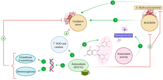
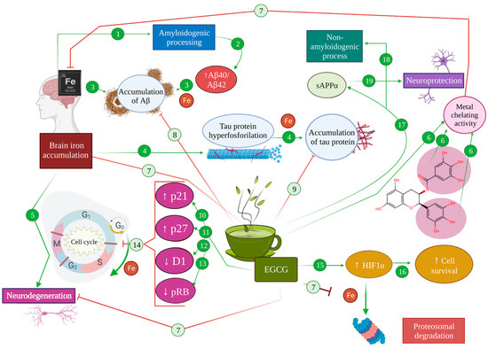
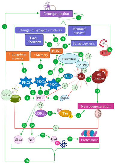
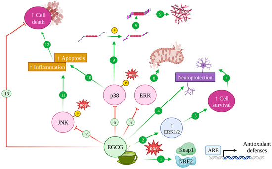
Alzheimer’s disease (AD) is the most common cause of dementia, characterised by a marked decline of both memory and cognition, along with pathophysiological hallmarks including amyloid beta peptide (Aβ) accumulation, tau protein hyperphosphorylation, neuronal loss and inflammation in the brain. Additionally, oxidative stress caused by an imbalance between free radicals and antioxidants is considered one of the main risk factors for AD, since it can result in protein, lipid and nucleic acid damage and exacerbate Aβ and tau pathology. Green tea, and its main bioactive compound, epigallocatechin-3-gallate (EGCG), have been targeted as a plausible option for the modulation of AD. Specifically, EGCG acts as an antioxidant by regulating inflammatory processes involved in neurodegeneration such as ferroptosis and microglia-induced cytotoxicity and by inducing signalling pathways related to neuronal survival. Furthermore, it reduces tau hyperphosphorylation and aggregation and promotes the non-amyloidogenic route of APP processing, thus preventing the formation of Aβ and its subsequent accumulation.

Alzheimer’s disease EGCG neuroprotection

1. Introduction

Dementia consists of a neurological syndrome characterized by memory, behaviour and language impairment that progressively builds up, leading to an inability to perform activities of daily living [1][2]. Alzheimer’s disease (AD), an eminently multifactorial neurodegenerative disorder, is considered to be the most frequent cause of dementia [2][3]. AD mainly manifests through short-term memory deficits, along with other cognitive, affective, psychosocial and, less frequently, motor impairments [2][3][4][5][6].
Current frameworks establish that, temporally, pathophysiological evidence of AD can be found 20–30 years before the beginning of symptoms that constitute the clinical phase of the disease, with molecular, cellular, biochemical, and functional alterations that have been linked to the development of symptoms later in life [7][8].
The main histopathological hallmarks of the disease are the accumulation of amyloid beta (Aβ) in the form of extracellular plaques, the aggregation of hyperphosphorylated tau protein, conforming intracellular neurofibrillary tangles, as well as evidence of neurodegeneration associated with amyloid and tau pathologies forming neuritic plaques [9][10][11]. All these are accompanied by other factors, including inflammation that implies astrocyte and microglial involvement and less specific mechanisms, such as Lewy bodies and vascular alteration [12][13][14]. The multifactorial nature of AD, together with the diffuse timeline differentiating preclinical and clinical stages and the fact that some of the predominant signs of the disease are shared by other, unrelated disorders, constitute the main causes that explain the current lack of efficient treatments to cure, delay or palliate the effects of this terrible disease [4][7].
Faced with this disheartening lack of therapeutic resources, several authors have proposed alternative approaches to the modulation of Alzheimer’s disease and other neurodegenerative disorders, by intervening in environmental and lifestyle factors such as encouraging physical exercise, avoiding alcohol and drug consumption, lowering stress levels, maintaining cognitive stimulation or promoting a healthy, balanced diet [15][16][17][18][19].
In this line, dietary modulation has been promoted as one of the more accessible interventions to promote and maintain health during people’s lifespan [17][20][21]. As one of the most extensively consumed beverages, only second to water, and due to the bioactive nature of its components, tea has acquired some popularity in recent years as a source of potentially beneficial compounds to help tackle complex pathophysiological processes [22][23].
Tea is obtained from the leaves of Camellia sinensis, a plant species native to East Asia that has been consumed for more than 4000 years [23][24]. Different varieties of tea can be obtained from the desiccated leaves of the plant, depending on the processing [23][25]. Green tea is obtained by steaming and drying freshly harvested leaves, which grants a composition virtually identical to that of the leaves and a higher concentration of bioactive molecules with potential benefits [24][25].
Green tea comprises several bioactive compounds, such as polyphenols, caffeine and amino acids [22][24]. Green tea polyphenols, often referred to as catechins, amount to around 30% of the dry weight of the leaves and have been proposed to mediate several health benefits [22][24][25]. Epigallocatechin gallate (EGCG), an ester of gallic acid and epigallocatechin, is the most abundant catechin within green tea, constituting up to 65% of total catechin content and displaying the highest biological activity [22], including neuroprotective and antioxidative effects reported in the literature [22][23][26][27].
In accordance with this, several epidemiological studies in the Asian population have linked green tea consumption and a decreased risk for neurodegenerative disorders, such as AD, Parkinson’s disease and other dementias. The effects of EGCG and its abundance within green tea, together with the fact that it can be efficiently absorbed in the intestine [28][29], constitute two key points of the potential therapeutic use of EGCG. In addition, since AD pathophysiology occurs mainly in the brain, the ability to cross the blood–brain barrier is a necessary feature of any potential therapeutic agent. In this regard, EGCG has been proven to cross the blood–brain barrier even at very low concentrations [30][31][32][33], which positions this catechin as a potential mediator with beneficial properties for Alzheimer’s disease and other forms of neurodegeneration [22][26][27][34].

2. Antioxidative Effects of EGCG

All the previous evidence allows people to conclude that there is a complex relationship between AD and oxidative stress that implies a positive feedback loop between both and poses oxidative stress and the mechanisms mentioned above as potential targets in efforts to mitigate AD pathology. Thus, antioxidants such as green tea catechins could potentially offer interesting incorporations to therapeutic arsenal. Assuredly, catechins have proven to be able to act as antioxidative systems, neutralising ROS and RNS and other free radicals, such as nitric oxide, peroxyl, peroxynitrite, carbon and lipidic radicals or 1,1-diphenyl-3-picrylhydrazine derivatives [35] (Figure 1 (8)).
Figure 1. Antioxidant activity of EGCG. The antioxidant compound EGCG binds to antioxidant regulatory elements (ARE), inducing the expression of stress response genes (1) such as haem oxygenase (2) and glutathione-S-transferase (3) enzymes, counteracting oxidative stress processes (4). Additionally, EGCG enhances the activity of SOD and catalase (5), which reduce oxidative stress (4). Thanks to its hydroxyl groups, EGCG harbours chelating properties (6) that exert antioxidant activity, promoting neuroprotective effects (7). Also, EGCG can inhibit the production of ROS/RNS and 3-Hydroxykynurenine effect due to this antioxidant capacity (8), avoiding oxidative stress (9).
EGCG has been proven to exert a greater antioxidative effect than other green tea catechins such as epicatechin or epigallocatechin, greater even than that of potent antioxidants such as vitamins E and C [35][36][37], linked to its structure that leaves several hydroxyl groups free to sequester radicals (Figure 1 (6)).
In addition, EGCG and other flavonoids, together with phenolic antioxidants found in green tea, can also activate endogenous antioxidative systems, thus exerting an indirect protective effect [34][35]. Namely, flavonoids promote the expression of stress response genes, including those coding for the enzymes haem oxygenase and glutathione-S-transferase [38], by binding to antioxidant regulatory elements (ARE) in the promoter of said genes [34][38] (Figure 1 (1–3)). Furthermore, under oxidative stress conditions, the activation of these genes seems to be accompanied by the modulation of MAPK’s function, promoting the activity of transcription factors Nrf1 and Nrf2 and increasing their nuclear binding to ARE sequences [38][39].
Moreover, EGCG showed an antioxidative effect in vitro in neuronal primary cultures, where it inhibited the toxic effects of 3-hydroxykynurenine, a metabolite of tryptophan that acts as a potent endogenous neurotoxin, increasing oxidative stress and promoting ROS production [35][40]. Upon EGCG-mediated inhibition, oxidative stress and ROS were reduced and caspase activation and apoptosis were diminished consequently [40] (Figure 1 (8)).
Synergically with all these effects, EGCG enhanced the activity of SOD and catalase, two of the most relevant endogenous antioxidative systems, which proved to be enough to reduce oxidative stress in C57BL mice [35][41] (Figure 1 (5)).

3. Iron-Chelating Effects of EGCG

The capability of EGCG and other catechins to bind metal ions poses another exciting overlap between oxidative stress, AD and the neuroprotective effects of catechins. More specifically, catechins have proven to be exceptional iron and copper chelators [35][42][43], which is especially relevant, given that iron metabolism alterations have been proposed to be a common link between several neurodegenerative disorders [44][45][46][47] (Figure 2 (16,17), Figure 1 (6,7) and Figure 2 (6,7)).
Figure 2. Effect of metal chelation mediated by EGCG in Alzheimer’s pathology. In an AD brain, iron accumulation promotes amyloidogenic processing (1), increasing Aβ40/Aβ42 levels (2), leading to the accumulation of Aβ (3). Moreover, brain iron accumulation produces an increase in tau hyperphosphorylation (4) that results in microtubule destabilization and the accumulation of tau protein (5). As a consequence, iron produces neurodegeneration (6). The metal chelator activity of EGCG (6) inhibits iron accumulation in the brain (7), therefore diminishing the accumulation of Aβ (8) and tau protein (9). Moreover, EGCG can directly potentiate the expression of p21 (10) and p27 (11), while diminishing the expression of cyclin D1 (12) and pRB (13), abolishing cell cycle re-entry (14). On the other hand, EGCG can promote the activation of HIF-1α (15), inducing the expression of cell survival genes (16). EGCG promotes the production of SAPPα (17) and the non-amyloidogenic processing of APP (18), generating neuroprotection effects (19).
Given the numerous pathways in which iron imbalance can modulate pathological mechanisms in Alzheimer’s disease, iron chelation promises an interesting research avenue to explore. In fact, iron chelators have been proposed as potential multi-target treatments for Alzheimer’s disease throughout the last decades [44][45][48]. In this regard, iron chelation exerts a direct neuroprotective effect in AD by avoiding the iron-mediated promotion of amyloid and tau pathology, through the mechanisms detailed above [48][49][50][51]. For instance, the use of iron chelators to assert a neuroprotective effect relies on both the elimination of excess iron in the brain and the prevention of its accumulation under oxidative stress conditions [48]. For instance, a prolonged administration of EGCG to C57BL mice in vivo was shown to diminish hippocampal APP without modifying APP mRNA, which suggests a post-transcriptional level of intervention, such as intracellular iron chelation [52] (Figure 2 (6,8,9)).
Apart from that, one of the main neuroprotective effects arising from iron chelation is associated with the activation of the hypoxia inducible factor 1α (HIF-1α) pathway that results in the stabilisation of the transcription factor HIF-1, involved in the transcription of cell survival and oxidative stress response genes [35][48][53]. A HIF-1α presence depends on the activity of HIF-prolyl-4-hydroxylases, which are iron-dependent enzymes. In the face of an overload of iron, these enzymes catalyse the hydroxylation of proline and asparagine residues within HIF that target its degradation via the proteasome. Thus, under excess iron conditions, those oxidative-stress-response and cell-survival genes’ expression is decreased [35][48]. On the contrary, EGCG has been proven to act as a direct HIF-1α activator, thus promoting cell survival genes and neuroprotection [34] (Figure 2 (7,15,16)).
Precisely, in relation to cell survival in neurons, intracellular iron modulation has been extensively proposed as a means to avoid apoptosis and stop the cell cycle [45][48][54]. In AD in particular, a dysregulation of the cell cycle has been equally characterised, with consequences such as cytoskeleton phosphorylation, mitochondrial abnormalities, and alteration in several transduction pathways, such as those of GSK3, CDK5 and ERK2 [54][55][56]. The activation of these pathways consequently produces aberrant tau phosphorylation, DNA replication and an increased expression of cell cycle proteins such as cyclins (A, B, D and E) [48][54]. Other reports show that control mechanisms on phases G1 and S of the cell cycle cannot be correctly performed in AD, allowing neurons to continue the cell cycle and progress to G2, even completing DNA replication, observing 3–4% of tetraploid cells [56][57]. In line with this, some genes related to the cell cycle have been proven to be altered under oxidative stress conditions such as the ones found in AD patients. For instance, PIN1, a gene with important implications in the correct regulation of the cell cycle, has been proven to regulate age-dependent neurodegenerative processes, including APP processing and tau dephosphorylation [58]. In the context of Alzheimer’s disease and oxidative stress, PIN1 can be inhibited, leading to either mitotic arrest or neuronal death [58]. In the same line, BRCA1, a gene with a role in cell growth and DNA repair, was overexpressed in neurons that possessed neurofibrillary tangles in AD, which would entail an increased genome instability [59].
Iron seems to carry out a regulatory function on the cell cycle, in that under iron deficiency conditions, cells cannot advance from the G1 phase to S, a regulatory role directly related to D1 cyclin degradation [60], while iron accumulation has been reported to disrupt the cell cycle, promoting abnormal progression through the cycle that leads to apoptosis [34]. All this is of vital importance due to the multiple cell cycle alterations found in AD commented on above [45][48]. In this regard, EGCG is capable of travelling across the blood–brain barrier and interferes with mitogenic signalling at the brain level, preventing the progression of an altered cell cycle in the presence of excess iron [34]. In addition, EGCG can directly potentiate the expression of p21 and p27, while diminishing the expression of D1 cyclin and pRB, abolishing re-entry to the cell cycle thanks to a primary antiproliferative action [27][48][61] (Figure 2 (10–14)).
It is worth mentioning that the antioxidant and iron chelating activity of EGCG may be useful to inhibit ferroptosis cell death that, as mentioned before, is increased in an AD brain. In fact, there is evidence that supports that the inhibition of brain ferroptosis protects from a brain haemorrhage [62]. Additionally, it has been shown that EGCG was able to inhibit ferroptosis after spinal cord injury through protein kinase D1 phosphorylation [63]. Therefore, several authors have suggested a potential protective role of EGCG by preventing ferroptosis in Alzheimer’s disease as a therapeutic strategy [64][65] (Figure 2 (14,18)).

4. Modulating Effect of EGCG in Cell Signalling, Survival and Death Pathways

As broadly commented on in previous sections, there are multiple intracellular signalling pathways related to neuroprotection and cell survival in which Alzheimer’s disease and oxidative stress mechanisms can converge, including protein kinase C, mitogen-activated protein kinases and phosphoinositide 3-kinase pathways [54]. All of them are related to several neuronal functions, such as plasticity, synaptic morphology, and protein synthesis, which can in turn affect memory and neurodegeneration [54][66][67].
Briefly, EGCG has been reported to exert direct neuroprotective effects through the modulation of cell survival and death, activating ERK, Akt/PKB, PI3K and PKC pathways that improve cell survival, while inhibiting p38 and JNK ones, thus avoiding apoptosis [35][67].

4.1. EGCG and the PKC Pathway: Implications in Alzheimer’s Disease

Protein kinase C constitutes a family of kinases whose function is phosphorylating serine/threonine residues of proteins, regulating their biological functions. Kinases from the PKC family are involved in the brain signalling network through the regulation of cell signalling, cell growth, differentiation, and apoptosis, with direct consequences on tumorigenesis, synaptic function, behaviour and cognition [68][69].
At least 12 isoforms can be found in mammals, classified in three subfamilies, according to their structure and specific requirements of the second messengers: classical PKC (cPKC), with isoforms α, βI, βII and γ; atypical PKC (aPKC), composed of isoforms ι, λ and ζ; and novel PKC (nPKC), which includes isoforms δ, ε, η, μ and θ [70]. Among these, α, γ, ε and ζ have been linked to signalling processes related to memory mechanisms and memory deficits, which has earned them the nickname of memory kinases [70]. PKC activators such as arachidonic acid, aplysiatoxins or bryostatins can improve memory [71][72] and restore synaptic and network functions [73][74], exerting anti-dementia effects [75][76]. In fact, PKC activation by arachidonic acid is one of the main mechanisms of astrocyte-induced synaptogenesis [77].
From a broad perspective, PKC activation has a wide range of biological effects. On synaptic transmission, for instance, it enhances the synthesis, vesicle replenishment and liberation of cholinergic, dopaminergic, glutaminergic and GABAergic neurotransmitters [78][79][80]. Relatedly, PKC has also been linked to synaptic plasticity, promoting long-term potentiation (LTP) phenomena, with PKCζ having proven to be necessary and sufficient to maintain hippocampal LTP [81] and having been associated with long-term memory [82][83], while PKCα, PKCγ and PKCε have been related to memory and learning processes [70] (Figure 3 (2–5,8–12)).
Figure 3. Modulation of PKC-mediated pathways by EGCG in Alzheimer’s disease. EGCG can induce the activation of PKC pathways (1), specifically the isoform PKCζ (2) that enhances long-term memory (3), PKCα (4) that results in an increase in memory function (5) and PKCε (6) that activates BDNF factor (7), promoting synaptogenesis (8), neuronal survival (9), Ca2+ liberation (10) and changes of synaptic structures (11), altogether promoting neuroprotection (12). The generation of Aβ produces Aβ plaques (13), promoting neurodegeneration processes (14). Aβ also inhibits the PKC pathway (15) and RACK (16), whose receptors are required to activate PKC (17), and blocks BDNF (18). Moreover, the isoforms PKCα and PKCε activate α-secretase (19), promoting the generation of sAPPα (20) that inhibits Aβ production (21). PKCε activates ECE1 (22), which degrades Aβ (21). The activation of the PKC pathway inhibits GSK3β (23), which is involved in tau hyperphosphorylation (24) that finally leads to neurodegeneration processes (14). EGCG produces the reduction in protein Bax (25) and promotes the degradation of protein Bad (26) through the proteasomal system (27).
In addition, different PKC isoforms have been reported to perform opposite functions regarding cell growth, differentiation and apoptosis. Specifically, PKCθ and PKCδ promote apoptosis, while PKCα, PKCβ, PKCε and PKCζ can avoid it and promote neurite growth instead [84][85]. In addition, PKCε has been proven to directly enhance the expression of brain-derived neurotrophic factor (BDNF), which can activate complex signalling pathways, resulting in the repair of the synaptic structure and function and production of new brain cells [70][86] (Figure 3 (6–12)).
Alterations in PKC signalling pathways have been found to contribute to Alzheimer’s disease pathogenesis and are associated with memory deficits and learning difficulties [70][87], possibly establishing reciprocal interactions with pathogenic mechanisms, since PKC isoforms are also sensitive to AD-related stress factors and amyloid plaques [87]. This can be linked to the anti-dementia effects mentioned for PKC activation [75]. In the context of AD, PKC activation has been proposed to stimulate LTP and cognitive improvement, by helping reduce the amyloid load [87][88]. On top of that, PKC activation also inhibits glycogen synthase kinase 3 (GSK3), the main kinase involved in tau phosphorylation, providing yet another confluence between PKC pathways and AD pathology [89] (Figure 3 (23,24,14)).
In turn, Aβ was shown to decrease PKC levels by directly binding PKC isoforms, effectively reducing their phosphorylation and translocation, blocking their activation and inducing their degradation [90]. Aβ peptides and oligomers also inhibit RACK and intracellular receptors required in PKC activation, and actively block BDNF, which specifically links it to PKCε [87][88][90]. Indeed, PKCε has been associated with the activation of endothelin converting enzyme 1 (ECE-1) [88], one of the main enzymes involved in amyloid β degradation and amyloid plaque reduction [91][92], rendering PKCε activation and overexpression as effective methods to reduce amyloid pathology [93][94]. Parallelly, both PKCε and PKCα act as activators of α-secretase that mediate the non-amyloidogenic processing of APP, contributing to alleviate amyloidogenic buildup and promoting the generation of sAPPα that also acts as an Aβ inhibitor and exerts neuroprotective functions [70][90][95][96]. In addition, PKCε may be able to contribute to amyloid degradation by activating circulating serine proteases that can cleave Aβ [88] (Figure 3 (15,17–22))
All these intricate pathways play a role on the neuroprotective function exerted by EGCG and other catechins. EGCG has been shown to contribute to PKC pathways through the direct activation of PKC by means of a fast phosphorylation that promotes the beneficial, neuroprotective effects previously detailed [27][87]. In vivo studies on C57BL mice under a 2 mg/kg/day consumption schedule of EGCG proved that this catechin is also able to induce a fast translocation of PKCα, which prevented PKCα depletion and counteracted the increase in the apoptotic protein Bax in neurons [34]. Synergically, EGCG has also been demonstrated to induce a rapid proteasomal degradation of Bad, another proapoptotic protein, via PKC activation [97] (Figure 3 (1,25–27)).
Moreover, the specific activation of PKCα and PKCε entails the stimulation of their anti-AD pathology pathways described above. Hence, several studies using an in vivo mice model of AD such as Tg2576 and APP695SWE proved that EGCG induces PKCε-mediated ECE-1 activation and promotes the non-amyloidogenic processing of APP through PKC activation, promoting sAPPα production and a significant reduction in Aβ and amyloid plaque levels in the brain [98] (Figure 3 (19–22)).

4.2. EGCG and the MAPK Pathway: Implications in Alzheimer’s Disease

The mitogen-activated protein kinases’ pathway constitutes another crucial signalling cascade in terms of cell proliferation, differentiation, apoptosis and survival, as well as inflammation and innate immunity. Mammal MAPK are grouped in three categories: c-Jun N-terminal kinases (JNK), with three different isoforms termed JNK1, JNK2 and JNK3; p38 kinases, with isoforms α, β, γ and δ; and extracellular signal-regulated kinases (ERK), composed of isoforms ERK1, ERK2 and ERK5 [99][100]. All of them act as transductors of extracellular stimuli by unfolding a phosphorylation cascade composed of, at least, three components: a MAPK kinase (MAP3K) that phosphorylates and activates another MAPK kinase (MAP2K), which in turn phosphorylates and activates a MAPK [99]. This activated MAPK is then able to phosphorylate several targets, including transcription factors and antiapoptotic and proapoptotic proteins [99][101].
Each individual MAPK signalling pathway is activated as a result of complex interactions between different kinase components or through a signalling complex composed of several kinases and a scaffold protein [99]. Broadly speaking, researchers  can assert that ERK1/2-mediated pathways are activated by growth factors and stimulate cell proliferation, migration, differentiation and survival [99][102], while p38 and JNK are activated by stress factors such as oxidative stress or inflammation, and are therefore responsible for inflammatory and stress responses, autophagy and apoptosis, although they can also participate in cell differentiation [103][104][105].
Given the myriad of pivotal processes in which these signalling pathways take part, it should not come as a surprise that MAPK signalling is altered in multifactorial pathogenic processes such as AD and other neurodegenerative disorders. In Alzheimer’s disease, Aβ-induced oxidative stress and microglial activation have both proven to be mediators of MAPK p38 signalling [106][107][108], which promotes apoptosis but also acts as a kinase of tau protein [109], further contributing to Alzheimer’s disease (Figure 4 (9,10)). In line with this, the inhibition of IL-1β signalling during neuroinflammation in AD ameliorated tau pathology and improved cognitive function in a p38-dependent manner [110]. Other AD-related pathological changes, such as mitochondrial dysfunction and mitochondrial dynamics alteration seem to be mediated by ERK pathways, since their blockage has been shown to improve mitochondrial morphology and function and reverse alteration in the expression and distribution of mitochondrial dynamics’ proteins such as DLP1 and Mfn2 [111] (Figure 4 (8)).
Figure 4. Modulation of MAPK pathway by EGCG in Alzheimer’s disease. Upon oxidative stress, EGCG induces antioxidant defences with the activation of the Keap1/Nrf2/ARE pathway (1) and increases ERK1/2 (2), promoting cell survival (3) and neuroprotective effects (4). Conversely, EGCG inhibits the ERK (5), p38 (6) and JNK (7) pathways whose effects contribute to mitochondrial disfunction and altered dynamics (8), induce tau protein hyperphosphorylation and aggregation (9) and increase apoptosis (10) and inflammation (11), leading to cell death (12). Therefore, EGCG can avoid this cell death (13).
As for the role of EGCG in relation to these signalling pathways, the green tea catechin was proven to preclude ERK1/2 downregulation mediated by oxidative stress, which results in increased cell survival, both in nervous and non-nervous tissue [105]. In accordance with the antioxidative effect discussed before, EGCG was also shown to induce antioxidant defence systems through the activation of the Keap1/Nrf2/ARE pathway and antioxidative enzymes through Akt and ERK1/2 activation [112][113]; albeit EGCG seemed to be unable to exert any activating effect on ERK1/2 in the absence of oxidative stress conditions [27] (Figure 4 (1–3)).
Additionally, EGCG delivers an orchestrated effect, by also inhibiting ROS-induced phosphorylation in MAPK from the JNK and p38 pathways, which rendered them inactive [105], while also inhibiting hydrogen-peroxide-dependent caspase 3 activation, thus avoiding apoptosis [105][114] (Figure 4 (6,10)).

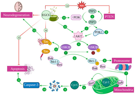
4.3. EGCG and the PI3K/Akt Pathway: Implications in Alzheimer’s Disease

Another signalling pathway of paramount importance for cell survival and cell cycle progression, as well as metabolism, cell motility and transcription, is the protein kinase B (PKB, also termed Akt) pathway. Mammals display three PKB isoforms: α, β and γ (Akt 1, 2 and 3) [115], which are activated by phosphoinositide 3 kinase (PI3K) to exert antiapoptotic functions [115][116] (Figure 5 (1)).
Figure 5. Modulation of PI3K/Akt pathway mediated by EGCG. Akt/protein kinase B (PKB) is activated by Phosphatidyl Inositol 3 Kinase (PI3K) (1), which catalyses the conversion of phosphatidyl inositol (4,5) biphosphate (PIP2) into phosphatidyl inositol (3,4,5) triphosphate (PIP3), a process that is reversed by phosphatidylinositol (3,4,5)-triphosphate 3-phosphatase (PTEM) (2). PIP3 also recruits phosphoinositide-dependent kinase 1 (PDK1) to the plasma membrane, activating Akt (3). Akt phosphorylates glycogen synthase kinase 3 (GSK3), inhibiting its function (4). Active GSK3 is able to phosphorylate Mcl-1 (5), which is targeted for proteasomal degradation (6), liberating Bax and Bak proapoptotic factors (7). This causes the permeabilization of the mitochondrial outer membrane, releasing cytochrome c (Cyt C) (8) that attaches to Apaf-1, generating the apoptosome (9), leading to the activation of caspase 3 (10) that induces apoptosis (11). GSK3 also phosphorylates tau, inducing its aggregation and subsequent neurodegeneration (12). EGCG can ultimately increase PI3K and Akt activity (13) and inhibit PTEM in the presence of ROS (14), inhibiting apoptosis (15), phospho-Tau aggregation (16) and, therefore, generating neuroprotection.
PI3K, in turn, can be activated by a number of stimuli, including trophic factors such as nerve growth factor (NGF), insulin-like growth factor (IGF-1) or BDNF [115][117]. Upon activation, PI3K catalyses the phosphorylation of phosphatidylinositol (4,5)-bisphosphate (PIP2) to phosphatidylinositol (3,4,5)-trisphosphate (PIP3), while the inverse reaction is catalysed by phosphatase PTEN (phosphatidylinositol (3,4,5)-triphosphate 3-phosphatase) [118][119]. The latter recruits Akt and the serine/threonine kinase PDK1 (phosphoinositide-dependent kinase 1) to the plasma membrane and promotes a signalling cascade that culminates with the activation of PKB/Akt [115][118] (Figure 5 (1–3)).
The Akt pathway regulates different proteins from the Bcl-2 family, which includes proapoptotic (Bax, Bad…) and antiapoptotic (Bcl-2) effectors. Akt can directly inhibit the apoptotic Bad proteins and caspases and indirectly inhibit the proapoptotic effects of GSK3 by increasing the levels of antiapoptotic proteins such as Bcl-2, effectively blocking neuronal apoptosis [115][120]. Relatedly, Akt is also a regulator of metabolism, with one of its main functions being the inhibition of GSK3 via phosphorylation, which prompts the storage of glucose and glycogen and seems to be in itself an antiapoptotic mechanism [120] (Figure 5 (4–11)).
In the context of Alzheimer’s disease, the Akt pathway plays a fundamental role as one of the most potent inhibitors of GSK3, the main kinase that drives tau phosphorylation [89][121]. Indeed, AD pathology seems to be mediated, at least partly, by the dysregulation of this pathway that allows GSK3 overactivation, which consequently causes tau hyperphosphorylation [121][122][123] (Figure 5 (12)).
Once again, EGCG is able to induce the activation of the PI3K/Akt pathway, consequently leading to increased cell survival and apoptosis inhibition [105], which seems to be the mechanism by which it prevented oxidative-stress-mediated cytotoxicity in PC12 cells in vitro [124]. In addition, under oxidative stress conditions, EGCG inactivated the phosphatase PTEN—what would prevent PIP3 transformation into PIP2—increasing PKB/Akt activation [124]. In any case, these mechanisms converge in the activation of Akt, which blocks GSK3, effectively inhibiting the proapoptotic caspases’ route, preventing the liberation of cytochrome c by avoiding mitochondrial damage and precluding tau hyperphosphorylation [122][124] (Figure 5 (13–16)).

References

  1. Sharma, P.; Srivastava, P.; Seth, A.; Tripathi, P.N.; Banerjee, A.G.; Shrivastava, S.K. Comprehensive review of mechanisms of pathogenesis involved in Alzheimer’s disease and potential therapeutic strategies. Prog. Neurobiol. 2019, 174, 53–89.
  2. Qiu, C.; Kivipelto, M.; Von Strauss, E. Epidemiology of Alzheimer’s disease: Occurrence, determinants, and strategies toward intervention. Dialogues Clin. Neurosci. 2022, 11, 111–128.
  3. Trejo-Lopez, J.A.; Yachnis, A.T.; Prokop, S. Neuropathology of Alzheimer’s disease. Neurotherapeutics 2022, 19, 173–185.
  4. Dubois, B.; Hampel, H.; Feldman, H.H.; Scheltens, P.; Aisen, P.; Andrieu, S.; Bakardjian, H.; Benali, H.; Bertram, L.; Blennow, K. Preclinical Alzheimer’s disease: Definition, natural history, and diagnostic criteria. Alzheimer’s Dement. 2016, 12, 292–323.
  5. Breijyeh, Z.; Karaman, R. Comprehensive review on Alzheimer’s disease: Causes and treatment. Molecules 2020, 25, 5789.
  6. Avila, J.; Perry, G. A multilevel view of the development of Alzheimer’s disease. Neuroscience 2021, 457, 283–293.
  7. Alzheimer’s-Association. 2022 Alzheimer’s disease facts and figures. Alzheimer’s Dement. 2022, 18, 700–789.
  8. Lloret, A.; Esteve, D.; Lloret, M.-A.; Cervera-Ferri, A.; Lopez, B.; Nepomuceno, M.; Monllor, P. When does Alzheimer′ s disease really start? The role of biomarkers. Int. J. Mol. Sci. 2019, 20, 5536.
  9. Knopman, D.S.; Petersen, R.C.; Jack, C.R. A brief history of “Alzheimer disease”: Multiple meanings separated by a common name. Neurology 2019, 92, 1053–1059.
  10. Gauthier, S.; Rosa-Neto, P.; Morais, J.; Webster, C. World Alzheimer Report 2021: Journey through the diagnosis of dementia. Alzheimer’s Dis. Int. 2021, 2022, 30.
  11. Chong, F.P.; Ng, K.Y.; Koh, R.Y.; Chye, S.M. Tau proteins and tauopathies in Alzheimer’s disease. Cell. Mol. Neurobiol. 2018, 38, 965–980.
  12. Li, T.; Lu, L.; Pember, E.; Li, X.; Zhang, B.; Zhu, Z. New Insights into Neuroinflammation Involved in Pathogenic Mechanism of Alzheimer’s Disease and Its Potential for Therapeutic Intervention. Cells 2022, 11, 1925.
  13. Lane, C.A.; Hardy, J.; Schott, J.M. Alzheimer’s disease. Eur. J. Neurol. 2018, 25, 59–70.
  14. Deschaintre, Y.; Richard, F.; Leys, D.; Pasquier, F. Treatment of vascular risk factors is associated with slower decline in Alzheimer disease. Neurology 2009, 73, 674–680.
  15. Reitz, C.; Mayeux, R. Alzheimer disease: Epidemiology, diagnostic criteria, risk factors and biomarkers. Biochem. Pharmacol. 2014, 88, 640–651.
  16. Armstrong, R.A. Risk factors for Alzheimer’s disease. Folia Neuropathol. 2019, 57, 87–105.
  17. Fernández-Sanz, P.; Ruiz-Gabarre, D.; García-Escudero, V. Modulating effect of diet on Alzheimer’s disease. Diseases 2019, 7, 12.
  18. Bellantuono, L.; Monaco, A.; Amoroso, N.; Lacalamita, A.; Pantaleo, E.; Tangaro, S.; Bellotti, R. Worldwide impact of lifestyle predictors of dementia prevalence: An eXplainable Artificial Intelligence analysis. Front. Big Data 2022, 5, 1027783.
  19. Graham, H.; White, P.C.L. Social determinants and lifestyles: Integrating environmental and public health perspectives. Public Health 2016, 141, 270–278.
  20. Duplantier, S.C.; Gardner, C.D. A critical review of the study of neuroprotective diets to reduce cognitive decline. Nutrients 2021, 13, 2264.
  21. Burns, S.; Selman, A.; Sehar, U.; Rawat, P.; Reddy, A.P.; Reddy, P.H. Therapeutics of Alzheimer’s Disease: Recent Developments. Antioxidants 2022, 11, 2402.
  22. Khalatbary, A.R.; Khademi, E. The green tea polyphenolic catechin epigallocatechin gallate and neuroprotection. Nutr. Neurosci. 2020, 23, 281–294.
  23. Farzaei, M.H.; Bahramsoltani, R.; Abbasabadi, Z.; Braidy, N.; Nabavi, S.M. Role of green tea catechins in prevention of age-related cognitive decline: Pharmacological targets and clinical perspective. J. Cell. Physiol. 2019, 234, 2447–2459.
  24. Hayat, K.; Iqbal, H.; Malik, U.; Bilal, U.; Mushtaq, S. Tea and its consumption: Benefits and risks. Crit. Rev. Food Sci. Nutr. 2015, 55, 939–954.
  25. Nabavi, S.M.; Silva, A.S. Nonvitamin and Nonmineral Nutritional Supplements; Academic Press: Cambridge, MA, USA, 2018.
  26. Urdzikova, L.M.; Ruzicka, J.; Karova, K.; Kloudova, A.; Svobodova, B.; Amin, A.; Dubisova, J.; Schmidt, M.; Kubinova, S.; Jhanwar-Uniyal, M. A green tea polyphenol epigallocatechin-3-gallate enhances neuroregeneration after spinal cord injury by altering levels of inflammatory cytokines. Neuropharmacology 2017, 126, 213–223.
  27. Mandel, S.A.; Amit, T.; Kalfon, L.; Reznichenko, L.; Youdim, M.B. Targeting multiple neurodegenerative diseases etiologies with multimodal-acting green tea catechins. J. Nutr. 2008, 138, 1578S–1583S.
  28. Moore, R.J.; Jackson, K.G.; Minihane, A.M. Green tea (Camellia sinensis) catechins and vascular function. Br. J. Nutr. 2009, 102, 1790–1802.
  29. Lee, M.-J.; Maliakal, P.; Chen, L.; Meng, X.; Bondoc, F.Y.; Prabhu, S.; Lambert, G.; Mohr, S.; Yang, C.S. Pharmacokinetics of tea catechins after ingestion of green tea and (−)-epigallocatechin-3-gallate by humans: Formation of different metabolites and individual variability. Cancer Epidemiol. Biomark. Prev. 2002, 11, 1025–1032.
  30. Pervin, M.; Unno, K.; Nakagawa, A.; Takahashi, Y.; Iguchi, K.; Yamamoto, H.; Hoshino, M.; Hara, A.; Takagaki, A.; Nanjo, F. Blood brain barrier permeability of (−)-epigallocatechin gallate, its proliferation-enhancing activity of human neuroblastoma SH-SY5Y cells, and its preventive effect on age-related cognitive dysfunction in mice. Biochem. Biophys. Rep. 2017, 9, 180–186.
  31. Pervin, M.; Unno, K.; Takagaki, A.; Isemura, M.; Nakamura, Y. Function of green tea catechins in the brain: Epigallocatechin gallate and its metabolites. Int. J. Mol. Sci. 2019, 20, 3630.
  32. Unno, K.; Pervin, M.; Nakagawa, A.; Iguchi, K.; Hara, A.; Takagaki, A.; Nanjo, F.; Minami, A.; Nakamura, Y. Blood–Brain Barrier Permeability of Green Tea Catechin Metabolites and their Neuritogenic Activity in Human Neuroblastoma SH-SY5Y Cells. Mol. Nutr. Food Res. 2017, 61, 1700294.
  33. Wyganowska-Świątkowska, M.; Matthews-Kozanecka, M.; Matthews-Brzozowska, T.; Skrzypczak-Jankun, E.; Jankun, J. Can EGCG Alleviate Symptoms Down Syndrome by Altering Proteolytic Act? Int. J. Mol. Sci. 2018, 19, 248.
  34. Mandel, S.; Weinreb, O.; Amit, T.; Youdim, M.B. Cell signaling pathways in the neuroprotective actions of the green tea polyphenol (-)-epigallocatechin-3-gallate: Implications for neurodegenerative diseases. J. Neurochem. 2004, 88, 1555–1569.
  35. Weinreb, O.; Amit, T.; Mandel, S.; Youdim, M.B. Neuroprotective molecular mechanisms of (−)-epigallocatechin-3-gallate: A reflective outcome of its antioxidant, iron chelating and neuritogenic properties. Genes Nutr. 2009, 4, 283–296.
  36. Pannala, A.; Rice-Evans, C.A.; Halliwell, B.; Singh, S. Inhibition of peroxynitrite-mediated tyrosine nitration by catechin polyphenols. Biochem. Biophys. Res. Commun. 1997, 232, 164–168.
  37. Nanjo, F.; Goto, K.; Seto, R.; Suzuki, M.; Sakai, M.; Hara, Y. Scavenging effects of tea catechins and their derivatives on 1, 1-diphenyl-2-picrylhydrazyl radical. Free Radic. Biol. Med. 1996, 21, 895–902.
  38. Chen, C.; Yu, R.; Owuor, E.D.; Tony Kong, A.-N. Activation of antioxidant-response element (ARE), mitogen-activated protein kinases (MAPKs) and caspases by major green tea polyphenol components during cell survival and death. Arch. Pharmacal Res. 2000, 23, 605–612.
  39. Owuor, E.D.; Kong, A.-N.T. Antioxidants and oxidants regulated signal transduction pathways. Biochem. Pharmacol. 2002, 64, 765–770.
  40. Jeong, J.H.; Kim, H.J.; Lee, T.J.; Kim, M.K.; Park, E.S.; Choi, B.S. Epigallocatechin 3-gallate attenuates neuronal damage induced by 3-hydroxykynurenine. Toxicology 2004, 195, 53–60.
  41. Levites, Y.; Weinreb, O.; Maor, G.; Youdim, M.B.; Mandel, S. Green tea polyphenol (–)-epigallocatechin-3-gallate prevents N-methyl-4-phenyl-1, 2, 3, 6-tetrahydropyridine-induced dopaminergic neurodegeneration. J. Neurochem. 2001, 78, 1073–1082.
  42. Thompson, M.; Williams, C.; Elliot, G. Stability of flavonoid complexes of copper (II) and flavonoid antioxidant activity. Anal. Chim. Acta 1976, 85, 375–381.
  43. Van Acker, S.A.; Tromp, M.N.; Griffioen, D.H.; Van Bennekom, W.P.; Van Der Vijgh, W.J.; Bast, A. Structural aspects of antioxidant activity of flavonoids. Free Radic. Biol. Med. 1996, 20, 331–342.
  44. Zecca, L.; Youdim, M.B.; Riederer, P.; Connor, J.R.; Crichton, R.R. Iron, brain ageing and neurodegenerative disorders. Nat. Rev. Neurosci. 2004, 5, 863–873.
  45. Wang, L.; Yin, Y.-L.; Liu, X.-Z.; Shen, P.; Zheng, Y.-G.; Lan, X.-R.; Lu, C.-B.; Wang, J.-Z. Current understanding of metal ions in the pathogenesis of Alzheimer’s disease. Transl. Neurodegener. 2020, 9, 10.
  46. Riederer, P.; Sofic, E.; Rausch, W.D.; Schmidt, B.; Reynolds, G.P.; Jellinger, K.; Youdim, M.B. Transition metals, ferritin, glutathione, and ascorbic acid in parkinsonian brains. J. Neurochem. 1989, 52, 515–520.
  47. Jellinger, K.; Paulus, W.; Grundke-Iqbal, I.; Riederer, P.; Youdim, M. Brain iron and ferritin in Parkinson’s and Alzheimer’s diseases. J. Neural Transm.-Park. Dis. Dement. Sect. 1990, 2, 327–340.
  48. Amit, T.; Avramovich-Tirosh, Y.; Youdim, M.B.; Mandel, S. Targeting multiple Alzheimer’s disease etiologies with multimodal neuroprotective and neurorestorative iron chelators. FASEB J. 2008, 22, 1296–1305.
  49. Wang, J.Z.; Grundke-Iqbal, I.; Iqbal, K. Kinases and phosphatases and tau sites involved in Alzheimer neurofibrillary degeneration. Eur. J. Neurosci. 2007, 25, 59–68.
  50. Rogers, J.T.; Randall, J.D.; Cahill, C.M.; Eder, P.S.; Huang, X.; Gunshin, H.; Leiter, L.; McPhee, J.; Sarang, S.S.; Utsuki, T. An iron-responsive element type II in the 5′-untranslated region of the Alzheimer’s amyloid precursor protein transcript. J. Biol. Chem. 2002, 277, 45518–45528.
  51. Sakaguchi, S.; Furusawa, S.; Yokota, K.; Sasaki, K.i.; Takayanagi, M.; Takayanagi, Y. The Enhancing Effect of Tumour Necrosis Factor-α on Oxidative Stress in Endotoxemia. Pharmacol. Toxicol. 1996, 79, 259–265.
  52. Mandel, S.A.; Avramovich-Tirosh, Y.; Reznichenko, L.; Zheng, H.; Weinreb, O.; Amit, T.; Youdim, M.B. Multifunctional activities of green tea catechins in neuroprotection. Neurosignals 2005, 14, 46–60.
  53. Siddiq, A.; Aminova, L.R.; Ratan, R.R. Hypoxia inducible factor prolyl 4-hydroxylase enzymes: Center stage in the battle against hypoxia, metabolic compromise and oxidative stress. Neurochem. Res. 2007, 32, 931–946.
  54. Kumari, S.; Dhapola, R.; Reddy, D.H. Apoptosis in Alzheimer’s disease: Insight into the signaling pathways and therapeutic avenues. Apoptosis 2023, 28, 943–957.
  55. Ma, Y.; Farny, N.G. Connecting the Dots: Neuronal Senescence, Stress Granules, and Neurodegeneration. Gene 2023, 871, 147437.
  56. Moh, C.; Kubiak, J.Z.; Bajic, V.P.; Zhu, X.; Smith, M.A.; Lee, H.-g. Cell cycle deregulation in the neurons of Alzheimer’s disease. Cell Cycle Dev. 2011, 53, 565–576.
  57. Yang, Y.; Geldmacher, D.S.; Herrup, K. DNA replication precedes neuronal cell death in Alzheimer’s disease. J. Neurosci. 2001, 21, 2661–2668.
  58. Pastorino, L.; Sun, A.; Lu, P.-J.; Zhou, X.Z.; Balastik, M.; Finn, G.; Wulf, G.; Lim, J.; Li, S.-H.; Li, X. The prolyl isomerase Pin1 regulates amyloid precursor protein processing and amyloid-β production. Nature 2006, 440, 528–534.
  59. Evans, T.A.; Raina, A.K.; Delacourte, A.; Aprelikova, O.; Lee, H.-g.; Zhu, X.; Perry, G.; Smith, M.A. BRCA1 may modulate neuronal cell cycle re-entry in Alzheimer disease. Int. J. Med. Sci. 2007, 4, 140.
  60. Nurtjahja-Tjendraputra, E.; Fu, D.; Phang, J.M.; Richardson, D.R. Iron chelation regulates cyclin D1 expression via the proteasome: A link to iron deficiency–mediated growth suppression. Blood 2007, 109, 4045–4054.
  61. Khan, N.; Afaq, F.; Saleem, M.; Ahmad, N.; Mukhtar, H. Targeting multiple signaling pathways by green tea polyphenol (−)-epigallocatechin-3-gallate. Cancer Res. 2006, 66, 2500–2505.
  62. Li, Q.; Han, X.; Lan, X.; Gao, Y.; Wan, J.; Durham, F.; Cheng, T.; Yang, J.; Wang, Z.; Jiang, C. Inhibition of neuronal ferroptosis protects hemorrhagic brain. JCI Insight 2017, 2, e90777.
  63. Wang, J.; Chen, Y.; Chen, L.; Duan, Y.; Kuang, X.; Peng, Z.; Li, C.; Li, Y.; Xiao, Y.; Jin, H. EGCG modulates PKD1 and ferroptosis to promote recovery in ST rats. Transl. Neurosci. 2020, 11, 173–181.
  64. Plascencia-Villa, G.; Perry, G. Preventive and therapeutic strategies in Alzheimer’s disease: Focus on oxidative stress, redox metals, and ferroptosis. Antioxid. Redox Signal. 2021, 34, 591–610.
  65. Ma, H.; Dong, Y.; Chu, Y.; Guo, Y.; Li, L. The mechanisms of ferroptosis and its role in Alzheimer’s disease. Front. Mol. Biosci. 2022, 9, 965064.
  66. Teleanu, D.M.; Niculescu, A.-G.; Lungu, I.I.; Radu, C.I.; Vladâcenco, O.; Roza, E.; Costăchescu, B.; Grumezescu, A.M.; Teleanu, R.I. An Overview of Oxidative Stress, Neuroinflammation and Neurodegenerative Diseases. Int. J. Mol. Sci. 2022, 23, 5938.
  67. Baptista, F.I.; Henriques, A.G.; Silva, A.M.; Wiltfang, J.; da Cruz e Silva, O.A. Flavonoids as therapeutic compounds targeting key proteins involved in Alzheimer’s disease. ACS Chem. Neurosci. 2014, 5, 83–92.
  68. Lipp, P.; Reither, G. Protein kinase C: The “masters” of calcium and lipid. Cold Spring Harb. Perspect. Biol. 2011, 3, a004556.
  69. Alkon, D.L.; Sun, M.-K.; Nelson, T.J. PKC signaling deficits: A mechanistic hypothesis for the origins of Alzheimer’s disease. Trends Pharmacol. Sci. 2007, 28, 51–60.
  70. Sun, M.-K.; Alkon, D.L. The “memory kinases”: Roles of PKC isoforms in signal processing and memory formation. Prog. Mol. Biol. Transl. Sci. 2014, 122, 31–59.
  71. Sun, M.-K.; Alkon, D.L. Dual effects of bryostatin-1 on spatial memory and depression. Eur. J. Pharmacol. 2005, 512, 43–51.
  72. Hongpaisan, J.; Alkon, D.L. A structural basis for enhancement of long-term associative memory in single dendritic spines regulated by PKC. Proc. Natl. Acad. Sci. USA 2007, 104, 19571–19576.
  73. Hongpaisan, J.; Sun, M.-K.; Alkon, D.L. PKC ε activation prevents synaptic loss, Aβ elevation, and cognitive deficits in Alzheimer’s disease transgenic mice. J. Neurosci. 2011, 31, 630–643.
  74. Sun, M.-K.; Hongpaisan, J.; Alkon, D.L. Postischemic PKC activation rescues retrograde and anterograde long-term memory. Proc. Natl. Acad. Sci. USA 2009, 106, 14676–14680.
  75. Pakaski, M.; Balaspiri, L.; Checler, F.; Kasa, P. Human amyloid-β causes changes in the levels of endothelial protein kinase C and its α isoform in vitro. Neurochem. Int. 2002, 41, 409–414.
  76. Desdouits, F.; Buxbaum, J.D.; Desdouits-Magnen, J.; Nairn, A.C.; Greengard, P. Amyloid β peptide formation in cell-free preparations: Regulation by protein kinase C, calmodulin, and calcineurin. J. Biol. Chem. 1996, 271, 24670–24674.
  77. Hama, H.; Hara, C.; Yamaguchi, K.; Miyawaki, A. PKC signaling mediates global enhancement of excitatory synaptogenesis in neurons triggered by local contact with astrocytes. Neuron 2004, 41, 405–415.
  78. Stevens, C.F.; Sullivan, J.M. Regulation of the readily releasable vesicle pool by protein kinase C. Neuron 1998, 21, 885–893.
  79. Okada, M.; Zhu, G.; Yoshida, S.; Hirose, S.; Kaneko, S. Protein kinase associated with gating and closing transmission mechanisms in temporoammonic pathway. Neuropharmacology 2004, 47, 485–504.
  80. Dobransky, T.; Doherty-Kirby, A.; Kim, A.-R.; Brewer, D.; Lajoie, G.; Rylett, R.J. Protein kinase C isoforms differentially phosphorylate human choline acetyltransferase regulating its catalytic activity. J. Biol. Chem. 2004, 279, 52059–52068.
  81. Meiri, N.; Sun, M.-K.; Segal, Z.; Alkon, D.L. Memory and long-term potentiation (LTP) dissociated: Normal spatial memory despite CA1 LTP elimination with Kv1. 4 antisense. Proc. Natl. Acad. Sci. USA 1998, 95, 15037–15042.
  82. Sacktor, T.C. Memory maintenance by PKMζ—An evolutionary perspective. Mol. Brain 2012, 5, 31.
  83. Shema, R.; Haramati, S.; Ron, S.; Hazvi, S.; Chen, A.; Sacktor, T.C.; Dudai, Y. Enhancement of consolidated long-term memory by overexpression of protein kinase Mζ in the neocortex. Science 2011, 331, 1207–1210.
  84. Basu, A.; Pal, D. Two faces of protein kinase Cδ: The contrasting roles of PKCδ in cell survival and cell death. Sci. J. 2010, 10, 2272–2284.
  85. Khadra, N.; Bresson-Bepoldin, L.; Penna, A.; Chaigne-Delalande, B.; Ségui, B.; Levade, T.; Vacher, A.-M.; Reiffers, J.; Ducret, T.; Moreau, J.-F. CD95 triggers Orai1-mediated localized Ca2+ entry, regulates recruitment of protein kinase C (PKC) β2, and prevents death-inducing signaling complex formation. Proc. Natl. Acad. Sci. USA 2011, 108, 19072–19077.
  86. Adasme, T.; Haeger, P.; Paula-Lima, A.C.; Espinoza, I.; Casas-Alarcón, M.M.; Carrasco, M.A.; Hidalgo, C. Involvement of ryanodine receptors in neurotrophin-induced hippocampal synaptic plasticity and spatial memory formation. Proc. Natl. Acad. Sci. USA 2011, 108, 3029–3034.
  87. Sun, M.-K.; Alkon, D.L. Activation of protein kinase C isozymes for the treatment of dementias. Adv. Pharmacol. 2012, 64, 273–302.
  88. Choi, D.-S.; Wang, D.; Yu, G.-Q.; Zhu, G.; Kharazia, V.N.; Paredes, J.P.; Chang, W.S.; Deitchman, J.K.; Mucke, L.; Messing, R.O. PKCε increases endothelin converting enzyme activity and reduces amyloid plaque pathology in transgenic mice. Proc. Natl. Acad. Sci. USA 2006, 103, 8215–8220.
  89. Hernandez, F.; Lucas, J.J.; Avila, J. GSK3 and tau: Two convergence points in Alzheimer’s disease. J. Alzheimer’s Dis. 2013, 33, S141–S144.
  90. Eckman, E.; Eckman, C. Aβ-degrading enzymes: Modulators of Alzheimer’s disease pathogenesis and targets for therapeutic intervention. Biochem. Soc. Trans. 2005, 33, 1101–1105.
  91. Kim, T.; Hinton, D.J.; Choi, D.-S. Protein kinase C-regulated Aβ production and clearance. Int. J. Alzheimer’s Dis. 2011, 2011, 857368.
  92. Eckman, E.A.; Watson, M.; Marlow, L.; Sambamurti, K.; Eckman, C.B. Alzheimer’s disease β-amyloid peptide is increased in mice deficient in endothelin-converting enzyme. J. Biol. Chem. 2003, 278, 2081–2084.
  93. Zhu, G.; Wang, D.; Lin, Y.-H.; McMahon, T.; Koo, E.H.; Messing, R.O. Protein kinase C ϵ suppresses Aβ production and promotes activation of α-secretase. Biochem. Biophys. Res. Commun. 2001, 285, 997–1006.
  94. Funalot, B.; Ouimet, T.; Claperon, A.; Fallet, C.; Delacourte, A.; Epelbaum, J.; Subkowski, T.; Leonard, N.; Codron, V.; David, J. Endothelin-converting enzyme-1 is expressed in human cerebral cortex and protects against Alzheimer’s disease. Mol. Psychiatry 2004, 9, 1122–1128.
  95. Behl, C.; Ziegler, C. Beyond amyloid–widening the view on Alzheimer’s disease. J. Neurochem. 2017, 143, 394–395.
  96. Zhang, T.; Chen, D.; Lee, T.H. Phosphorylation signaling in APP processing in Alzheimer’s disease. Int. J. Mol. Sci. 2019, 21, 209.
  97. Kalfon, L.; Youdim, M.B.; Mandel, S.A. Green tea polyphenol (–)-epigallocatechin-3-gallate promotes the rapid protein kinase C-and proteasome-mediated degradation of Bad: Implications for neuroprotection. J. Neurochem. 2007, 100, 992–1002.
  98. Rezai-Zadeh, K.; Shytle, D.; Sun, N.; Mori, T.; Hou, H.; Jeanniton, D.; Ehrhart, J.; Townsend, K.; Zeng, J.; Morgan, D. Green tea epigallocatechin-3-gallate (EGCG) modulates amyloid precursor protein cleavage and reduces cerebral amyloidosis in Alzheimer transgenic mice. J. Neurosci. 2005, 25, 8807–8814.
  99. Kim, E.K.; Choi, E.-J. Compromised MAPK signaling in human diseases: An update. Arch. Toxicol. 2015, 89, 867–882.
  100. Bogoyevitch, M.A.; Ngoei, K.R.; Zhao, T.T.; Yeap, Y.Y.; Ng, D.C. c-Jun N-terminal kinase (JNK) signaling: Recent advances and challenges. Biochim. Biophys. Acta (BBA)-Proteins Proteom. 2010, 1804, 463–475.
  101. Takeda, K.; Naguro, I.; Nishitoh, H.; Matsuzawa, A.; Ichijo, H. Apoptosis signaling kinases: From stress response to health outcomes. Antioxid. Redox Signal. 2011, 15, 719–761.
  102. Newlaczyl, A.U.; Hood, F.E.; Coulson, J.M.; Prior, I.A. Decoding RAS isoform and codon-specific signalling. Biochem. Soc. Trans. 2014, 42, 742–746.
  103. Sabio, G.; Davis, R.J. TNF and MAP kinase signalling pathways. Semin. Immunol. 2014, 26, 237–245.
  104. Arthur, J.S.C.; Ley, S.C. Mitogen-activated protein kinases in innate immunity. Nat. Rev. Immunol. 2013, 13, 679–692.
  105. Mansuri, M.L.; Parihar, P.; Solanki, I.; Parihar, M.S. Flavonoids in modulation of cell survival signalling pathways. Genes Nutr. 2014, 9, 400.
  106. Chlan-Fourney, J.; Zhao, T.; Walz, W.; Mousseau, D. The increased density of p38 mitogen-activated protein kinase-immunoreactive microglia in the sensorimotor cortex of aged TgCRND8 mice is associated predominantly with smaller dense-core amyloid plaques. Eur. J. Neurosci. 2011, 33, 1433–1444.
  107. Maezawa, I.; Zimin, P.I.; Wulff, H.; Jin, L.-W. Amyloid-β protein oligomer at low nanomolar concentrations activates microglia and induces microglial neurotoxicity. J. Biol. Chem. 2011, 286, 3693–3706.
  108. Gruendler, R.; Hippe, B.; Sendula Jengic, V.; Peterlin, B.; Haslberger, A.G. Nutraceutical approaches of autophagy and neuroinflammation in Alzheimer’s disease: A systematic review. Molecules 2020, 25, 6018.
  109. Giraldo, E.; Lloret, A.; Fuchsberger, T.; Viña, J. Aβ and tau toxicities in Alzheimer’s are linked via oxidative stress-induced p38 activation: Protective role of vitamin E. Redox Biol. 2014, 2, 873–877.
  110. Kitazawa, M.; Cheng, D.; Tsukamoto, M.R.; Koike, M.A.; Wes, P.D.; Vasilevko, V.; Cribbs, D.H.; LaFerla, F.M. Blocking IL-1 signaling rescues cognition, attenuates tau pathology, and restores neuronal β-catenin pathway function in an Alzheimer’s disease model. J. Immunol. 2011, 187, 6539–6549.
  111. Gan, X.; Huang, S.; Wu, L.; Wang, Y.; Hu, G.; Li, G.; Zhang, H.; Yu, H.; Swerdlow, R.H.; Chen, J.X. Inhibition of ERK-DLP1 signaling and mitochondrial division alleviates mitochondrial dysfunction in Alzheimer’s disease cybrid cell. Biochim. Biophys. Acta (BBA)-Mol. Basis Dis. 2014, 1842, 220–231.
  112. Na, H.-K.; Kim, E.-H.; Jung, J.-H.; Lee, H.-H.; Hyun, J.-W.; Surh, Y.-J. (−)-Epigallocatechin gallate induces Nrf2-mediated antioxidant enzyme expression via activation of PI3K and ERK in human mammary epithelial cells. Arch. Biochem. Biophys. 2008, 476, 171–177.
  113. Kensler, T.W.; Wakabayashi, N.; Biswal, S. Cell survival responses to environmental stresses via the Keap1-Nrf2-ARE pathway. Annu. Rev. Pharmacol. Toxicol. 2007, 47, 89–116.
  114. Choi, Y.-J.; Jeong, Y.-J.; Lee, Y.-J.; Kwon, H.-M.; Kang, Y.-H. (-) Epigallocatechin gallate and quercetin enhance survival signaling in response to oxidant-induced human endothelial apoptosis. J. Nutr. 2005, 135, 707–713.
  115. Rai, S.N.; Dilnashin, H.; Birla, H.; Singh, S.S.; Zahra, W.; Rathore, A.S.; Singh, B.K.; Singh, S.P. The role of PI3K/Akt and ERK in neurodegenerative disorders. Neurotox. Res. 2019, 35, 775–795.
  116. Brazil, D.P.; Yang, Z.-Z.; Hemmings, B.A. Advances in protein kinase B signalling: AKTion on multiple fronts. Trends Biochem. Sci. 2004, 29, 233–242.
  117. Bayascas, J.R.; Alessi, D.R. Regulation of Akt/PKB Ser473 phosphorylation. Mol. Cell 2005, 18, 143–145.
  118. Czech, M.P. PIP2 and PIP3: Complex roles at the cell surface. Cell 2000, 100, 603–606.
  119. Eldeeb, M.A.; Esmaili, M.; Hassan, M.; Ragheb, M.A. The role of PTEN-L in modulating PINK1-Parkin-mediated mitophagy. Neurotox. Res. 2022, 40, 1103–1114.
  120. Song, G.; Ouyang, G.; Bao, S. The activation of Akt/PKB signaling pathway and cell survival. J. Cell. Mol. Med. 2005, 9, 59–71.
  121. Beurel, E.; Grieco, S.F.; Jope, R.S. Glycogen synthase kinase-3 (GSK3): Regulation, actions, and diseases. Pharmacol. Ther. 2015, 148, 114–131.
  122. Sayas, C.L.; Ávila, J. GSK-3 and tau: A key duet in alzheimer’s disease. Cells 2021, 10, 721.
  123. Kitagishi, Y.; Nakanishi, A.; Ogura, Y.; Matsuda, S. Dietary regulation of PI3K/AKT/GSK-3β pathway in Alzheimer’s disease. Alzheimer’s Res. Ther. 2014, 6, 35.
  124. Koh, S.-H.; Kim, S.H.; Kwon, H.; Park, Y.; Kim, K.S.; Song, C.W.; Kim, J.; Kim, M.-H.; Yu, H.-J.; Henkel, J.S. Epigallocatechin gallate protects nerve growth factor differentiated PC12 cells from oxidative-radical-stress-induced apoptosis through its effect on phosphoinositide 3-kinase/Akt and glycogen synthase kinase-3. Mol. Brain Res. 2003, 118, 72–81.
More
Information
Contributors MDPI registered users' name will be linked to their SciProfiles pages. To register with us, please refer to https://encyclopedia.pub/register : , ,
View Times: 439
Entry Collection: Neurodegeneration
Revisions: 2 times (View History)
Update Date: 20 Oct 2023
1000/1000
Video Production Service