Your browser does not fully support modern features. Please upgrade for a smoother experience.
Submitted Successfully!
 +1 credit
Thank you for your contribution! You can also upload a video entry or images related to this topic.
For video creation, please contact our Academic Video Service.
| Version | Summary | Created by | Modification | Content Size | Created at | Operation |
|---|---|---|---|---|---|---|
| 1 | Elena Moretti | -- | 3318 | 2023-05-24 11:13:01 | | | |
| 2 | Rita Xu | Meta information modification | 3318 | 2023-05-24 12:01:38 | | | | |
| 3 | Rita Xu | Meta information modification | 3318 | 2023-05-24 12:03:20 | | |
Video Upload Options
We provide professional Academic Video Service to translate complex research into visually appealing presentations. Would you like to try it?
Cite
If you have any further questions, please contact Encyclopedia Editorial Office.
Moretti, E.; Signorini, C.; Collodel, G. Human Sperm as Model and Relationship with ROS. Encyclopedia. Available online: https://encyclopedia.pub/entry/44765 (accessed on 26 July 2026).
Moretti E, Signorini C, Collodel G. Human Sperm as Model and Relationship with ROS. Encyclopedia. Available at: https://encyclopedia.pub/entry/44765. Accessed July 26, 2026.
Moretti, Elena, Cinzia Signorini, Giulia Collodel. "Human Sperm as Model and Relationship with ROS" Encyclopedia, https://encyclopedia.pub/entry/44765 (accessed July 26, 2026).
Moretti, E., Signorini, C., & Collodel, G. (2023, May 24). Human Sperm as Model and Relationship with ROS. In Encyclopedia. https://encyclopedia.pub/entry/44765
Moretti, Elena, et al. "Human Sperm as Model and Relationship with ROS." Encyclopedia. Web. 24 May, 2023.
Copy Citation
Spermatozoa are highly differentiated cells that produce reactive oxygen species (ROS) due to aerobic metabolism. Below a certain threshold, ROS are important in signal transduction pathways and cellular physiological processes, whereas ROS overproduction damages spermatozoa. Sperm manipulation and preparation protocols during assisted reproductive procedures—for example, cryopreservation—can result in excessive ROS production, exposing these cells to oxidative damage. Human spermatozoa represent an ideal cell model to test numerous compounds in vitro, including antioxidants that can be used as supplements to improve the outcome of these procedures.
antioxidants
human sperm in vitro
oxidative stress
1. Introduction
Infertility is a global health issue. It is defined as the failure to achieve a pregnancy after 12 months or more of regular, unprotected sexual intercourse [1]. It is estimated that 8–12% of couples worldwide are infertile [2] and resort to fertility medical treatment. Both of the partners of a couple can be responsible for non-conception. In at least 50% of cases, a male infertility factor is involved, isolated or in combination with a female factor [3][4]. There are numerous causes and risk factors contributing to the increasing incidence of male infertility [2]. Many of them share oxidative stress (OS) as a common pathway, including varicocele [5], genitourinary infection and inflammation [6][7]. In this scenario, assisted fertilisation technologies (ART) represent the treatment of choice for many couples facing infertility problems. The use of these techniques implies gamete handling in the laboratory and the exposure of gametes to atmospheric oxygen. In addition, semen laboratory processing such as centrifugation; cryopreservation; exposure to visible light; and the variation in oxygen tension, pH and temperature [8][9] enhance the production of reactive oxygen species (ROS).
Spermatozoa are highly differentiated cells that produce ROS as consequence of aerobic metabolism. Below a certain threshold, ROS play a key role in signal transduction pathways and cellular physiological processes. In particular, ROS are necessary for sperm motility, capacitation, the acrosome reaction and oocyte interaction [10][11]. On the contrary, ROS overproduction and the consequent antioxidant imbalance can damage the cellular structure [12], including the membrane, particularly rich in polyunsaturated fatty acids (PUFA), and molecules such as DNA and proteins [13][14][15][16].
Assuming that external laboratory conditions are optimal, a strategy to minimise OS during sperm manipulation can include the treatment of the patients with antioxidants even though the real efficacy of in vivo supplementation is still debated [16][17][18]. Another very interesting strategy includes the supplementation of media with antioxidants. Many in vitro studies have investigated the scavenging ability of antioxidant compounds against OS induced in human sperm [9] and many other studies have successfully used antioxidants to supplement media applied in semen cryopreservation [19][20].
Human spermatozoa represent a model for an in vitro study because they are motile, and the motility is a parameter that is easy to evaluate; they are nearly transcriptionally and translationally silenced; they do not have DNA-repair activity; and they lack intracellular antioxidant protection. Thus, they depend on and are deeply influenced by the external environment.
2. Structure of Human Spermatozoa
Human spermatozoa are highly differentiated and polarised cells made up of a head and a flagellum joined by a connecting piece, all of which is enveloped by the plasma membrane (Figure 1).
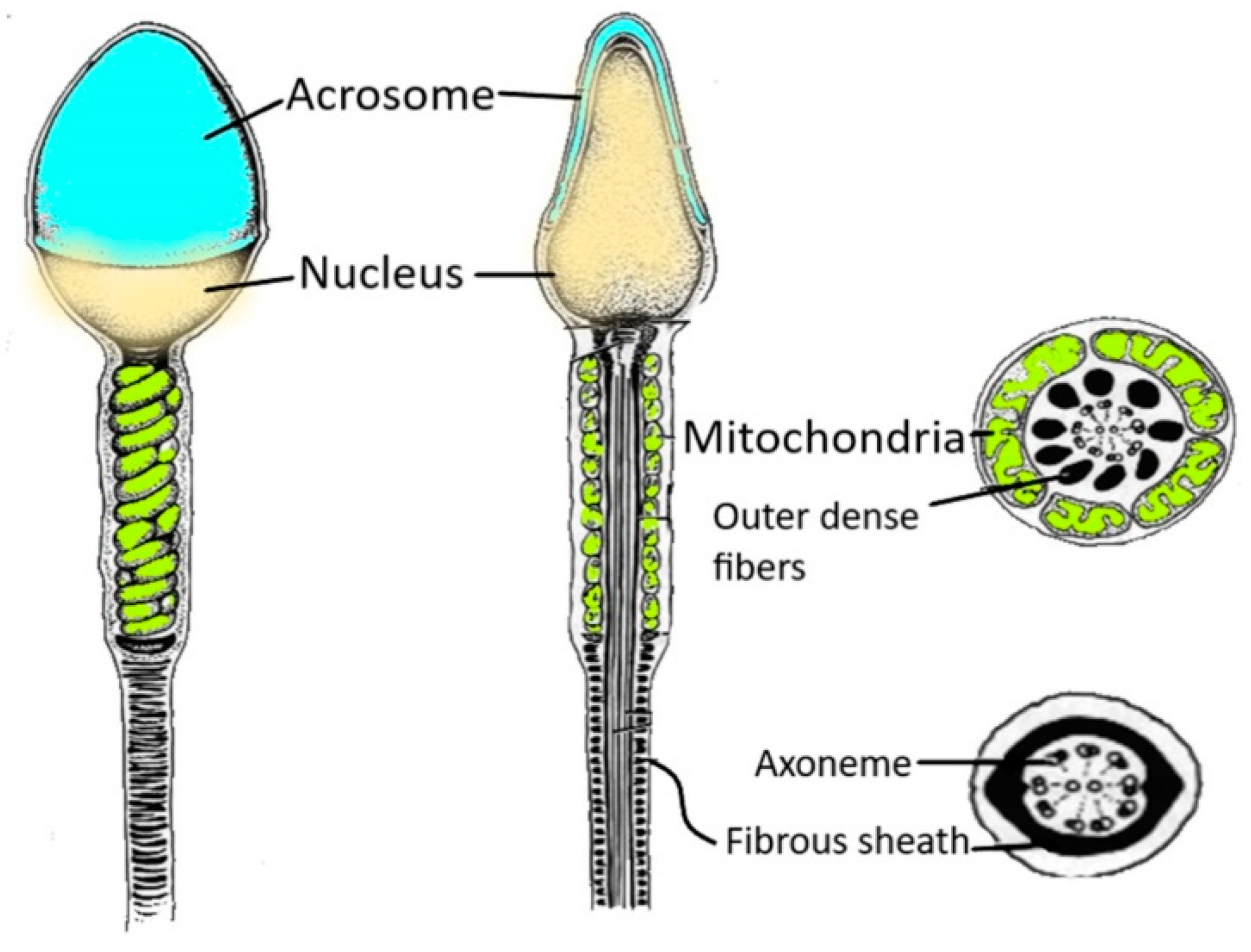
Figure 1. Spermatozoon structure. The figure shows the various regions of a human spermatozoon. Starting from the head region, it is possible to distinguish the acrosome and the nucleus. The sperm flagellum contains the axoneme and periaxonemal structures such as outer dense fibres and a fibrous sheath.
The head has an oval and flattened shape, and it comprises the acrosome and the nucleus. The acrosome is a cap-like Golgi-derived vesicle delimited by inner and outer acrosomal membranes and covers about 75% of the head. The acrosome contains lytic enzymes such as hyaluronidase and acrosine, which are necessary for the fertilisation process [21]. Only capacitated sperm can interact with the cumulus oophorous and the zona pellucida of an oocyte, resulting in the acrosome reaction and allowing for sperm to penetrate and fertilise an egg. Sperm can undergo a spontaneous acrosomal reaction before reaching the egg, preventing successful fertilisation. Bowker et al. [22] demonstrated the involvement of a mechanism based on protein acetylation that protects bovine spermatozoa from a spontaneous acrosomal reaction.
The region located at the posterior edge of the acrosome is named the equatorial segment; it plays an important role in the fusion between sperm and egg membranes [23]. The head area underneath the acrosomal cap is called the post-acrosomal region. It is particularly important because it contains the phospholipase Cζ (PLCζ), which is widely considered to be the sperm oocyte activation factor through the stimulation of Ca2+ oscillations in the oocyte [24][25].
The core of the sperm head is the nucleus, which contains highly condensed chromatin. During the sperm maturation process, about 85% of histones are replaced by protamines (small proteins rich in arginine) characterised by disulphide bridges between the sulfhydryl groups of cysteines [26][27]. The replacement of histones with protamines allows for dense packaging of chromatin; this organisation plays a pivotal role in protecting the genetic material devoted to the perpetuation of the species.
The head is linked to the tail by the connecting piece, a sort of neck made up of nine striated columns closed by the capitulum, a protein structure that interacts with the head [28]. This protein box encases the proximal centriole, under the nucleus, that shows a typical barrel shape with nine triplets of microtubules and an atypical distal centriole composed of splayed microtubules [29] and originates the axoneme, which forms the core of the entire flagellum (Figure 1). The axonemal structure is regulated by hundreds of microtubule-associated proteins and motor proteins, some of which form helical structures within the microtubule lumen that have been hypothesised to be involved in controlling the direction of sperm motion [30].
The midpiece is the flagellar region surrounded by a mitochondrial helix (Figure 1) that envelopes the axoneme and the nine outer dense fibres (ODFs). Mitochondria play a central role in sperm metabolism and are involved in energy production, redox balance, calcium regulation and apoptotic pathways, all of which are necessary for flagellar motility, capacitation, the acrosome reaction and fertilisation [31]. The midpiece ends with the annulus, a septin-based ring structure located beneath the plasma membrane that connects the midpiece and the principal piece of the mammalian sperm flagellum [32]. The annulus is probably involved in the flagellar assembly during spermiogenesis and confines proteins to define the different regions of the tail in mature sperm.
The principal piece, the longest part of the flagellum, contains the axoneme, ODFs and the fibrous sheath (FS) that wraps the axoneme along the entire length of the segment. The FS is a sperm-specific cytoskeletal structure that acts as a scaffold for enzymes involved in signal transduction and glycolytic pathway [33]. Therefore, the human sperm tail shows a complex anatomy, and every structure (proximal and distal centrioles, mitochondria, the axoneme, ODFs and the FS) plays a significant role in motility. Testicular spermatozoa are immature cells that are unable to fertilize an oocyte. After leaving the testis, spermatozoa transit along the epididymis to acquire motility and fertilizing abilities. During post-testicular maturation, many changes that regard a sperm membrane and sperm proteome profile occur [34]. At this purpose, a peculiar role is played by the epididymosomes, exosomes produced by epididymis that prime spermatozoa with a large amount of proteins and RNAs [35].
3. Sperm and ROS: An Ambivalent Relationship
Spermatozoa represent a perfect example of the oxygen paradox [36][37]. Indeed, oxygen homeostasis and the maintenance of a redox steady state are critical for spermatozoa. Spermatozoa generate ROS because they need them for several physiological processes (Figure 2). On the other hand, when ROS production overcomes the antioxidant defences, there are detrimental effects to the sperm membrane, proteins and DNA (Figure 2), which have a negative impact on male fertility [38].
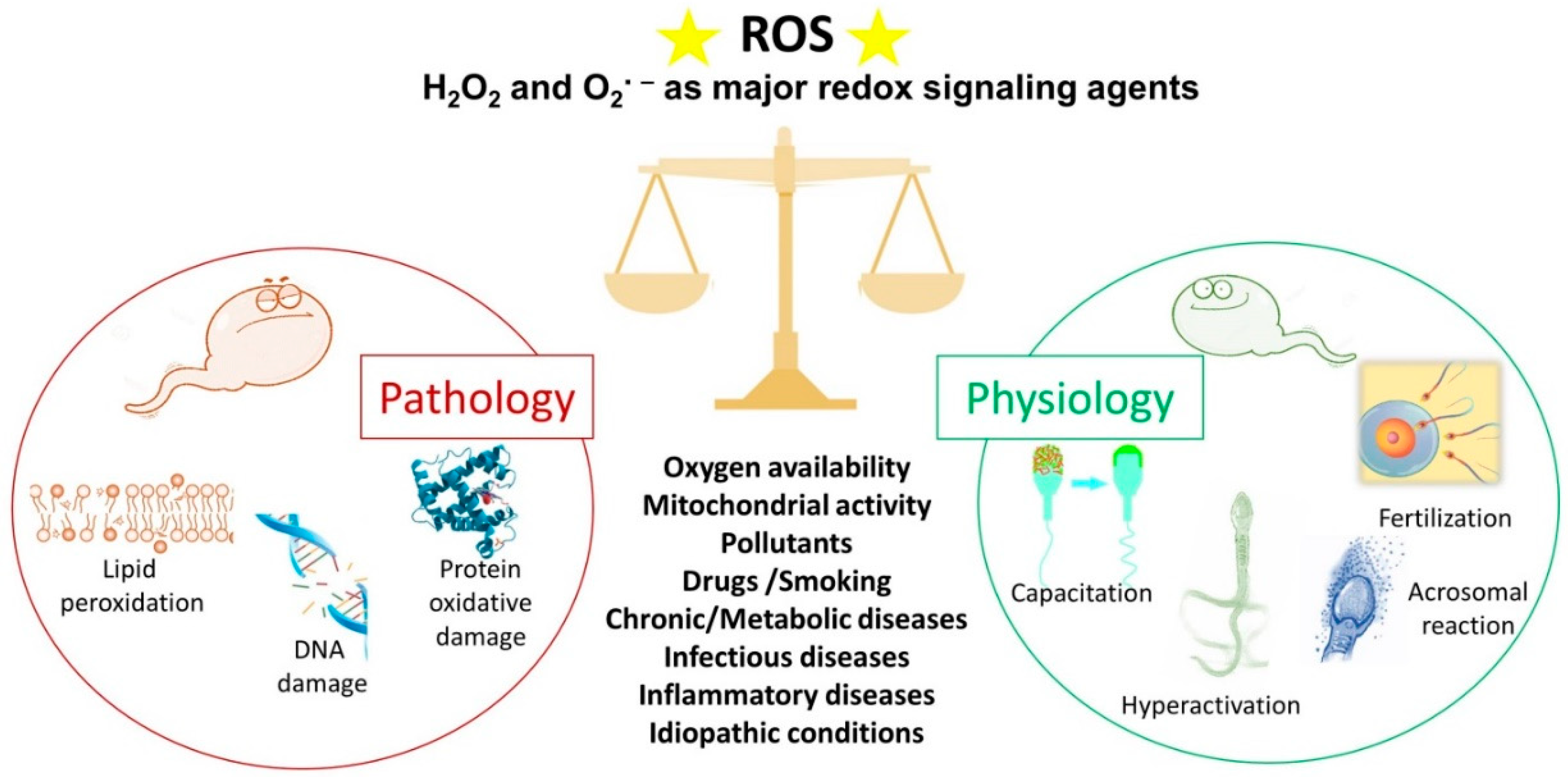
Figure 2. The double face of reactive oxygen species (ROS). Under physiological conditions, ROS regulate the sperm mechanisms involved in capacitation, hyperactivation, the acrosome reaction and fertilisation. On the contrary, some pathophysiological conditions can increase ROS levels over the physiological threshold, generating a condition of oxidative stress that leads to lipid peroxidation, DNA damage and protein oxidative damage. The stars represent ROS.
3.1. ROS and Sperm Physiology
ROS play an important role in sperm physiology because they regulate several intracellular pathways, thus modulating the activation of different transcription factors [38][39][40].
First, ROS are responsible for the stability and compaction of sperm chromatin during epididymal transit and storage. They act as oxidising agents and allow for the formation of disulphide bonds between cysteine residues of protamines, arginine-rich proteins that replace histones during spermiogenesis. Chromatin folding is a crucial event in protecting the paternal genome as the spermatozoa travel through the female reproductive tract [27].
One of the most important roles of ROS in sperm physiology (Figure 2) concerns the process of capacitation by which spermatozoa undergo dramatic changes in the membrane composition and acquire hyperactivated motility, leading to the acrosome reaction and fertilisation [41][42]. The molecular processes behind capacitation include an increase in pH, Ca2+ and HCO3− influx, efflux of cholesterol from the plasma membrane, an increase in cyclic adenosine monophosphate (cAMP) concentration; and protein hyper-phosphorylation [43][44]. During capacitation, the concentration of O2•−, H2O2, nitric oxide and peroxynitrite increase progressively. These ROS stimulate the activation of adenylate cyclase that drives the increase in cAMP. This second messenger activates protein kinase A (PKA) that triggers a massive tyrosine phosphorylation cascade, inhibiting tyrosine phosphatase [45][46]. Redox signalling occurs alongside modification of thiol groups of proteins of the plasma membrane and the inner sperm compartments [41]. As a part of the capacitation process, the hyperactivated motility triggered by calcium signalling [47] is characterised by a high amplitude and extremely asymmetrical beating pattern of sperm flagellum, as well as lateral head displacement. These motility modifications are dependent on ROS-mediated tyrosine phosphorylation of flagellar protein and are needed to make spermatozoa that can penetrate the cumulus oophorous and zona pellucida and fertilise the oocyte [48][49]. In addition, the acrosome reaction, the universal requisite for sperm–egg fusion, is influenced by ROS [50] that increase membrane fluidity. In fact, spermatozoa exposed to H2O2 show an enhanced acrosome reaction and an increased ability to fuse with oocyte [51]. Finally, the involvement of ROS in sperm function and physiology is supported by the observations that antioxidants can alter sperm maturation and, in particular, catalase or superoxide dismutase can inhibit sperm capacitation and the acrosome reaction [52].
3.2. ROS and Sperm Pathology
In recent decades, OS has emerged as a one of the main causes of altered sperm function [53]. Spermatozoa represent an easy target for free radical attack due to the high content of unsaturated fatty acids in their membranes, the limited ability to repair DNA damage and the virtual lack of cytoplasm. Moreover, spermatozoa are poor in antioxidant enzymes such as superoxide dismutase, catalase and glutathione peroxidase, as well as peroxiredoxins [54] that protect most cells from oxidative damage. To compensate the scarce presence of cellular antioxidants, the seminal plasma is rich in antioxidant enzymes and radical scavengers, among them glutathione peroxidase, glutathione-S-transferase, catalase and superoxide dismutase [55]. In addition, other hydrophilic compounds as uric acid, hypotaurine, tyrosine, polyphenols, vitamin C, ergothioneine and glutathione, and hydrophobic scavengers as trans-retinoic acids, trans-retinols, α-tocopherol, carotenoids and coenzyme Q10 are present [56].
The antioxidant properties of seminal plasma are important to balance the presence of ROS that are not just from the physiological production by spermatozoa. Human semen contain various amounts of immature spermatozoa, germinal cells, leucocytes, macrophages and epithelial cells. Among them, the main contributors to OS are leucocytes and immature spermatozoa with large cytoplasmic residues, but in physiological conditions, the redox balance is maintained by antioxidants contained in seminal plasma [57]. However, leucocytospermia occurs when the peroxidase-positive leucocyte concentration exceeds 1 × 10⁶/mL [1]. In this pathology, leucocytes produce a hundred times as much ROS as what they would in physiological conditions and the antioxidant power of the seminal plasma is not sufficient to counteract free radicals, leading to OS [58].
Immature spermatozoa fail to extrude the cytoplasm during maturation and the residual cytoplasm allows for the production of NADPH from glucose-6-phosphate (G6PDH) via the hexose monophosphate shunt [59][60]. NADPH generates ROS by using two different pathways: via NADPH oxidase, a membrane bound enzyme that produces the O2•− by oxygen, and via NADPH dehydrogenase, which is responsible for redox reactions in the mitochondria. The enhanced ROS production triggered by immature spermatozoa is responsible for OS propagation to maturing normal spermatozoa during epididymal transit [61].
During recent decades, a growing body of evidence has revealed the role of altered redox balance in seminal plasma, sperm alterations and male infertility [12][60][62]. OS is enhanced in situations when non-physiological ROS levels overwhelm the natural scavenger systems. These situations can be represented by primary pathologies affecting the male reproductive system, including varicocele [63], bacterial and viral infections, inflammation and leucocytospermia; chronic pathologies such as diabetes and cancer [60][64]; and environmental and lifestyle factors such as use of drugs, smoking, pollution and radiation (Figure 2). In these conditions, the unconjugated double bonds of PUFA in the sperm membrane are attacked by ROS, producing lipid hydroperoxides and its secondary decomposition product, aldehydes [59]. These highly reactive by-products produced by lipid peroxidation react with proteins and DNA and alter the proteins of the electron transport chain to induce mitochondrial dysfunction, enhancing the production of mitochondrial ROS in a self-perpetrating mechanism [9][59][65]. The most evident effect of OS and lipid peroxidation on spermatozoa is the loss of motility by inhibiting energy generation and the decrease in vitality as observed when sperm are frozen and thawed, two processes that boost ROS production [59]. The relevance of lipid oxidative damage to sperm conditions has been highlighted by the detection, quantification or immunolocalization of the specific end products of lipid peroxidation (aldehydes and oxygenated metabolites of PUFA) [66][67].
OS is a major cause of DNA damage in mammalian spermatozoa. Aitken and De Iuliis [68] proposed a two-step mechanism for the origin of oxidative DNA damage in spermatozoa. The first phase occurs during spermiogenesis and leads to defective protamination and compaction of sperm chromatin, making DNA more vulnerable to ROS attack. In normal conditions, chromatin compaction is mandatory to protect paternal DNA—this stabilisation makes the spermatozoa resistant to oxidative damage. The second phase is referred to a direct oxidative insult on the DNA due to increased ROS generation by sperm and a loss of extracellular antioxidant protection. The evident ROS effects on sperm nuclear DNA include DNA fragmentation, chromatin cross-linking, base-pair modifications and chromosomal microdeletions [11].
In addition, paternal aging plays a negative effect on sperm parameters and induces a ROS-related DNA fragmentation. These alterations negatively influence the reproductive outcome and offspring health documented for cancer, genetic and congenital diseases, chromosomal alterations and others [69].
4. Human Spermatozoa as a Model for In Vitro Studies
There are many reasons why human spermatozoa represent a model for in vitro studies (Figure 3). First, the easy collection of spermatozoa from fertile men can guarantee abundant cellular material. Spermatozoa are differentiated cells with features and specific functions that enable them to reach the oocyte and to fertilise it. The first peculiar characteristic of spermatozoa is their motility, which enables them to reach and fertilise the oocyte. This motility is guaranteed by the ability to obtain energy by both mitochondrial oxidative phosphorylation and glycolysis, the enzymes for which are located along the FS [33].
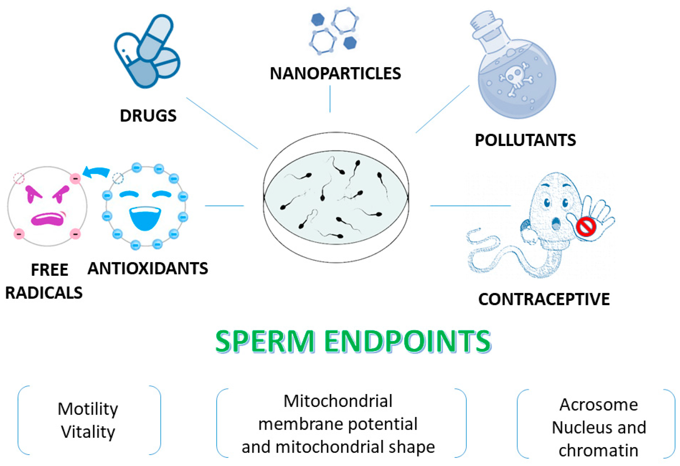
Figure 3. Human sperm as an in vitro model to test various compounds. Sperm endpoints such as motility, vitality, mitochondrial membrane potential, the acrosome status and DNA integrity can be evaluated when human spermatozoa are used as an in vitro model to test effects of toxic compounds, natural extracts and molecules.
Spermatozoa were supposed to be incapable of transcription and translation; however, several types of coding and non-coding RNAs have been identified. These RNAs derive from the testis, epididymis, but also by spermatozoa themselves, meaning that also the sperm condensed chromatin can be partially transcribed [70].
However, due to the low level of transcription, sperm are deeply influenced by the external environment [71]. Human spermatozoa do not possess DNA-repair activity. When fertilisation occurs, DNA-repair activity depends on the oocyte transcripts that had been stored during maturation [72]. Finally, due to an intrinsic lack of intracellular antioxidant protection, human spermatozoa have a limited capacity to repair oxidative damage [12].
These aforementioned characteristics make spermatozoa a general and ideal cell model to test in vitro many compounds at different concentrations related and unrelated to the reproductive field (Figure 3). Indeed, many antioxidants [73][74][75][76][77][78][79] have been tested using this model. In addition, spermatozoa have been used as an in vitro monitor of toxicity due to many compounds including natural substances that have potential contraceptive activity; thus, this research has aimed to identify promising products that could be used as vaginal contraceptive agents [80][81][82][83].
There is a broad group of studies dealing with human sperm as model for testing the potential toxic effects of pollutants as heavy metals and phthalates [84][85][86], nanoparticles [87][88][89], herbicides [90] and drugs [91][92][93]. When these protocols to study the effects of spermatozoa exposure to compounds of interest are applied, it is important to evaluate how the different sperm structures react to the treatment (Figure 3).
The functional status of the acrosome can be tested by assessing several molecular biomarkers such as acrosin, equatorin, A disintegrin and metalloprotease 3 (ADAM3) and others [94]. However, fluorescently labelled lectins, such as Arachis hypogaea agglutinin (PNA, peanut agglutinin), Pisum sativum (PSA) and Canavalia ensiformis (Con A), represent an easy and inexpensive method to visualise the morphology of the acrosome and the acrosome reaction to determine the percentage of spermatozoa undergoing exocytosis upon stimulation [95]. Lectins interact with specific carbohydrates and provide information on the morphology of the acrosome rather than on the molecules that function during the fertilisation process. The analysis can be performed with flow cytometry or by scoring cells using a light microscope equipped with a fluorescence apparatus.
Another endpoint that can be evaluated after in vitro treatment of spermatozoa is DNA integrity. The World Health Organization (WHO) guidelines [1] report the most common tests to assess sperm DNA integrity. The direct techniques are terminal deoxynucleotidyl transferase deoxyuridine triphosphate (dUTP) nick-end labelling (TUNEL) and a single-cell gel electrophoresis assay (comet assay). TUNEL enables detecting DNA fragmentation by labelling the 3′-OH generated by the breaks with fluorescent nucleotides. The comet assay is a gel electrophoresis-based method that can be used to measure DNA damage in individual spermatozoa. An indirect test based on acridine orange measures the susceptibility of the DNA to denaturation under acidic conditions. The evaluation can be performed by flow cytometry (sperm chromatin structure assay [SCSA]) or by fluorescence microscopy [96].
Mitochondria, the hallmark of the sperm tail midpiece, control motility, ROS production, redox equilibrium and calcium regulation, represent an important indicator of the health status of spermatozoa. An easy and fast way to measure the mitochondrial membrane potential is the use of the fluorescent cationic dye 5,5,6,6′-tetrachloro-1,1′,3,3′-tetraethylbenzimi-dazoylcarbocyanine iodide (JC-1) that shows membrane potential-dependent accumulation in the mitochondria [97]. JC-1 forms J-aggregates and fluoresces red when the mitochondrial membrane potential is high, while it remains in its monomeric state, emitting green fluorescence, when the membrane mitochondrial potential is low.
Finally, sperm motility is one of the most important indices of cell function. A reduction in motility indicates the presence of damage caused by the compounds used in in vitro studies. Analysis of sperm motility is easy and cheap: it requires a light microscope and a cell-counting chamber [1]. In addition, a sophisticated method such as computer-assisted sperm analysis (CASA) systems represents a great tool that has the ability to provide the rapid, reliable and objective quantitative assessment of sperm kinematic characteristics [98].
Vitality should be assessed concomitantly with motility, particularly when the in vitro treatment reduces this parameter drastically. The evaluation of both motility and vitality allows for clarifying whether the immotile spermatozoa are dead or alive, and this is important to understand whether the substance under study has an impact on sperm motility or if it is able to kill the spermatozoa. Eosin-nigrosin stain and the hypoosmotic swelling test are the most commonly applied assays. Eosin-nigrosin staining is based on the integrity of the plasma membrane: dead spermatozoa are stained because the membrane is disrupted. The hypoosmotic swelling test is based on the semi-permeable properties of the plasma membrane: living spermatozoa with intact membranes swell in hypotonic solutions [99].
References
- World Health Organization. WHO Laboratory Manual for the Examination and Processing of Human Semen, 6th ed.; WHO Press: Geneva, Switzerland, 2021.
- Agarwal, A.; Baskaran, S.; Parekh, N.; Cho, C.L.; Henkel, R.; Vij, S.; Arafa, M.; Panner Selvam, M.K.; Shah, R. Male infertility. Lancet 2021, 397, 319–333.
- Nieschlag, E.; Lenzi, A. The conventional management of male infertility. Int. J. Gynaecol. Obstet. 2013, 123 (Suppl. S2), S31–S35.
- Leslie, S.W.; Soon-Sutton, T.L.; Khan, M.A.B. Male infertility. In StatPearls; StatPearls Publishing: Treasure Island, FL, USA, 2022.
- Wang, K.; Gao, Y.; Wang, C.; Liang, M.; Liao, Y.; Hu, K. Role of oxidative stress in varicocele. Front. Genet. 2022, 13, 850114.
- Pellati, D.; Mylonakis, I.; Bertoloni, G.; Fiore, C.; Andrisani, A.; Ambrosini, G.; Armanini, D. Genital tract infections and infertility. Eur. J. Obstet. Gynecol. Reprod. Biol. 2008, 140, 3–11.
- Wang, S.; Zhang, K.; Yao, Y.; Li, J.; Deng, S. Bacterial infections affect male fertility: A focus on the oxidative stress-autophagy axis. Front. Cell Dev. Biol. 2021, 9, 727812.
- Walczak-Jedrzejowska, R.; Wolski, J.K.; Slowikowska-Hilczer, J. The role of oxidative stress and antioxidants in male fertility. Cent. Eur. J. Urol. 2013, 66, 60–67.
- Gualtieri, R.; Kalthur, G.; Barbato, V.; Longobardi, S.; Di Rella, F.; Adiga, S.K.; Talevi, R. Sperm oxidative stress during in vitro manipulation and its effects on sperm function and embryo development. Antioxidants 2021, 10, 1025.
- Aitken, R.J.; Gibb, Z.; Baker, M.A.; Drevet, J.; Gharagozloo, P. Causes and consequences of oxidative stress in spermatozoa. Reprod. Fertil. Dev. 2016, 28, 1–10.
- Dutta, S.; Majzoub, A.; Agarwal, A. Oxidative stress and sperm function: A systematic review on evaluation and management. Arab J. Urol. 2019, 17, 87–97.
- Aitken, R.J.; Drevet, J.R.; Moazamian, A.; Gharagozloo, P. Male infertility and oxidative stress: A focus on the underlying mechanisms. Antioxidants 2022, 11, 306.
- Ford, W.C. Regulation of sperm function by reactive oxygen species. Hum. Reprod. Update 2004, 10, 387–399.
- Wright, C.; Milne, S.; Leeson, H. Sperm DNA damage caused by oxidative stress: Modifiable clinical, lifestyle and nutritional factors in male infertility. Reprod. Biomed. Online 2014, 28, 684–703.
- Lone, S.A.; Mohanty, T.K.; Baithalu, R.K.; Yadav, H.P. Sperm protein carbonylation. Andrologia 2019, 51, e13233.
- Agarwal, A.; Maldonado Rosas, I.; Anagnostopoulou, C.; Cannarella, R.; Boitrelle, F.; Munoz, L.V.; Finelli, R.; Durairajanayagam, D.; Henkel, R.; Saleh, R. Oxidative stress and assisted reproduction: A comprehensive review of its pathophysiological role and strategies for optimizing embryo culture environment. Antioxidants 2022, 11, 477.
- Martin-Hidalgo, D.; Bragado, M.J.; Batista, A.R.; Oliveira, P.F.; Alves, M.G. Antioxidants and male fertility: From molecular studies to clinical evidence. Antioxidants 2019, 8, 89.
- Symeonidis, E.N.; Evgeni, E.; Palapelas, V.; Koumasi, D.; Pyrgidis, N.; Sokolakis, I.; Hatzichristodoulou, G.; Tsiampali, C.; Mykoniatis, I.; Zachariou, A.; et al. Redox balance in male infertility: Excellence through moderation—“Μέτρον ἄριστον”. Antioxidants 2021, 10, 1534.
- Amidi, F.; Pazhohan, A.; Shabani Nashtaei, M.; Khodarahmian, M.; Nekoonam, S. The role of antioxidants in sperm freezing: A review. Cell Tissue Bank 2016, 17, 745–756.
- Pasquariello, R.; Verdile, N.; Brevini, T.A.L.; Gandolfi, F.; Boiti, C.; Zerani, M.; Maranesi, M. The role of resveratrol in mammalian reproduction. Molecules 2020, 25, 4554.
- Abou-Haila, A.; Tulsiani, D.R. Mammalian sperm acrosome: Formation, contents, and function. Arch. Biochem. Biophys. 2000, 379, 173–182.
- Bowker, Z.; Goldstein, S.; Breitbart, H. Protein acetylation protects sperm from spontaneous acrosome reaction. Theriogenology 2022, 191, 231–238.
- Fujihara, Y.; Murakami, M.; Inoue, N.; Satouh, Y.; Kaseda, K.; Ikawa, M.; Okabe, M. Sperm equatorial segment protein 1, SPESP1, is required for fully fertile sperm in mouse. J. Cell Sci. 2010, 123 Pt 9, 1531–1536.
- Kashir, J.; Heindryckx, B.; Jones, C.; De Sutter, P.; Parrington, J.; Coward, K. Oocyte activation, phospholipase C zeta and human infertility. Hum. Reprod. Update 2010, 16, 690–703.
- Saleh, A.; Kashir, J.; Thanassoulas, A.; Safieh-Garabedian, B.; Lai, F.A.; Nomikos, M. Essential role of sperm-specific PLC-zeta in egg activation and male factor infertility: An update. Front. Cell Dev. Biol. 2020, 8, 28.
- Ward, W.S. Organization of sperm DNA by the nuclear matrix. Am. J. Clin. Exp. Urol. 2018, 6, 87–92.
- Ribas-Maynou, J.; Nguyen, H.; Wu, H.; Ward, W.S. Functional aspects of sperm chromatin organization. Results Probl. Cell Differ. 2022, 70, 295–311.
- Chemes, H.E.; Rawe, V.Y. The making of abnormal spermatozoa: Cellular and molecular mechanisms underlying pathological spermiogenesis. Cell Tissue Res. 2010, 341, 349–357.
- Avidor-Reiss, T.; Carr, A.; Fishman, E.L. The sperm centrioles. Mol. Cell Endocrinol. 2020, 518, 110987.
- Zabeo, D.; Heumann, J.M.; Schwartz, C.L.; Suzuki-Shinjo, A.; Morgan, G.; Widlund, P.O.; Höög, J.L. A lumenal interrupted helix in human sperm tail microtubules. Sci. Rep. 2018, 8, 2727.
- Boguenet, M.; Bouet, P.E.; Spiers, A.; Reynier, P.; May-Panloup, P. Mitochondria: Their role in spermatozoa and in male infertility. Hum. Reprod. Update 2021, 27, 697–719.
- Toure, A.; Rode, B.; Hunnicutt, G.R.; Escalier, D.; Gacon, G. Septins at the annulus of mammalian sperm. Biol. Chem. 2011, 392, 799–803.
- Kim, Y.H.; Haidl, G.; Schaefer, M.; Egner, U.; Mandal, A.; Herr, J.C. Compartmentalization of a unique ADP/ATP carrier protein SFEC (Sperm Flagellar Energy Carrier, AAC4) with glycolytic enzymes in the fibrous sheath of the human sperm flagellar principal piece. Dev. Biol. 2007, 302, 463–476.
- James, E.R.; Carrell, D.T.; Aston, K.I.; Jenkins, T.G.; Yeste, M.; Salas-Huetos, A. The Role of the Epididymis and the Contribution of Epididymosomes to Mammalian Reproduction. Int. J. Mol. Sci. 2020, 21, 5377.
- Barrachina, F.; Battistone, M.A.; Castillo, J.; Mallofré, C.; Jodar, M.; Breton, S.; Oliva, R. Sperm acquire epididymis-derived proteins through epididymosomes. Hum. Reprod. 2022, 37, 651–668.
- Sies, H. Strategies of antioxidant defense. Eur. J. Biochem. 1993, 215, 213–219.
- Davies, K.J. Oxidative stress: The paradox of aerobic life. Biochem. Soc. Symp. 1995, 61, 1–31.
- Mannucci, A.; Argento, F.R.; Fini, E.; Coccia, M.E.; Taddei, N.; Becatti, M.; Fiorillo, C. The impact of oxidative stress in male infertility. Front. Mol. Biosci. 2022, 8, 799294.
- Burton, G.J.; Jauniaux, E. Oxidative stress. Best Pract. Res. Clin. Obstet. Gynaecol. 2011, 25, 287–299.
- Takei, G.L.; Tourzani, D.A.; Paudel, B.; Visconti, P.E. Activation of cAMP-dependent phosphorylation pathways is independent of ROS production during mouse sperm capacitation. Mol. Reprod. Dev. 2021, 88, 544–557.
- O’Flaherty, C. Redox regulation of mammalian sperm capacitation. Asian J. Androl. 2015, 17, 583–590.
- De Jonge, C. Biological basis for human capacitation-revisited. Hum. Reprod. Update 2017, 23, 289–299.
- Carrasquel Martínez, G.; Aldana, A.; Carneiro, J.; Treviño, C.L.; Darszon, A. Acrosomal alkalinization occurs during human sperm capacitation. Mol. Hum. Reprod. 2022, 28, gaac005.
- Delgado-Bermúdez, A.; Yeste, M.; Bonet, S.; Pinart, E. A review on the role of bicarbonate and proton transporters during sperm capacitation in mammals. Int. J. Mol. Sci. 2022, 23, 6333.
- Herrero, M.B.; Chatterjee, S.; Lefièvre, L.; de Lamirande, E.; Gagnon, C. Nitric oxide interacts with the cAMP pathway to modulate capacitation of human spermatozoa. Free Radic. Biol. Med. 2000, 29, 522–536.
- O’Flaherty, C.; de Lamirande, E.; Gagnon, C. Positive role of reactive oxygen species in mammalian sperm capacitation: Triggering and modulation of phosphorylation events. Free Radic. Biol. Med. 2006, 41, 528–540.
- Cordero-Martínez, J.; Jimenez-Gutierrez, G.E.; Aguirre-Alvarado, C.; Alacántara-Farfán, V.; Chamorro-Cevallos, G.; Roa-Espitia, A.L.; Hernández-González, E.O.; Rodríguez-Páez, L. Participation of signaling proteins in sperm hyperactivation. Syst. Biol. Reprod. Med. 2022, 68, 315–330.
- de Lamirande, E.; Gagnon, C. Human sperm hyperactivation and capacitation as parts of an oxidative process. Free Radic. Biol. Med. 1993, 14, 157–166.
- Suarez, S.S. Control of hyperactivation in sperm. Hum. Reprod. Update 2008, 14, 647–657.
- de Lamirande, E.; Tsai, C.; Harakat, A.; Gagnon, C. Involvement of reactive oxygen species in human sperm arcosome reaction induced by A23187, lysophosphatidylcholine, and biological fluid ultrafiltrates. J. Androl. 1998, 19, 585–594.
- Rivlin, J.; Mendel, J.; Rubinstein, S.; Etkovitz, N.; Breitbart, H. Role of hydrogen peroxide in sperm capacitation and acrosome reaction. Biol. Reprod. 2004, 70, 518–522.
- Wagner, H.; Cheng, J.W.; Ko, E.Y. Role of reactive oxygen species in male infertility: An updated review of literature. Arab J. Urol. 2017, 16, 35–43.
- Aitken, R.J.; Drevet, J.R. The importance of oxidative stress in determining the functionality of mammalian spermatozoa: A two-edged sword. Antioxidants 2020, 9, 111.
- O’Flaherty, C. Peroxiredoxin 6: The protector of male fertility. Antioxidants 2018, 7, 173.
- Micheli, L.; Cerretani, D.; Collodel, G.; Menchiari, A.; Moltoni, L.; Fiaschi, A.I.; Moretti, E. Evaluation of enzymatic and non-enzymatic antioxidants in seminal plasma of men with genitourinary infections, varicocele and idiopathic infertility. Andrology 2016, 4, 456–464.
- Lazzarino, G.; Listorti, I.; Bilotta, G.; Capozzolo, T.; Amorini, A.M.; Longo, S.; Caruso, G.; Lazzarino, G.; Tavazzi, B.; Bilotta, P. Water- and fat-soluble antioxidants in human seminal plasma and serum of fertile males. Antioxidants 2019, 8, 96.
- Aitken, R.J.; Baker, M.A. Oxidative stress, spermatozoa and leukocytic infiltration: Relationships forged by the opposing forces of microbial invasion and the search for perfection. J. Reprod. Immunol. 2013, 100, 11–19.
- Henkel, R.R. Leukocytes and oxidative stress: Dilemma for sperm function and male fertility. Asian J. Androl. 2011, 13, 43–52.
- Aitken, R.J. Reactive oxygen species as mediators of sperm capacitation and pathological damage. Mol. Reprod. Dev. 2017, 84, 1039–1052.
- Agarwal, A.; Rana, M.; Qiu, E.; AlBunni, H.; Bui, A.D.; Henkel, R. Role of oxidative stress, infection and inflammation in male infertility. Andrologia 2018, 50, e13126.
- Gil-Guzman, E.; Ollero, M.; Lopez, M.C.; Sharma, R.K.; Alvarez, J.G.; Thomas, A.J., Jr.; Agarwal, A. Differential production of reactive oxygen species by subsets of human spermatozoa at different stages of maturation. Hum. Reprod. 2001, 16, 1922–1930.
- Villaverde, A.I.S.B.; Netherton, J.; Baker, M.A. From past to present: The link between reactive oxygen species in sperm and male infertility. Antioxidants 2019, 8, 616.
- Wood, G.J.A.; Cardoso, J.P.G.; Paluello, D.V.; Nunes, T.F.; Cocuzza, M. Varicocele-associated infertility and the role of oxidative stress on sperm DNA fragmentation. Front. Reprod. Health 2021, 3, 695992.
- Signorini, C.; Moretti, E.; Noto, D.; Micheli, L.; Ponchia, R.; Collodel, G. Fatty acid oxidation and pro-resolving lipid mediators are related to male infertility. Antioxidants 2022, 11, 107.
- Aitken, R.J.; Whiting, S.; De Iuliis, G.N.; McClymont, S.; Mitchell, L.A.; Baker, M.A. Electrophilic aldehydes generated by sperm metabolism activate mitochondrial reactive oxygen species generation and apoptosis by targeting succinate dehydrogenase. J. Biol. Chem. 2012, 287, 33048–33060.
- Moretti, E.; Cerretani, D.; Noto, D.; Signorini, C.; Iacoponi, F.; Collodel, G. Relationship between semen IL-6, IL-33 and malondialdehyde generation in human seminal plasma and spermatozoa. Reprod. Sci. 2021, 28, 2136–2143.
- Moretti, E.; Signorini, C.; Ferretti, F.; Noto, D.; Collodel, G. A study to validate the relevance of semen F2-isoprostanes on human male infertility. Int. J. Environ. Res. Public Health 2022, 19, 1642.
- Aitken, R.J.; De Iuliis, G.N. On the possible origins of DNA damage in human spermatozoa. Mol. Hum. Reprod. 2010, 16, 3–13.
- Jimbo, M.; Kunisaki, J.; Ghaed, M.; Yu, V.; Flores, H.A.; Hotaling, J.M. Fertility in the aging male: A systematic review. Fertil. Steril. 2022, 118, 1022–1034.
- Santiago, J.; Silva, J.V.; Howl, J.; Santos, M.A.S.; Fardilha, M. All you need to know about sperm RNAs. Hum. Reprod. Update 2021, 28, 67–91.
- Vollmer, T.; Ljungberg, B.; Jankowski, V.; Jankowski, J.; Glorieux, G.; Stegmayr, B.G. An in-vitro assay using human spermatozoa to detect toxicity of biologically active substances. Sci. Rep. 2019, 9, 14525.
- Setti, A.S.; Braga, D.P.A.F.; Provenza, R.R.; Iaconelli, A., Jr.; Borges, E., Jr. Oocyte ability to repair sperm DNA fragmentation: The impact of maternal age on intracytoplasmic sperm injection outcomes. Fertil. Steril. 2021, 116, 123–129.
- Collodel, G.; Federico, M.G.; Geminiani, M.; Martini, S.; Bonechi, C.; Rossi, C.; Figura, N.; Moretti, E. Effect of trans-resveratrol on induced oxidative stress in human sperm and in rat germinal cells. Reprod. Toxicol. 2011, 31, 239–246.
- Eskandari, F.; Momeni, H.R. Silymarin protects plasma membrane and acrosome integrity in sperm treated with sodium arsenite. Int. J. Reprod. Biomed. 2016, 14, 47–52.
- Zhang, L.; Diao, R.Y.; Duan, Y.G.; Yi, T.H.; Cai, Z.M. In vitro antioxidant effect of curcumin on human sperm quality in leucocytospermia. Andrologia 2017, 49, e12760.
- Noto, D.; Collodel, G.; Cerretani, D.; Signorini, C.; Gambera, L.; Menchiari, A.; Moretti, E. Protective effect of chlorogenic acid on human sperm: In vitro studies and frozen-thawed protocol. Antioxidants 2021, 10, 744.
- Amirjannaty, S.; Gashti, N.G.; Mojtahedi, A.; Ashouri, A.; Bahadori, M.H. An in vitro study on the protective effect of melatonin on human sperm parameters treated by cadmium. J. Hum. Reprod. Sci. 2022, 15, 21–26.
- Mottola, F.; Iovine, C.; Carannante, M.; Santonastaso, M.; Rocco, L. In vitro combination of ascorbic and ellagic acids in sperm oxidative damage inhibition. Int. J. Mol. Sci. 2022, 23, 14751.
- Munir, N.; Mahmood, Z.; Shahid, M.; Afzal, M.N.; Jahangir, M.; Ali Shah, S.M.; Tahir, I.M.; Riaz, M.; Hussain, S.; Akram, M.; et al. Withania somnifera chemical constituents’ in vitro antioxidant potential and their response on spermatozoa parameters. Dose Response 2022, 20, 15593258221074936.
- Paul, S.; Kang, S.C. In vitro determination of the contraceptive spermicidal activity of essential oil of Trachyspermum ammi (L.) Sprague ex Turrill fruits. New Biotechnol. 2011, 28, 684–690.
- Fan, Y.; Chen, N.; Wang, M.; Rao, M.; Su, P. In vitro study evaluating the instantaneous treatment of ozonised olive oil on human sperm. Eur. J. Contracept. Reprod. Health Care 2018, 23, 147–153.
- Sha, W.; Chang, X.; Han, Y.; Miao, H.; Yang, Y. In vitro effect of ulipristal acetate on human sperm parameters and function. Pak. J. Pharm. Sci. 2019, 32, 1419–1422.
- Mondal, P.; Maity, R.; Mallick, C. In vitro spermicidal effect of Thevetia Peruviana leaves on human spermatozoa. Andrologia 2022, 54, e14323.
- Marchiani, S.; Tamburrino, L.; Farnetani, G.; Muratori, M.; Vignozzi, L.; Baldi, E. Acute effects on human sperm exposed in vitro to cadmium chloride and diisobutyl phthalate. Reproduction 2019, 158, 281–290.
- Xie, F.; Chen, X.; Weng, S.; Xia, T.; Sun, X.; Luo, T.; Li, P. Effects of two environmental endocrine disruptors di-n-butyl phthalate (DBP) and mono-n-butyl phthalate (MBP) on human sperm functions in vitro. Reprod. Toxicol. 2019, 83, 1–7.
- Chen, C.; Li, B.; Huang, R.; Dong, S.; Zhou, Y.; Song, J.; Zeng, X.; Zhang, X. Involvement of Ca2+ and ROS signals in nickel-impaired human sperm function. Ecotoxicol. Environ. Saf. 2022, 231, 113181.
- Moretti, E.; Terzuoli, G.; Renieri, T.; Iacoponi, F.; Castellini, C.; Giordano, C.; Collodel, G. In vitro effect of gold and silver nanoparticles on human spermatozoa. Andrologia 2013, 45, 392–396.
- Préaubert, L.; Tassistro, V.; Auffan, M.; Sari-Minodier, I.; Rose, J.; Courbiere, B.; Perrin, J. Very low concentration of cerium dioxide nanoparticles induce DNA damage, but no loss of vitality, in human spermatozoa. Toxicol. Vitr. 2018, 50, 236–241.
- Santonastaso, M.; Mottola, F.; Iovine, C.; Cesaroni, F.; Colacurci, N.; Rocco, L. In vitro effects of titanium dioxide nanoparticles (TiO2NPs) on cadmium chloride (CdCl2) genotoxicity in human sperm cells. Nanomaterials 2020, 10, 1118.
- Tan, Z.; Zhou, J.; Chen, H.; Zou, Q.; Weng, S.; Luo, T.; Tang, Y. Toxic effects of 2,4-dichlorophenoxyacetic acid on human sperm function in vitro. J. Toxicol. Sci. 2016, 41, 543–549.
- Xu, B.; Wang, Z.P.; Wang, Y.J.; Lu, P.H.; Wang, L.J.; Wang, X.H. The toxic effect of opioid analgesics on human sperm motility in vitro. Drug Chem. Toxicol. 2013, 36, 205–208.
- Pascarelli, N.A.; Fioravanti, A.; Moretti, E.; Guidelli, G.M.; Mazzi, L.; Collodel, G. The effects in vitro of TNF-α and its antagonist ‘etanercept’ on ejaculated human sperm. Reprod. Fertil. Dev. 2017, 29, 1169–1177.
- Ali Banihani, S.; Al-Khawalde, A.A. Omeprazole does not alter human sperm motility, viability or DNA integrity in vitro. Andrologia 2019, 51, e13260.
- Ito, C.; Toshimori, K. Acrosome markers of human sperm. Anat. Sci. Int. 2016, 91, 128–142.
- Zoppino, F.C.; Halón, N.D.; Bustos, M.A.; Pavarotti, M.A.; Mayorga, L.S. Recording and sorting live human sperm undergoing acrosome reaction. Fertil. Steril. 2012, 97, 1309–1315.
- Farkouh, A.; Salvio, G.; Kuroda, S.; Saleh, R.; Vogiatzi, P.; Agarwal, A. Sperm DNA integrity and male infertility: A narrative review and guide for the reproductive physicians. Transl. Androl. Urol. 2022, 11, 1023–1044.
- Carrageta, D.F.; Freire-Brito, L.; Oliveira, P.F.; Alves, M.G. Evaluation of human spermatozoa mitochondrial membrane potential using the JC-1 dye. Curr. Protoc. 2022, 2, e531.
- van der Horst, G.; Maree, L.; du Plessis, S.S. Current perspectives of CASA applications in diverse mammalian spermatozoa. Reprod. Fertil. Dev. 2018, 30, 875–888.
- Agarwal, A.; Sharma, R.K.; Gupta, S.; Boitrelle, F.; Finelli, R.; Parekh, N.; Durairajanayagam, D.; Saleh, R.; Arafa, M.; Cho, C.L.; et al. Sperm vitality and necrozoospermia: Diagnosis, management, and results of a global survey of clinical practice. World J. Mens Health 2022, 40, 228–242.
More
Information
Subjects:
Reproductive Biology
Contributors
MDPI registered users' name will be linked to their SciProfiles pages. To register with us, please refer to https://encyclopedia.pub/register
:
View Times:
1.8K
Revisions:
3 times
(View History)
Update Date:
24 May 2023
Table of Contents


Notice
You are not a member of the advisory board for this topic. If you want to update advisory board member profile, please contact office@encyclopedia.pub.
OK
Confirm
Only members of the Encyclopedia advisory board for this topic are allowed to note entries. Would you like to become an advisory board member of the Encyclopedia?
Yes
No
${ textCharacter }/${ maxCharacter }
Submit
Cancel
Back
Comments
${ item }
|
${ item.createdUser.fullName }
${ item.createdAt }
${ item.vote }
${ item.reply }
Delete
${ reply.createdUser.fullName }
${ reply.createdAt }
${ reply.vote }
Delete
There is no reply to this comment~
${ item.replyTextCharacter }/${ item.replyMaxCharacter }
Submit
Cancel
More
No more~
There is no comment~
${ textCharacter }/${ maxCharacter }
Submit
Cancel
${ selectedItem.replyTextCharacter }/${ selectedItem.replyMaxCharacter }
Submit
Cancel
Confirm
Are you sure to Delete?
Yes
No