Submitted Successfully!
To reward your contribution, here is a gift for you: A free trial for our video production service.
Thank you for your contribution! You can also upload a video entry or images related to this topic.
Version Summary Created by Modification Content Size Created at Operation
1 -- 2870 2023-05-03 16:47:21 |
2 layout Meta information modification 2870 2023-05-04 02:54:06 |

Video Upload Options

Do you have a full video?

Confirm

Are you sure to Delete?
Cite
If you have any further questions, please contact Encyclopedia Editorial Office.
Grochau-Wright, Z.I.; Nedelcu, A.M.; Michod, R.E. Volvocine regA Gene Model for Cellular Differentiation Evolution. Encyclopedia. Available online: https://encyclopedia.pub/entry/43708 (accessed on 14 May 2024).
Grochau-Wright ZI, Nedelcu AM, Michod RE. Volvocine regA Gene Model for Cellular Differentiation Evolution. Encyclopedia. Available at: https://encyclopedia.pub/entry/43708. Accessed May 14, 2024.
Grochau-Wright, Zachariah I., Aurora M. Nedelcu, Richard E. Michod. "Volvocine regA Gene Model for Cellular Differentiation Evolution" Encyclopedia, https://encyclopedia.pub/entry/43708 (accessed May 14, 2024).
Grochau-Wright, Z.I., Nedelcu, A.M., & Michod, R.E. (2023, May 03). Volvocine regA Gene Model for Cellular Differentiation Evolution. In Encyclopedia. https://encyclopedia.pub/entry/43708
Grochau-Wright, Zachariah I., et al. "Volvocine regA Gene Model for Cellular Differentiation Evolution." Encyclopedia. Web. 03 May, 2023.
Volvocine regA Gene Model for Cellular Differentiation Evolution
Edit

A group of green algae in the order of Volvocales provides an ideal model system for studying the transition from unicellular to differentiated multicellularity. This group—known as the volvocine algae—evolved multicellularity relatively recently (~240 million years ago) and contains extant relatives that span a range of complexities from unicellularity, to undifferentiated multicellularity, to differentiated multicellularity. The regA-like gene family within the volvocine algae serves as a model for the evolution of the genetic basis of cellular differentiation.

multicellularity cellular differentiation life history individuality gene co-option Volvox Chlamydomonas volvocine algae regA

1. The Volvocine Model System

The volvocine lineage is a group of freshwater haploid bi-flagellated chlorophyte green algae that reproduce asexually in optimal environments but can undergo rounds of sexual reproduction under stressful conditions [1]. This group has been developed as a model system for the evolution of multicellularity and cellular differentiation because its species span a range of morphological and developmental traits from single-celled organisms (e.g., Chlamydomonas), to multicellular forms without cell specialization (e.g., Gonium and Eudorina), to multicellular organisms with complex embryonic development and germ–soma differentiation (i.e., Volvox) [2][3][4].
The multicellular volvocine species are included in three families: Tetrabaenaceae, Goniaceae, and Volvocaceae. In addition, within the Volvocaceae family, two sub-clades have been defined: the “Eudorina Group” and the Euvolvox (or section Volvox) [5] (Figure 1). The Tetrabaenaceae family contains two species, Tetrabaena socialis and Basichlamys sacculifera. These are the simplest multicellular volvocine algae with four Chlamydomonas-like cells arranged like a four-leaf clover [6][7]. The polyphyletic Goniaceae family includes several species in two genera, Gonium and Astrephomene. The Gonium species have 8–16 Chlamydomonas-like cells arranged as flat plates, whereas the Astrephomene species are 32- or 64-celled spheroidal colonies with 2 to 4 sterile somatic cells in the posterior of the colony [1][8][9]. The Volvocaceae is the largest and most diverse family of volvocine green algae with many polyphyletic genera. Algae in the genera Eudorina, Pandorina, Volvulina, Yamagishiella, and Colemanosphaera all have spheroidal body plans with between 16 and 64 cells (cell numbers vary between genera) with no germ–soma cellular differentiation under standard growth conditions. Species in the Pleodorina genus have 32 to 128 cells with specialized somatic cells in the anterior portion of the colony, except for one species, Pleodorina sphaerica, that has somatic cells distributed in both the anterior and posterior of the colony [1][10]. Finally, species in the genus Volvox are the largest and most complex members of the Volvocaceae, with several hundred to several thousand cells and two distinct cell types, specialized germ and specialized soma [1].
Figure 1. Origin and evolution of the regA gene cluster in volvocine algae. Phylogenetic tree shows relationships between selected volvocine algae species based on topology from the study by Lindsey et al. [11], with families and major clades of volvocine species indicated using brackets on the right. Species in green have obligate somatic cells, while species in black are undifferentiated. The numbers and positions of origins of somatic cells are consistent with the studies by Grochau-Wright et al. [12] and Lindsey et al. [11]. Typical cell numbers for specific species or lineages are indicated above the branches. Currently available regA gene cluster sequences and assemblies are shown to the right. regA and rls genes are shown in green, while nearby syntenic marker gene ACK2/ackB is shown in blue. Gene cluster diagrams show assembly status and completeness but are drawn to maintain alignment of homologs not to scale of actual genomic distances. Note that P. californica is assumed to possess an rlsC gene that has not yet been sequenced. Major events in the evolutionary history of the regA gene cluster marked on phylogeny: R = origin of regA gene cluster, 1 = loss of rlsO, 2 = loss and reduplication of regA, rlsO, and/or rlsB in Y. unicocca, 3 = inversion of regA cluster relative to nearby syntenic genes in P. caudata, and 4 = transformation of rlsO into rlsN through domain duplication.
Multicellular volvocine algae evolved from unicellular ancestors related to species in the Chlamydomonas and Vitreochlamys genera. Historically, analyses based on single or small sets of genes have indicated that multicellularity has arisen only once in the volvocine green algae clade [13]. However, a recent phylotranscriptomic analysis that more than quadrupled the number of single-copy nuclear genes used for phylogenetic reconstruction suggests that (i) multicellularity possibly evolved twice in this group, and (ii) the Goniaceae family is not monophyletic [11]; but these conclusions need additional verification through future studies. The current tree topology (Figure 1) also implies that cellular differentiation independently evolved four to six times in volvocine green algae, which is consistent with past analyses [3][12].
The species V. carteri forma nagariensis has served as the primary model organism for studying the developmental and genetic mechanisms that underlie cellular differentiation [2][14][15]. An asexual V. carteri individual contains 1000–2000 small Chlamydomonas-like flagellated somatic cells and up to 16 large unflagellated germ cells known as gonidia. The somatic cells are terminally differentiated and have no cell division potential. The lack of cell division ensures that the motility of the individual is maintained because flagellar activity is compromised during cell division in volvocine algae due to the so-called “flagellation constraint” and the presence of a rigid cell wall [16]. Juvenile V. carteri develop from gonidia, which grow to up to ~1000 times the volume of somatic cells before they start dividing. The development of a juvenile V. carteri begins with a series of 5 symmetric divisions resulting in a 32-celled embryo. Then, during the sixth division cycle, the sixteen cells in the anterior of the embryo divide asymmetrically with one daughter cell inheriting a larger volume of cytoplasm than the other. These large cells go through 2 additional asymmetric divisions and then cease dividing, while all other cells go through a series of 11 to 12 symmetric divisions. At the end of cleavage, the embryo contains ~2000 cells, most of which are small soma initial cells, except the 16 large germ cell initials generated through asymmetric division. At this stage, the embryo is effectively inside-out relative to the adult organization, with the flagella of the somatic cells pointing inward. To gain the adult configuration, the embryo goes through an inversion process.
Cytodifferentiation occurs just after inversion; all cells of <8 µm terminally differentiate into somatic cells, while larger cells become germ cells [15]. Cell size has been shown to be sufficient for determining cell fate in V. carteri [17], though this is not the case in all Volvox species [18][19][20][21]. In V. carteri, a gene known as regA is turned on in small cells, which results in the suppression of germ cell development and the differentiation of somatic cells. On the other hand, a set of lag genes are thought to be specifically induced in large cells, which suppresses somatic cell development and initiates the differentiation of germ cells [15].

2. regA Gene Structure and Function

Early investigations into cellular differentiation in V. carteri identified a class of mutants called “somatic regenerators” in which somatic cells appear to first develop normally but then dedifferentiate and become reproductive [22][23]. Linkage analysis found that all such regenerator mutants map onto a single locus which was named regA (from “regenerator”) [14][24][25]. Huskey & Griffin [25] originally described a second regB locus based on linkage group analysis of regenerator mutants, but reexamination of regB mutants by members of the same research lab determined that they are not regenerator mutants and have a different mutant phenotype [26]. Thus, in retrospect, all regenerator mutants can be mapped onto the regA locus [14]. However, it is worth noting that the annotation “RegA” or “Reg genes” has been used multiple times independently in species from other groups (e.g., bacteria and animals) to refer to different genes coding for distinct unrelated proteins. Such similarities in name are due to historical and linguistic coincidence rather than any shared function or homology. In this entry, researchers are strictly discussing the regA gene and its gene family that is restricted to volvocine algae.
Based on the link between somatic regeneration and the regA locus, the regA gene was deemed the master regulatory gene that controls somatic cell development in V. carteri [14][15][27]. Kirk et al. [27] used transposon tagging to identify the regA gene and went on to determine that the RegA protein is localized in the nuclei of somatic cells. In V. carteri f. nagariensis, regA is expressed exclusively in somatic progenitor cells, with its transcription beginning early in development shortly after inversion [27][28][29][30]. regA transcript levels appear to persist and fluctuate throughout the life cycle [27], but see the study by König and Nedelcu [31] for an alternative possibility and discussion.
The functional role of RegA, its amino acid composition, and the presence of a DNA-binding SAND domain in the RegA protein [32] helped establish the current working model in which RegA acts as a transcriptional repressor of genes needed for gonidial development [15]. A long-standing hypothesis is that regA suppresses the expression of nuclear-encoded chloroplast proteins required for chloroplast biogenesis and turn-over [33][34][35]. These negative effects on the chloroplasts would be reflected in the inability of the somatic cells to photosynthesize, grow, and divide. However, Matt and Umen [30] cast some doubt on this idea. They used whole transcriptome analysis to compare the expression profiles of germ cells and somatic cells. While photosynthetic genes were expressed at around two-fold higher levels in germ cells, photosynthetic genes were nevertheless highly abundant in somatic cells as well. Matt and Umen [30] propose that both germ cells and somatic cells maintain active photosynthesis, but germ cells are specialized in anabolic processes such as starch, fatty acid, and amino acid biosynthesis, while somatic cells break down starch and lipids to provide the substrates needed to synthesize ECM glycoproteins. Therefore, while it remains plausible that regA downregulates photosynthetic genes, it is also possible that regA downregulates other genes related to germ cell growth such as starch synthesis.
The structure of the regA gene has been well described for V. carteri and serves as the basic template for the gene structures of many other homologs of regA in the VARL (volvocine algae regA-like) gene family. The minimal promoter of regA consists of only 42 nt found directly upstream of the transcription start site with a plausible TATA box with the sequence TAATTGA beginning at −28 and an initiator region with the sequence CACTCAT beginning -1 relative to the transcription start site [36]. The transcriptional unit of regA is 12,477 nt long and contains 7 introns and 8 exons. After the introns are spliced out, the mature regA mRNA is 6725 nt long and consists of a 940 nt 5′UTR (exons 1–5), a 3147 nt coding region (exons 5–8), and a 2638 nt 3′UTR with a UGUAA polyadenylation signal [27].
However, a splice variant that retains intron 7 (1194 bp) is expressed at low levels in V. carteri f. nagariensis as well. The donor splice site of intron 7 is GC instead of the typical GU, which may explain the variation in splicing. Remarkably, intron 7 encodes an ORF in the same frame as the rest of the regA coding region and, therefore, is likely to be translated, resulting in two different RegA protein products. However, experiments using modified regA transformation constructs to alter the splicing and translation of intron 7 have demonstrated that the presence or absence of intron 7 splicing has no detectable effect on the phenotypic rescue of regenerator mutants, despite the retention of intron 7 adding nearly 400 more amino acid residues to the RegA protein [36]. Interestingly, the homologous region to intron 7 is not spliced out in the closely related V. carteri f. kawasakiensis, and protein-level homology has been described in the intron 7 region across a wide variety of volvocine algae species [12][32]. Thus, it appears likely that splicing out intron 7 is a quirk specific to V. carteri f. nagariensis, while homologous regions are exonic in other species.
In addition to the promoter, the differential transcription of regA is regulated by two enhancers found in introns 3 and 5 and a silencer found in intron 7 [36]. Eight possible AUG start codons are found in the 5′UTR of mature regA mRNA and are thought to be bypassed via a ribosome shunting mechanism so that translation begins at the ninth AUG sequence of the mRNA [37].
Following translation, the predicted RegA protein is 1049-amino-acids-long without the inclusion of intron 7 or 1447 with intron 7 and contains a high proportion of glutamine, alanine, and proline residues [27][36]. A key structural region within the RegA protein is the VARL domain, which is the distinguishing feature of the VARL gene family [32][38]. The VARL domain is located between amino acids 444 and 558 in the RegA of V. carteri f. nagariensis and is composed of a highly conserved core VARL region (sites 484–558), a short but highly conserved N-terminal extension region (sites 444–455), and a less conserved linker region between these two [12][32][38]. In addition, two short motifs of high amino acid conservation have been identified that are shared across the predicted RegA proteins of numerous volvocine algae species: a “LALRP” motif upstream of the VARL domain and an “FLQ” motif found within the intron 7 region downstream of the VARL domain [12].
The core VARL domain appears to encode a DNA-binding SAND domain [32]. The SAND domain (IPR000770/PF01342)—named after Sp100, AIRE-1, NucP41/75, and DEAF-1—is a DNA-binding domain found in animal and plant proteins that function in chromatin-dependent transcriptional control or bind-specific DNA sequences (e.g., [39]). SAND-containing proteins are involved in multiple distinct processes, both general and lineage/tissue-specific. However, most of the SAND-containing proteins with known functions are involved in multicellular development, including cell differentiation, cell proliferation, tissue homeostasis, and organ formation. For instance, DEAF-1 (Deformed Epidermal Autoregulatory Factor-1) is involved in breast epithelial cell differentiation in mammals [40] and is necessary for embryonic development in Drosophila melanogaster [41]. GMEB (Glucocorticoid Modulatory Element Binding) regulates neural apoptosis in the nematode Caenorhabditis elegans [42]. Spe44 (Speckled protein 44 kDa) is a master switch for germ cell fate in C. elegans and, like the mammalian AIRE1 (Autoimmune Regulator 1), plays a role in sperm cell differentiation [43][44][45]. In land plants, SAND domains are associated with ATX (the Arabidopsis homolog of trithorax) and ULTRAPETALA (ULT) proteins, which are involved in cell proliferation, cell differentiation, and tissue patterning. Specifically, ATX1 in Arabidopsis thaliana is required for root, leaf, and floral development through its histone methyltransferase activity [46], and ULT is a negative regulator that influences shoot and floral meristem size by controlling cell accumulation [47][48][49].

3. regA-like Gene Family Evolution

The VARL gene family is defined by the presence of a homologous VARL domain within the predicted protein (note that volvocine algae possess additional SAND-containing proteins outside the VARL family). Although all VARL genes contain the VARL domain, the sequence level conservation outside of the VARL domain is very low. Thus, entire gene sequences cannot be aligned and used for phylogenetic analyses. The VARL domain itself is very short (~86 amino acids) and not highly conserved, such that its utility for inferring evolutionary relationships between the members of the VARL gene family is also limited. Nevertheless, information from gene synteny, sequence signatures outside of the VARL domain, and the locations of conserved introns can help draw more robust conclusions regarding the evolution of the VARL family [25].

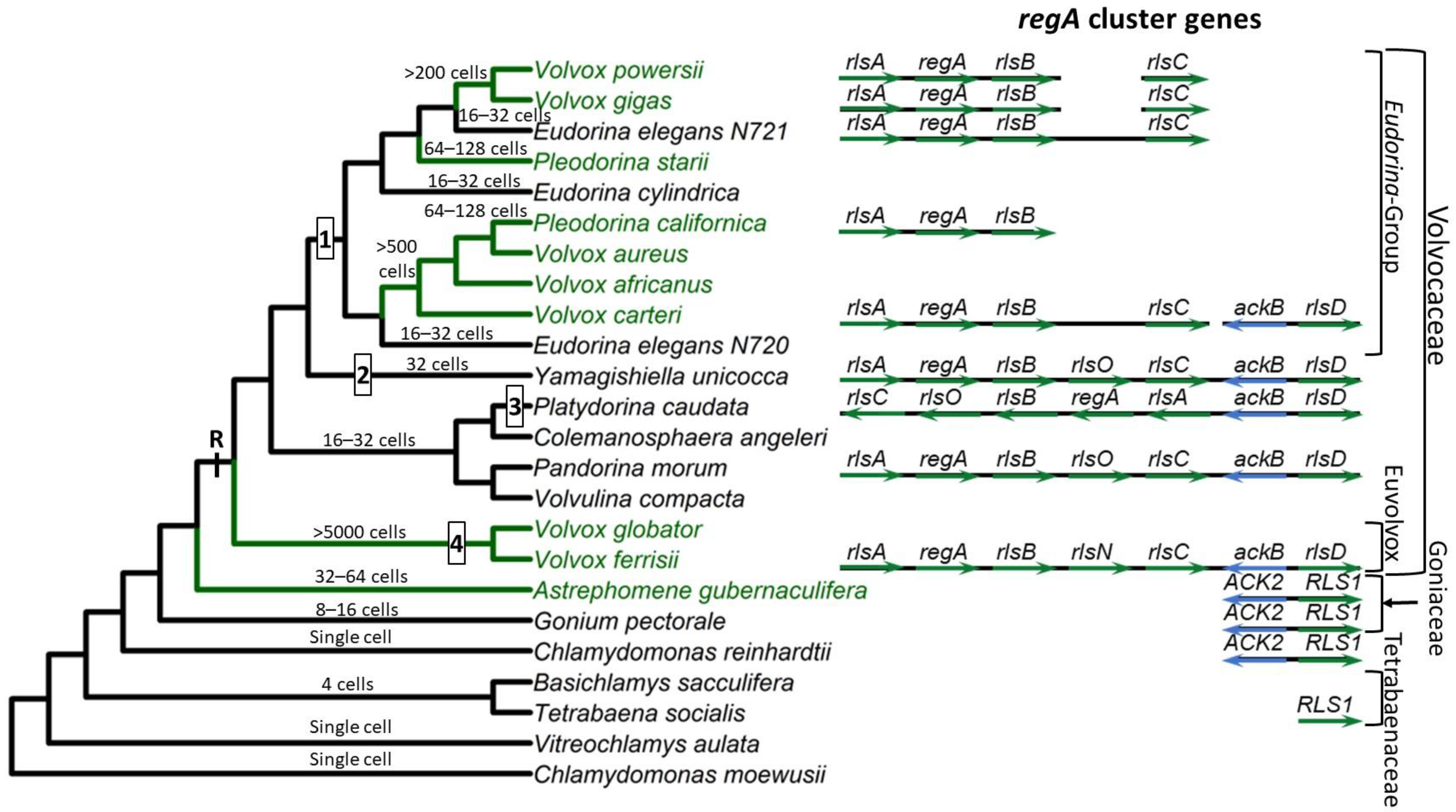
Based on currently available whole genome sequence data, the VARL gene family contains 12 members in C. reinhardtii [38], 8 in G. pectorale [8] and T. socialis [50], 6 in A. gubernaculifera [9], and 14 in V. carteri [38]. With the exception of regA orthologs (when present), all other regA homologs are known as regA-like sequences, annotated as RLS1-12 in Chlamydomonas and Goniaceae or rlsA-O in Volvocaceae. C. reinhardtii and other volvocine algae outside the Volvocaceae lack orthologs of any of the regA cluster genes. The closest homolog to the regA cluster genes found in these species is RLS1. This gene is an ortholog of the Volvocaceaen rlsD, which is the closest rls paralog of the regA cluster. Currently it is thought that the VARL gene family comprising several paralogs including RLS1/rlsD was already present in the common ancestor of all volvocine green algae. RLS1/rlsD underwent one or more duplication events in the common ancestor of the Volvocaceae family to give rise to a five-gene regA gene cluster comprising rlsA, regA, rlsB, rlsO, and rlsC. After the lineage leading to V. ferrisii diverged from the rest of the Volvocaceae, its rlsO gene gained a second VARL domain and evolved into rlsN. Meanwhile, the common ancestor of the Eudorina group lost rlsO. In addition, Y. unicocca lost two internal regA cluster genes (regA, rlsB, or rlsO) but restored the five-gene cluster via gene duplication, and the regA cluster of P. caudata became inverted relative to nearby syntenic markers (Figure 1).

Based on its role in suppressing reproduction in somatic cells, it has been hypothesized that regA evolved from a gene that was involved in trading off reproduction for survival (i.e., a life history trade-off gene) in the single-celled ancestors of V. carteri. Specifically, such a gene could have been co-opted by changing its expression from a temporal context (in response to an environmental cue) into a spatial context (in response to a developmental cue) [51].The common ancestor of V. carteri and C. reinhardtii likely had several VARL gene family members, one of which was RLS1. The RLS1 gene duplicated several times to give rise to the regA gene cluster in the common ancestor of the Volvocaceae, setting the stage for the functional co-option of regA during the evolution of cellular differentiation as well as other lineage-specific changes to regA cluster genes (Figure 1). The co-option of RLS1′s functions into a regA-like gene responsible for somatic cell differentiation likely involved the simulation of the ancestral environmentally induced signal in a developmental context.

References

  1. Coleman, A.W. A Comparative Analysis of the Volvocaceae (Chlorophyta). J. Phycol. 2012, 48, 491–513.
  2. Umen, J.G. Volvox and volvocine green algae. EvoDevo 2020, 11, 7–15.
  3. Herron, M.D.; Michod, R.E. Evolution of complexity in the volvocine algae: Transitions in individuality through Darwin’s eye. Evol. Int. J. Org. Evol. 2008, 62, 436–451.
  4. Kirk, D.L. A twelve-step program for evolving multicellularity and a division of labor. BioEssays News Rev. Mol. Cell. Dev. Biol. 2005, 27, 299–310.
  5. Nozaki, H.; Misawa, K.; Kajita, T.; Kato, M.; Nohara, S.; Watanabe, M.M. Origin and evolution of the colonial volvocales (Chlorophyceae) as inferred from multiple, chloroplast gene sequences. Mol. Phylogenet. Evol. 2000, 17, 256–268.
  6. Arakaki, Y.; Kawai-Toyooka, H.; Hamamura, Y.; Higashiyama, T.; Noga, A.; Hirono, M.; Olson, B.J.S.C.; Nozaki, H. The simplest integrated multicellular organism unveiled. PLoS ONE 2013, 8, e81641.
  7. Nozaki, H.; Itoh, M.; Watanabe, M.M.; Kuroiwa, T. Ultrastructure of the vegetative colonies and systematic position of basichlamys (Volvocales, Chlorophyta). Eur. J. Phycol. 1996, 31, 67–72.
  8. Hanschen, E.R.; Marriage, T.N.; Ferris, P.J.; Hamaji, T.; Toyoda, A.; Fujiyama, A.; Neme, R.; Noguchi, H.; Minakuchi, Y.; Suzuki, M.; et al. The Gonium pectorale genome demonstrates co-option of cell cycle regulation during the evolution of multicellularity. Nat. Commun. 2016, 7, 11370.
  9. Yamashita, S.; Yamamoto, K.; Matsuzaki, R.; Suzuki, S.; Yamaguchi, H.; Hirooka, S.; Minakuchi, Y.; Miyagishima, S.-Y.; Kawachi, M.; Toyoda, A.; et al. Genome sequencing of the multicellular alga Astrephomene provides insights into convergent evolution of germ-soma differentiation. Sci. Rep. 2021, 11, 22231.
  10. Nozaki, H.; Mahakham, W.; Athibai, S.; Yamamoto, K.; Takusagawa, M.; Misumi, O.; Herron, M.D.; Rosenzweig, F.; Kawachi, M. Rediscovery of the species of ‘ancestral Volvox’: Morphology and phylogenetic position of Pleodorina sphaerica (Volvocales, Chlorophyceae) from Thailand. ’ Phycologia 2017, 56, 469–475.
  11. Lindsey, C.R.; Rosenzweig, F.; Herron, M.D. Phylotranscriptomics points to multiple independent origins of multicellularity and cellular differentiation in the volvocine algae. BMC Biol. 2021, 19, 182.
  12. Grochau-Wright, Z.I.; Hanschen, E.R.; Ferris, P.J.; Hamaji, T.; Nozaki, H.; Olson, B.J.S.C.; Michod, R.E. Genetic Basis for Soma is Present in Undifferentiated Volvocine Green Algae. J. Evol. Biol. 2017, 30, 1205–1218.
  13. Herron, M.D.; Hackett, J.D.; Aylward, F.O.; Michod, R.E. Triassic origin and early radiation of multicellular volvocine algae. Proc. Natl. Acad. Sci. USA 2009, 106, 3254–3258.
  14. Kirk, D.L. Volvox: Molecular-Genetic Origins of Multicellularity and Cellular Differentiation; Cambridge University Press: Cambridge, UK, 1998.
  15. Kirk, D.L. Germ-soma differentiation in Volvox. Dev. Biol. 2001, 238, 213–223.
  16. Koufopanou, V. The Evolution of Soma in the Volvocales. Am. Nat. 1994, 143, 907–931.
  17. Kirk, M.M.; Ransick, A.; McRae, S.E.; Kirk, D.L. The Relationship between Cell Size and Cell Fate in Volvox carteri. J. Cell Biol. 1993, 123, 191–208.
  18. Grochau-Wright, Z.I. The Origin and Evolution of the Reg Cluster in the Volvocine Green Algae: A Model System for the Evolution of Cellular Differentiation. Ph.D. Dissertation, University of Arizona, Tucson, AZ, USA, 2019.
  19. Grochau-Wright, Z.I.; Ferris, P.J.; Tumberger, J.; Jiménez-Marin, B.; Olson, B.J.S.C.; Michod, R.E. Characterization and Transformation of reg Cluster Genes in Volvox powersii Enable Investigation of Convergent Evolution of Cellular Differentiation in Volvox. Protist 2021, 172, 125834.
  20. Ransick, A. Reproductive cell specification during Volvox obversus development. Dev. Biol. 1991, 143, 185–198.
  21. Ransick, A. Specification of reproductive cells in Volvox. In Evolutionary Conservation of Developmental Mechanisms, Proceedings of the 50th Symposium of the Society for Developmental Biology, Marquette University, Milwaukee, WI, USA, 20–23 June 1991; Spradling, A., Ed.; Wiley-Liss: Hoboken, NJ, USA, 1993; pp. 55–70.
  22. Sessoms, A.H.; Huskey, R.J. Genetic Control of Development in Volvox: Isolation and Characterization of Morphogenetic Mutants. Proc. Natl. Acad. Sci. USA 1973, 70, 1335–1338.
  23. Starr, R.C. Control of differentiation in Volvox. Dev. Biol. 1970, 4, 59–100.
  24. Huskey, R.J.; Griffin, B.E.; Cecil, P.O.; Callahan, A.M. A Preliminary Genetic Investigation of Volvox carteri. Genetics 1979, 91, 229–244.
  25. Huskey, R.J.; Griffin, B.E. Genetic Control of Somatic Cell Differentiation in Volvox. Dev. Biol. 1979, 72, 226–235.
  26. Baran, G. Analysis of Somatic Cell Differentiation in Volvox carteri f. nagariensis; University of Virginia: Charlottesville, VA, USA, 1984.
  27. Kirk, M.M.; Stark, K.; Miller, S.M.; Müller, W.; Taillon, B.E.; Gruber, H.; Schmitt, R.; Kirk, D.L. regA, a Volvox gene that plays a central role in germ-soma differentiation, encodes a novel regulatory protein. Development 1999, 126, 639–647.
  28. Harryman, A. Investigating the Roles of regA and Related Genes in the Evolution of Multicellularity in the Volvocine Green Algae. Ph.D. Thesis, University of Maryland, Baltimore, MD, USA, 2012.
  29. Klein, B.; Wibberg, D.; Hallmann, A. Whole transcriptome RNA-Seq analysis reveals extensive cell type-specific compartmentalization in Volvox carteri. BMC Biol. 2017, 15, 1–22.
  30. Matt, G.Y.; Umen, J.G. Cell-Type Transcriptomes of the Multicellular Green Alga Volvox carteri Yield Insights into the Evolutionary Origins of Germ and Somatic Differentiation Programs. G3 Genes|Genomes|Genet. 2018, 8, 531–550.
  31. König, S.G.; Nedelcu, A.M. The genetic basis for the evolution of soma: Mechanistic evidence for the co-option of a stress-induced gene into a developmental master regulator. Proc. R. Soc. B Biol. Sci. 2020, 287, 20201414.
  32. Duncan, L.; Nishii, I.; Howard, A.; Kirk, D.; Miller, S.M. Orthologs and paralogs of regA, a master cell-type regulatory gene in Volvox carteri. Curr. Genet. 2006, 50, 61–72.
  33. Choi, G.; Przybylska, M.; Straus, D. Three abundant germ line-specific transcripts in Volvox carteri encode photosynthetic proteins. Curr. Genet. 1996, 30, 347–355.
  34. Meissner, M.; Stark, K.; Cresnar, B.; Kirk, D.L.; Schmitt, R. Volvox germline-specific genes that are putative targets of RegA repression encode chloroplast proteins. Curr. Genet. 1999, 36, 363–370.
  35. Tam, L.; Kirk, D.L. Identification of Cell-Type-Specific Characterization of their Expression Genes of Volvox carteri and during the Asexual Life Cycle. Dev. Biol. 1991, 145, 51–66.
  36. Stark, K.; Kirk, D.L.; Schmitt, R. Two enhancers and one silencer located in the introns of regA control somatic cell differentiation in Volvox carteri. Genes Dev. 2001, 15, 1449–1460.
  37. Babinger, K.; Hallmann, A.; Schmitt, R. Translational control of regA, a key gene controlling cell differentiation in Volvox carteri. Development 2006, 133, 4045–4051.
  38. Duncan, L.; Nishii, I.; Harryman, A.; Buckley, S.; Howard, A.; Friedman, N.R.; Miller, S.M. The VARL gene family and the evolutionary origins of the master cell-type regulatory gene, regA, in Volvox carteri. J. Mol. Evol. 2007, 65, 1–11.
  39. Bottomley, M.J.J.; Collard, M.W.W.; Huggenvik, J.I.I.; Liu, Z.; Gibson, T.J.J.; Sattler, M. The SAND domain structure defines a novel DNA-binding fold in transcriptional regulation. Nat. Struct. Biol. 2001, 8, 626–633.
  40. Barker, H.E.; Smyth, G.K.; Wettenhall, J.; Ward, T.A.; Bath, M.L.; Lindeman, G.J.; Visvader, J.E. Deaf-1 regulates epithelial cell proliferation and side-branching in the mammary gland. BMC Dev. Biol. 2008, 8, 94.
  41. Veraksa, A.; Kennison, J.; McGinnis, W. DEAF-1 function is essential for the early embryonic development of Drosophila. Genesis 2002, 33, 67–76.
  42. Nakagawa, T.; Tsuruma, K.; Uehara, T.; Nomura, Y. GMEB1, a novel endogenous caspase inhibitor, prevents hypoxia- and oxidative stress-induced neuronal apoptosis. Neurosci. Lett. 2008, 438, 34–37.
  43. Kulkarni, M.; Shakes, D.C.; Guevel, K.; Smith, H.E. SPE-44 Implements Sperm Cell Fate. PLoS Genet. 2012, 8, e1002678.
  44. Radhakrishnan, K.; Bhagya, K.P.; Kumar, A.T.; Devi, A.N.; Sengottaiyan, J.; Kumar, P.G. Autoimmune Regulator (AIRE) Is Expressed in Spermatogenic Cells, and It Altered the Expression of Several Nucleic-Acid-Binding and Cytoskeletal Proteins in Germ Cell 1 Spermatogonial (GC1-spg) Cells. Mol. Cell. Proteom. 2016, 15, 2686–2698.
  45. Schaller, C.E.; Wang, C.L.; Beck-engeser, G.; Goss, L.; Scott, H.S.; Anderson, M.S.; Wabl, M. Expression of Aire and the Early Wave of Apoptosis in Spermatogenesis. J. Immunol. 2008, 180, 1338–1343.
  46. Chen, L.-Q.; Luo, J.-H.; Cui, Z.-H.; Xue, M.; Wang, L.; Zhang, X.-Y.; Pawlowski, W.P.; He, Y. ATX3, ATX4, and ATX5 Encode Putative H3K4 Methyltransferases and Are Critical for Plant Development. Plant Physiol. 2017, 174, 1795–1806.
  47. Carles, C.C. ULTRAPETALA1 encodes a SAND domain putative transcriptional regulator that controls shoot and floral meristem activity in Arabidopsis. Development 2005, 132, 897–911.
  48. Carles, C.C.; Fletcher, J.C. The SAND domain protein ULTRAPETALA1 acts as a trithorax group factor to regulate cell fate in plants. Genes Dev. 2009, 23, 2723–2728.
  49. Fletcher, J.C. The ULTRAPETALA gene controls shoot and floral meristem size in Arabidopsis. Development 2001, 128, 1323–1333.
  50. Featherston, J.; Arakaki, Y.; Hanschen, E.R.; Ferris, P.J.; Michod, R.E.; Olson, B.J.S.C.; Nozaki, H.; Durand, P.M. The 4-celled tetrabaena socialis nuclear genome reveals the essential components for genetic control of cell number at the origin of multicellularity in the volvocine lineage. Mol. Biol. Evol. 2018, 35, 855–870.
  51. Nedelcu, A.M.; Michod, R.E. The evolutionary origin of an altruistic gene. Mol. Biol. Evol. 2006, 23, 1460–1464.
More
Information
Contributors MDPI registered users' name will be linked to their SciProfiles pages. To register with us, please refer to https://encyclopedia.pub/register : , ,
View Times: 248
Revisions: 2 times (View History)
Update Date: 04 May 2023
1000/1000