Submitted Successfully!
To reward your contribution, here is a gift for you: A free trial for our video production service.
Thank you for your contribution! You can also upload a video entry or images related to this topic.
Version Summary Created by Modification Content Size Created at Operation
1 -- 1471 2023-04-28 04:43:39 |
2 format correct Meta information modification 1471 2023-04-28 05:42:24 |

Video Upload Options

Do you have a full video?

Confirm

Are you sure to Delete?
Cite
If you have any further questions, please contact Encyclopedia Editorial Office.
Yamasaki, H.; Imai, H.; Tanaka, A.; Otaki, J.M. Antiviral Activity of Nitric Oxide. Encyclopedia. Available online: https://encyclopedia.pub/entry/43599 (accessed on 26 April 2024).
Yamasaki H, Imai H, Tanaka A, Otaki JM. Antiviral Activity of Nitric Oxide. Encyclopedia. Available at: https://encyclopedia.pub/entry/43599. Accessed April 26, 2024.
Yamasaki, Hideo, Hideyuki Imai, Atsuko Tanaka, Joji M. Otaki. "Antiviral Activity of Nitric Oxide" Encyclopedia, https://encyclopedia.pub/entry/43599 (accessed April 26, 2024).
Yamasaki, H., Imai, H., Tanaka, A., & Otaki, J.M. (2023, April 28). Antiviral Activity of Nitric Oxide. In Encyclopedia. https://encyclopedia.pub/entry/43599
Yamasaki, Hideo, et al. "Antiviral Activity of Nitric Oxide." Encyclopedia. Web. 28 April, 2023.
Antiviral Activity of Nitric Oxide
Edit

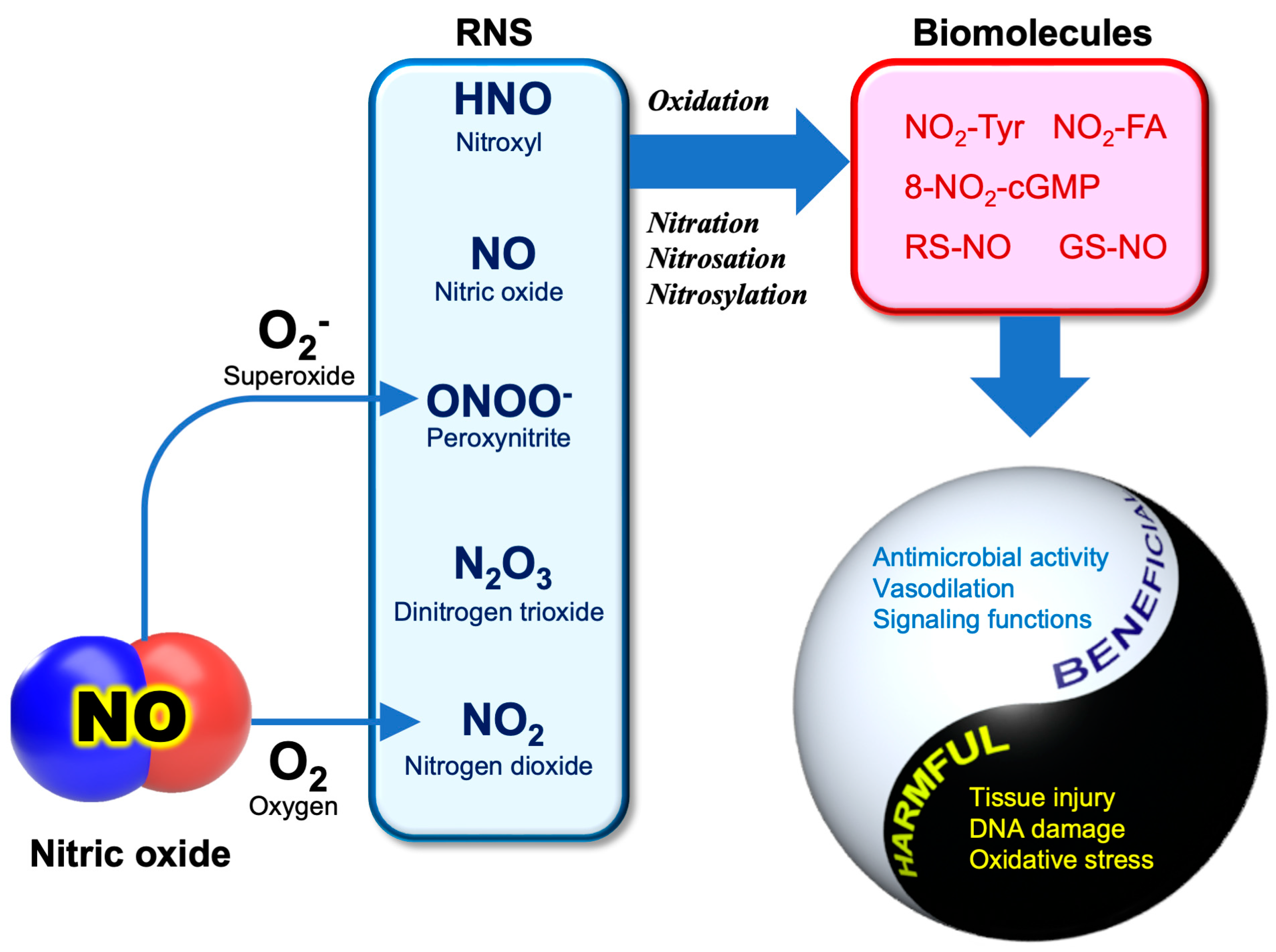
Nitric oxide (NO) is a gaseous free radical that is largely produced by the enzyme NO synthase (NOS) in cells. NO produced by upper epidermal cells contributes to the inactivation of viruses and bacteria contained in air or aerosols. In addition to enzymatic production, NO can be generated by the chemical reduction of inorganic nitrite (NO2), an alternative mechanism for NO production in living organisms. Dietary vitamin C, largely contained in fruits and vegetables, can reduce the nitrite in saliva to produce NO in the oral cavity when chewing foods. In the stomach, salivary nitrite can also be reduced to NO by vitamin C secreted from the epidermal cells of the stomach. The strong acidic pH of gastric juice facilitates the chemical reduction of salivary nitrite to produce NO. It is evident that NO exhibits substantial antiviral activity against many types of viruses, including SARS-CoV-2.

antiviral activity nitric oxide nitrite SARS-CoV-2

1. Smokers’ Paradox

Asthma and cigarette smoking, as well as obesity, diabetes, and chronic heart disease, are considered high-risk factors for acquiring COVID-19 or poorer outcomes. Early in the pandemic, however, there were few asthma patients with severe cases of COVID-19 [1][2]. A recent meta-analysis study also supported that people with asthma have a lower risk of SARS-CoV-2 infection than those without asthma [3]. The use of inhaled corticosteroids might partly account for a protective effect against SARS-CoV-2 infection, due to decreased ACE2 in asthma patients [4]. It is important to note that NO emission is generally high due to eosinophilic airway inflammation in asthmatic patients. In fact, the fraction exhaled NO (FENO) has been adopted as a non-invasive indicator of the type 2 airway inflammation of asthma [5]. Although non-allergic asthma (non-type 2) seems to have a greater risk, there were fewer asthma patients with COVID-19 in many countries [2], implying that high NO emission from the airway may protect against SARS-CoV-2 infection.
Cigarette smoking has also been listed as a risk factor for contracting COVID-19. In general, cigarette smoking is associated with relatively poor outcomes in respiratory infectious diseases [6]. In 2020, the WHO and the FDA released statements warning that smoking may increase the risk and severity of COVID-19. However, only a low proportion of smokers suffered from SARS-CoV-2 infection [7]. This is referred to as the “smokers’ paradox” [8]. Although smoking cannot be recommended as a protective measure for COVID-19, the underlying mechanism for the smokers’ paradox may give a clue for the consideration of preventing SARS-CoV-2 infection. Farsalinos et al. proposed that nicotine intake could be the reason for the low prevalence of smoking among hospitalized patients [6], whereas Hedenstierna et al. hypothesized that the short burst of concentrated NO (approximately 250 to 1350 ppm per puff) contained in cigarette smoke may prevent SARS-CoV-2 infection [9], an explanation similar to that given for why asthmatic patients are less likely to contract COVID-19.

2. RNS Biochemistry

Apart from its potent actions on cardiovascular systems, NO is involved in innate immunological host defense. The innate immune response is also mediated by reactive oxygen species (ROS), including O2, H2O2, and hypochlorite anion (OCl), which are produced by phagocytic cells such as neutrophils and activated macrophages [10]. Its action is non-specific, and potentially inactivates a broad range of pathogens, including parasites, fungi, bacteria, and viruses [11]. As ROS is the term for a group of reactive molecules derived from O2, reactive molecules originating from NO are referred to as “reactive nitrogen species (RNS)” [12]. Figure 1 illustrates the pathophysiological conditions associated with the major RNS.
Figure 1. NO and RNS in COVID-19. NO and its derived reactive molecules are frequently referred to as “reactive nitrogen species” (RNS). Peroxynitrite (ONOO) is a reaction product between NO and superoxide (O2). RNS potentially mediate the oxidation, nitration, nitrosation and nitrosylation of biomolecules. Those reactions exhibit both beneficial and harmful effects. NO2-Tyr, nitro-tyrosine; 8-NO2-cGMP, 8-nitroguanosine 3′,5′-cyclic monophosphate; NO2-FA, nitro-fatty acids; RS-NO, S-nitrosothiol; GS-NO, S-nitrosoglutathine.
Like ROS, RNS are highly reactive oxidants that are associated with oxidative stress in living organisms. Compared with ROS, the chemistry of RNS-related reactions is much more complicated, especially under in vivo conditions, and most of them are not fully understood. Basically, the reactions of NO are involved in the oxidation, nitration (the addition of NO2), nitrosation (the addition of NO+), and nitrosylation (the addition of NO) of biomolecules [13]. An uncontrolled situation of these reactions may cause “nitrative” or “nitrosative stress”, leading to cellular damage or cell death.
Most classes of biomolecules, including proteins, nucleic acids, and lipids, can be nitrated, generating products such as nitro-tyrosine (NO2-Tyr) [14], 8-nitroguanosine 3′,5′-cyclclic monophosphate (8-nitro-cGMP) [15], nitro-fatty acids (NO2-FA) [16], and nitro-phenolics [17]. NO+ could directly react with the thiols (RSH) of cysteine residues or the reduced form of glutathione (GSH) to produce S-nitrosothiols (RS-NO, GS-NO). Regarding RS-NO chemical generation in biological systems, several possible mechanisms have been proposed, but currently none of them have reached a consensus [18]. The antipathogenic activity of NO relies on these unique RNS reactions (nitration, nitrosation, and nitrosylation) that are capable of inactivating or killing pathogens through the modification of biomolecules, including enzyme proteins. It is important to remember that these reactions are non-specific, thereby also causing cellular damage to the host cells during inflammation [19]. It appears that the use of RNS as a countermeasure against pathogens is a risky business for hosts.
Under oxidative stress conditions where ROS are overproduced, such as during inflammation, peroxynitrite (ONOO) can be produced as the reaction product between NO and O2, an important interplay between ROS and RNS [20].
The rate constant for the reaction between NO and O2 is near diffusion controlled (Equation (1)), which is faster than the superoxide dismutase (SOD) reaction that removes O2. The product ONOO is stable at pH 12 in the absence of target molecules. At physiological pH, ONOO is in rapid equilibrium with its conjugated acid peroxynitrous acid (ONOOH, pKa 6.8) (Equation (2)), which is a short-lived molecule that spontaneously decays to nitrate (Equation (3)) [21]. Due to its high reactivity, ONOO- is considered the major cytotoxic agent in RNS.
NO + O2 → ONOO
ONOO + H+ → ONOOH
ONOOH → NO3 + H+
ONOO and ONOOH are strong oxidants capable of oxidizing various molecules, such as thiols, sulfides, ascorbate, and phenols [22]. In addition to the oxidation of molecules, ONOO can chemically nitrate aromatics, with a reaction being facilitated in the presence of bicarbonate anion (HCO3) [23]. The dysfunction of proteins or enzymes may occur due to the formation of nitro-tyrosine residues (NO2-Tyr). ONOO- is also involved in DNA fragmentation [22] and RNA viral mutation [24] through deamination of the bases.

3. Anti-SARS-CoV-2 Activity of NO

The antiviral activity of NO has been reported for many types of viruses, most typically, DNA viruses such as murine poxvirus, herpesviruses, and some RNA viruses [10]. The direct action of NO as an antiviral agent involves the inhibition of viral replication and viral entry into the host [25][26]. In 1999, Saura et al. demonstrated that the in vitro replication of the RNA virus coxsackievirus is suppressed by NO-dependent S-nitrosylation that causes the inactivation of viral cysteine protease, an enzyme necessary for replication [27]. The S-nitrosylation of the cysteine-containing enzymes of viruses is thought to be a general mechanism for the antiviral activity of NO [28].
SARS-CoV-2 is a positive-sense RNA virus belonging to the family Coronaviridae, which includes severe acute respiratory syndrome coronavirus (SARS-CoV), the pathogen that caused the SARS outbreak. In 2005, Akerstrom et al. reported that the NO chemical donor SNAP inhibits the in vitro replication cycle and the protein and RNA synthesis of SARS-CoV [29]. This inhibitory effect was not observed with SNP (sodium nitroprusside), another chemical NO donor [30]. Likewise, NO released from SNAP was reported to inhibit the replication of SARS-CoV-2 in Vero E6 cells through the inhibition of the SARS-CoV-2 3CL cysteine protease [31].
Macrophages are multifunctional innate immune cells that play an essential role in the clearance of pathogens and control inflammatory responses. Recent studies have suggested that S-palmitoylation is a key reaction for control macrophages in the processes of endocytosis [32]. Interestingly, NO was reported to suppress the palmitoylation of the spike (S) proteins that is needed for their binding to ACE2 [33]. The spike (S) proteins of coronaviruses are receptor-binding proteins that are synthesized in the endoplasmic reticulum (ER), followed by complex post-translational modification in the host Golgi apparatus [34][35][36]. S-Palmitoylation is one of the post-translational modifications in the Golgi apparatus where palmitoyl acyltransferase (PAT) adds the saturated fatty acid palmitate (C16:0) to the cysteine thiol group (-SH) of proteins [34][36]. Protein modification causes an increase in the hydrophobicity of the proteins, which is essential for cell-cell fusion activity [35][37]. Endothelial NO synthase (eNOS), an isoform of the host’s NO-producing enzyme, can be modified by palmitoylation, and its activity is decreased by the modification [38]. S-nitrosylation of the SARS-CoV spike (S) protein with NO may reduce cell-cell fusion activity through decreased amounts of spike (S) protein palmitoylation [33]. It is presumable that the disturbance of the cysteine palmitoylation of the spike (S) proteins is also involved in the mechanism for antiviral activity of NO against the coronavirus [39].

References

  1. Zhang, J.J.; Dong, X.; Cao, Y.Y.; Yuan, Y.D.; Yang, Y.B.; Yan, Y.Q.; Akdis, C.A.; Gao, Y.D. Clinical characteristics of 140 patients infected with SARS-CoV-2 in Wuhan, China. Allergy 2020, 75, 1730–1741.
  2. Lombardi, C.; Gani, F.; Berti, A.; Comberiati, P.; Peroni, D.; Cottini, M. Asthma and COVID-19: A dangerous liaison? Asthma Res. Pract. 2021, 7, 9.
  3. Sunjaya, A.P.; Allida, S.M.; Di Tanna, G.L.; Jenkins, C. Asthma and risk of infection, hospitalization, ICU admission and mortality from COVID-19: Systematic review and meta-analysis. J. Asthma 2022, 59, 866–879.
  4. Morais-Almeida, M.; Aguiar, R.; Martin, B.; Ansotegui, I.J.; Ebisawa, M.; Arruda, L.K.; Caminati, M.; Canonica, G.W.; Carr, T.; Chupp, G.; et al. COVID-19, asthma, and biologic therapies: What we need to know. World Allergy Organ. J. 2020, 13, 100126.
  5. Sandrini, A.; Taylor, D.R.; Thomas, P.S.; Yates, D.H. Fractional exhaled nitric oxide in asthma: An update. Respirology 2010, 15, 57–70.
  6. Farsalinos, K.; Barbouni, A.; Poulas, K.; Polosa, R.; Caponnetto, P.; Niaura, R. Current smoking, former smoking, and adverse outcome among hospitalized COVID-19 patients: A systematic review and meta-analysis. Ther. Adv. Chronic Dis. 2020, 11, 2040622320935765.
  7. Miyara, M.; Tubach, F.; Pourcher, V.; Morelot-Panzini, C.; Pernet, J.; Haroche, J.; Lebbah, S.; Morawiec, E.; Gorochov, G.; Caumes, E.; et al. Low rate of daily active tobacco smoking in patients with symptomatic COVID-19. Qeios 2020, WPP19W.4.
  8. Usman, M.S.; Siddiqi, T.J.; Khan, M.S.; Patel, U.K.; Shahid, I.; Ahmed, J.; Kalra, A.; Michos, E.D. Is there a smoker’s paradox in COVID-19? BMJ Evidence-Based Med. 2021, 26, 279–284.
  9. Hedenstierna, G.; Chen, L.; Hedenstierna, M.; Lieberman, R.; Fine, D.H. Nitric oxide dosed in short bursts at high concentrations may protect against COVID 19. Nitric Oxide 2020, 103, 1–3.
  10. Akaike, T.; Maeda, H. Nitric oxide and virus infection. Immunology 2000, 101, 300–308.
  11. DeGroote, M.A.; Fang, F.C. Antimicrobial properties of nitric oxide. In Nitric Oxide and Infection; Fang, F.C., Ed.; Kluwer Academic/Plenum Publishers: New York, NY, USA, 2002; pp. 231–261.
  12. Cortese-Krott, M.M.; Koning, A.; Kuhnle, G.G.C.; Nagy, P.; Bianco, C.L.; Pasch, A.; Wink, D.A.; Fukuto, J.M.; Jackson, A.A.; van Goor, H.; et al. The reactive rpecies Iinteractome: Evolutionary emergence, biological significance, and opportunities for redox metabolomics and personalized medicine. Antioxid. Redox Signal. 2017, 27, 684–712.
  13. Patel, R.P.; McAndrew, J.; Sellak, H.; White, C.R.; Jo, H.; Freeman, B.A.; Darley-Usmar, V.M. Biological aspects of reactive nitrogen species. Biochim. Biophys. Acta 1999, 1411, 385–400.
  14. Nag, T.C.; Kathpalia, P.; Gorla, S.; Wadhwa, S. Localization of nitro-tyrosine immunoreactivity in human retina. Ann. Anat. 2019, 223, 8–18.
  15. Akaike, T.; Okamoto, S.; Sawa, T.; Yoshitake, J.; Tamura, F.; Ichimori, K.; Miyazaki, K.; Sasamoto, K.; Maeda, H. 8-nitroguanosine formation in viral pneumonia and its implication for pathogenesis. Proc. Natl. Acad. Sci. USA 2003, 100, 685–690.
  16. Villacorta, L.; Gao, Z.; Schopfer, F.J.; Freeman, B.A.; Chen, Y.E. Nitro-fatty acids in cardiovascular regulation and diseases: Characteristics and molecular mechanisms. Front. Biosci. 2016, 21, 873.
  17. Sakihama, Y.; Tamaki, R.; Shimoji, H.; Ichiba, T.; Fukushi, Y.; Tahara, S.; Yamasaki, H. Enzymatic nitration of phytophenolics: Evidence for peroxynitrite-independent nitration of plant secondary metabolites. FEBS Lett. 2003, 553, 377–380.
  18. Fukuto, J.M.; Perez-Ternero, C.; Zarenkiewicz, J.; Lin, J.; Hobbs, A.J.; Toscano, J.P. Hydropersulfides (RSSH) and nitric oxide (NO) signaling: Possible effects on S-nitrosothiols (RS-NO). Antioxidants 2022, 11, 169.
  19. Dedon, P.C.; Tannenbaum, S.R. Reactive nitrogen species in the chemical biology of inflammation. Arch. Biochem. Biophys. 2004, 423, 12–22.
  20. Yamasaki, H. Nitrite-dependent nitric oxide production pathway: Implications for involvement of active nitrogen species in photoinhibition in vivo. Philos. Trans. R. Soc. B Biol. Sci. 2000, 355, 1477–1488.
  21. Toda, N.; Ayajiki, K.; Okamura, T. Control of systemic and pulmonary blood pressure by nitric oxide formed through neuronal nitric oxide synthase. J. Hypertens. 2009, 27, 1929–1940.
  22. Szabo, C. Multiple pathways of peroxynitrite cytotoxicity. Toxicol. Lett. 2003, 140-141, 105–112.
  23. Sanchez, A.G.; Ibargoyen, M.N.; Mastrogiovanni, M.; Radi, R.; Keszenman, D.J.; Peluffo, R.D. Fast and biphasic 8-nitroguanine production from guanine and peroxynitrite. Free Radic. Biol. Med. 2022, 193, 474–484.
  24. Akaike, T.; Fujii, S.; Kato, A.; Yoshitake, J.; Miyamoto, Y.; Sawa, T.; Okamoto, S.; Suga, M.; Asakawa, M.; Nagai, Y.; et al. Viral mutation accelerated by nitric oxide production during infectionin vivo. FASEB J. 2000, 14, 1447–1454.
  25. Oza, P.P.; Kashfi, K. Utility of NO and H2S donating platforms in managing COVID-19: Rationale and promise. Nitric Oxide 2022, 128, 72–102.
  26. Klingstrom, J.; Akerstrom, S.; Hardestam, J.; Stoltz, M.; Simon, M.; Falk, K.I.; Mirazimi, A.; Rottenberg, M.; Lundkvist, A. Nitric oxide and peroxynitrite have different antiviral effects against hantavirus replication and free mature virions. Eur. J. Immunol. 2006, 36, 2649–2657.
  27. Saura, M.; Zaragoza, C.; McMillan, A.; Quick, R.A.; Hohenadl, C.; Lowenstein, J.M.; Lowenstein, C.J. An antiviral mechanism of nitric oxide: Inhibition of a viral protease. Immunity 1999, 10, 21–28.
  28. Colasanti, M.; Persichini, T.; Venturini, G.; Ascenzi, P. S-nitrosylation of viral proteins: Molecular bases for antiviral effect of nitric oxide. IUBMB Life 1999, 48, 25–31.
  29. Akerstrom, S.; Mousavi-Jazi, M.; Klingstrom, J.; Leijon, M.; Lundkvist, A.; Mirazimi, A. Nitric oxide inhibits the replication cycle of severe acute respiratory syndrome coronavirus. J. Virol. 2005, 79, 1966–1969.
  30. Keyaerts, E.; Vijgen, L.; Chen, L.; Maes, P.; Hedenstierna, G.; Van Ranst, M. Inhibition of SARS-coronavirus infection in vitro by S-nitroso-N-acetylpenicillamine, a nitric oxide donor compound. Int. J. Infect. Dis. 2004, 8, 223–226.
  31. Akaberi, D.; Krambrich, J.; Ling, J.; Luni, C.; Hedenstierna, G.; Jarhult, J.D.; Lennerstrand, J.; Lundkvist, A. Mitigation of the replication of SARS-CoV-2 by nitric oxide in vitro. Redox Biol. 2020, 37, 101734.
  32. Guns, J.; Vanherle, S.; Hendriks, J.J.A.; Bogie, J.F.J. Protein lipidation by palmitate controls macrophage function. Cells 2022, 11, 565.
  33. Akerstrom, S.; Gunalan, V.; Keng, C.T.; Tan, Y.J.; Mirazimi, A. Dual effect of nitric oxide on SARS-CoV replication: Viral RNA production and palmitoylation of the S protein are affected. Virology 2009, 395, 1–9.
  34. Wong, N.A.; Saier, M.H., Jr. The SARS-coronavirus infection cycle: A survey of viral membraneproteins, their functional interactions and pathogenesis. Int. J. Mol. Sci. 2021, 22, 1308.
  35. Li, D.; Liu, Y.; Lu, Y.; Gao, S.; Zhang, L. Palmitoylation of SARS-CoV-2 S protein is critical for S-mediated syncytia formation and virus entry. J. Med. Virol. 2022, 94, 342–348.
  36. Main, A.; Fuller, W. Protein S-palmitoylation: Advances and challenges in studying a therapeutically important lipid modification. FEBS J. 2022, 289, 861–882.
  37. Gordon, D.E.; Jang, G.M.; Bouhaddou, M.; Xu, J.; Obernier, K.; White, K.M.; O’Meara, M.J.; Rezelj, V.V.; Guo, J.Z.; Swaney, D.L. A SARS-CoV-2 protein interaction map reveals targets for drug repurposing. Nature 2020, 583, 459–468.
  38. Fernandez-Hernando, C.; Fukata, M.; Bernatchez, P.N.; Fukata, Y.; Lin, M.I.; Bredt, D.S.; Sessa, W.C. Identification of Golgi-localized acyl transferases that palmitoylate and regulate endothelial nitric oxide synthase. J. Cell Biol. 2006, 174, 369–377.
  39. Li, X.; Yuan, H.; Li, X.; Wang, H. Spike protein mediated membrane fusion during SARS-CoV-2 infection. J. Med. Virol. 2022, 95, e28212.
More
Information
Contributors MDPI registered users' name will be linked to their SciProfiles pages. To register with us, please refer to https://encyclopedia.pub/register : , , ,
View Times: 483
Entry Collection: COVID-19
Revisions: 2 times (View History)
Update Date: 28 Apr 2023
1000/1000