Submitted Successfully!
To reward your contribution, here is a gift for you: A free trial for our video production service.
Thank you for your contribution! You can also upload a video entry or images related to this topic.
Version Summary Created by Modification Content Size Created at Operation
1 -- 2098 2023-04-13 11:40:05 |
2 Reference format revised. Meta information modification 2098 2023-04-14 02:49:03 |

Video Upload Options

Do you have a full video?

Confirm

Are you sure to Delete?
Cite
If you have any further questions, please contact Encyclopedia Editorial Office.
Hager, J.; Sergi, C.M. Pediatric Echinococcosis of the Liver in Austria. Encyclopedia. Available online: https://encyclopedia.pub/entry/43023 (accessed on 27 July 2024).
Hager J, Sergi CM. Pediatric Echinococcosis of the Liver in Austria. Encyclopedia. Available at: https://encyclopedia.pub/entry/43023. Accessed July 27, 2024.
Hager, Josef, Consolato M. Sergi. "Pediatric Echinococcosis of the Liver in Austria" Encyclopedia, https://encyclopedia.pub/entry/43023 (accessed July 27, 2024).
Hager, J., & Sergi, C.M. (2023, April 13). Pediatric Echinococcosis of the Liver in Austria. In Encyclopedia. https://encyclopedia.pub/entry/43023
Hager, Josef and Consolato M. Sergi. "Pediatric Echinococcosis of the Liver in Austria." Encyclopedia. Web. 13 April, 2023.
Pediatric Echinococcosis of the Liver in Austria
Edit

Echinococcosis is considered a neglected disease in most European countries. However, migratory flows of populations, long-term stays in endemic areas, uninterrupted tourism (travel to Echinococcus-endemic countries), traveling dogs and dog translocations from endemic areas, and inappropriate hygiene practices are potential factors that alarm public health officials. Identifying a cyst-like mass in the liver or lung of an individual with a travel history of likely exposure to sheepdogs in an area where the parasite Echinococcus (E.) granulosus (sive cysticus) is endemic advocates for a prompt preliminary diagnosis of cystic echinococcosis (CE), no matter the age of the affected individuals. 

Echinococcosis liver cysts

1. Introduction

Echinococcosis is rarely diagnosed in children/adolescents because the incubation period can be relatively long. Echinococcosis is not a single disease; a few agents can cause this disease, and the plural term of echinococcosis is typically used. Since children rarely manifest echinococcosis due to age, these diseases, which primarily affect the liver, are often not included in differential diagnoses of abdominal diseases, and missing the diagnosis can have devastating consequences. Indeed, this aspect is not trivial because echinococcosis is a potentially life-threatening disease [1][2][3][4][5].

2. Echinococcosis

Echinococcosis is an infectious disease. It is a zoonosis often caused by ingesting tapeworm larvae from the Echinococcus genus. Thus far, five echinococcus species with different geographical distributions have been described, some of which may cause very other diseases. The most widespread worldwide is the dog tapeworm, E. granulosus, which is the causative agent of cystic echinococcosis (the genetic variation within E. granulosus is relatively large due to adaptations to various intermediate hosts, which is why new genotypes are constantly being added to the E. granulosus (Geno) complex), and the fox tapeworm, E. multilocularis, the causative agent of alveolar echinococcosis. In contrast, E. vogeli and E. oligarthrus are found only in rural areas of South America. They also cause polycystic echinococcosis, similar to the effects caused by E. multilocularis. They are rarely observed in humans, and imported infections to Europe have not yet been described [6]. Another species, E. shiquicus, first described in 2005, is native to Tibet. The pathogenic potential of this species to humans is still unclear [7].

3. Epidemiology

The dog tapeworm, as the etiology of cystic echinococcosis (CE, hydatid cyst disease), is found worldwide. It occurs mostly in eastern Europe, the Mediterranean area, and the Balkans or Balkan Peninsula, a geographical region located in southeastern Europe with different geographical and historical definitions [4].
In contrast, alveolar echinococcosis (AE) is mainly observed in central Europe, primarily in eastern France, southern Germany (Bavaria), eastern Switzerland, and western Austria. UK, Norway, Finland, Malta, and Ireland are officially free of the lesser fox tapeworm [8].

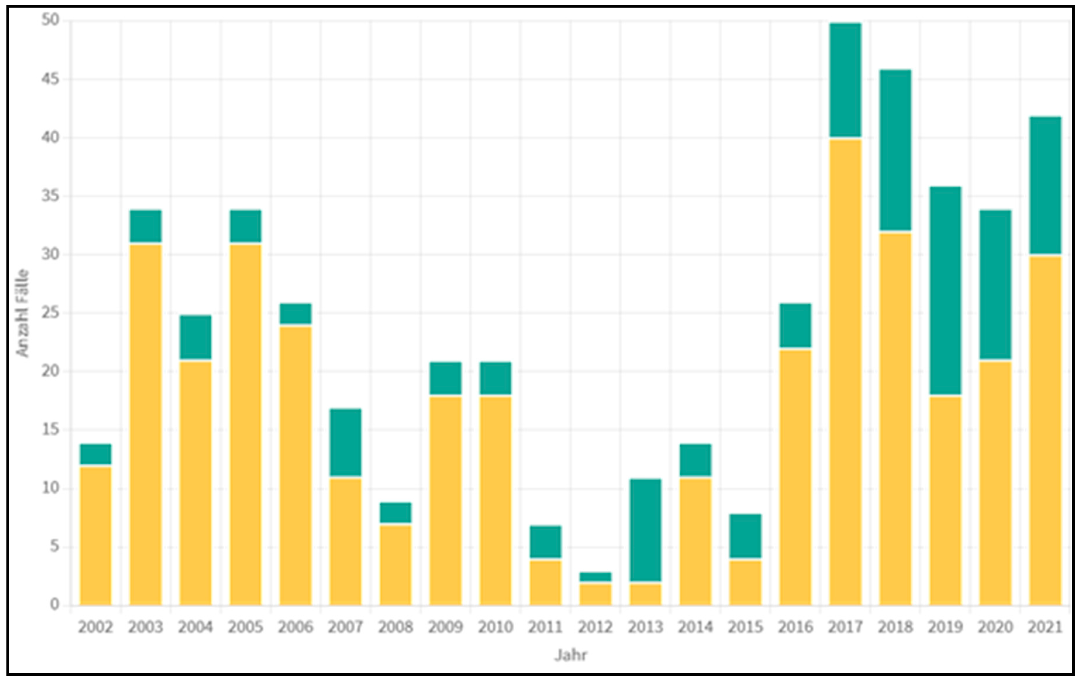
4. Statistical Data from Austria

In Austria, the dog tapeworm is considered extinct. Between 2 and 40 cases of CE per year have been documented over the past 20 years (Figure 1), whereas the annual incidence should be between 50 and 100 cases. During the last three decades, two-thirds of the patients were from abroad, and one-third were from Austria. Today, almost all CE cases are in imported patients. Molecular biological studies have shown that all Turkish CE patients and nearly all patients from other countries are infected with E. granulosus genotype 1 (“sheep strain”). In contrast, the patients of the Austrian birthplace are predominantly infected with E. granulosus genotype 7 (“pig strain”) [9].
Figure 1. Echinococcus cases in Austria from 2002 through 2021 (yellow–E. granulosus, green–E. multilocularis). The original report of the Austrian Public Health Office in Tyrol, Innsbruck (Tiroler Gesundheitsamt): “Anzahl Fälle”: Number of Cases (Y-axis) and “Jahr”: Year (X-axis). This data have been collected directly from the Austrian Public Health Office from the first author (JH).
In Austria, an average of two to three cases of AE per year have been registered over the last 20 years (Figure 1); since 2013, the incidence has increased significantly, sometimes to more than 12 cases per year. The main endemic areas are in the country’s west (Salzburg, Tyrol, Vorarlberg). In Tyrol, the fox tapeworm can be detected on average in every third to the fourth fox. In Vorarlberg, it occurs almost in every second fox. In Austria (especially in Tyrol and Vorarlberg), despite the high incidence of E. multilocularis in foxes, more patients suffer from CE than AE. E. granulosus is mainly transmitted by immigrants from South-East Europe and Europe–Asian boundaries, mainly Turkey and the Balkan countries. Despite the rarity of the disease in schools, beginning in elementary school in Austria an extensive education is provided to the learners regarding E. multilocularis (e.g., do not eat berries found in the forest immediately but only after washing, do not put grass in your mouth, after collecting mushrooms, but wash hands thoroughly after petting mouse-catching cats, etc.). Nevertheless, an inevitable increase in AE can still be observed.

5. The Parasite Biology

Humans become infected with echinococcosis by consuming contaminated food (such as mushrooms or wild berries with eggs from infected animal excrement). The eggs hatch in the duodenum and a released oncosphere reaches the common locations (liver and/or lungs) via the bloodstream. E. granulosus causes unilocular hydatid cysts, whereas E. multilocularis causes alveolar hydatid cysts. The larval stage is decisive for the development of echinococcosis. The larvae encapsulate themselves like a tumor and, depending on the echinococcal species, build different forms of cystic structures that cause two completely different clinical presentations.
Adult E. granulosus may be found in dogs, while larval stages in livestock include goats, pigs, sheep, and cattle. These tapeworms measure 2 to 7 mm and have three to four proglottids, depending on their maturity. E. granulosus usually forms a solitary cyst, compressing the surrounding tissue, especially in a delicate position like the liver hilum [10]. In rare cases, multiple cysts with smooth borders are seen. Cysts are commonly seen in the liver (70%) and the lungs (20%) but can also be found in other organs, including the brain, skeleton, heart, and even the female genital tract [4][11]. The incubation time ranges from months to years. Precise information is lacking in the literature, not least because the slow growth rate of the cysts is highly variable (1–30 mm and rarely even more per year) [12].

6. Clinical Symptomatology

The liver is the most common site of E. multilocularis infection. It has been suggested that it occurs in more than 90% of patients with infected livers. Like E. granulosus, there are no typical early symptoms in E. multilocularis, which rarely happens in children [13]. Non-specific symptoms such as abdominal pressure, upper abdominal pain, nausea, and possibly fever usually only appear later. What is striking is the discrepancy between absent or minimal or unspecific symptoms in the case of parasitic liver tumors that are already large on imaging (often >10 cm in diameter). Since the parasite usually involves the bile ducts and liver vessels, cholestasis, jaundice, and/or portal vein thrombosis/portal hypertension typically occur during the disease. If the focus of the liver is in the hilum area, cholestasis or jaundice can be expected much earlier [14]. Children with CE usually have a cyst in the right lobe of the liver (Figure 2a,b). Multiple cysts are not uncommon [15] and can be asymptomatic for years. However, since the lesions, apart from a few exceptions [16], grow slowly, they typically only become symptomatic in older children or adolescents due to local effects, e.g., a feeling of pressure or pain in the right hypochondrium; jaundice is rare.
Figure 2. (a,b). Hydatid cyst in segment 5 of the Liver (EC) (Arrow). Operation site before (a) and after preservation of the gallbladder (b) (Arrow) EC: E. cysticus.
Of the nine children, four were symptom-free (including the 15-year-old with the ruptured cyst), three complained about pressure in the upper abdomen, one had pain in the left flank (involvement of the spleen; Figure 3), and one became severely ill with jaundice.
Figure 3. Spleen with massive involvement of E. granularis (EC) in a six-year-old girl. The remaining splenic tissue is marked (spleen).

7. Diagnostics

The diagnostic procedures include sonography (better contrast-enhanced sonography (CEUS)), a CT scan, and, if possible, an MRI of the abdomen [14][17][18]. In addition, a lung X-ray is indicated because lung involvement—usually asymptomatic—is possible [19]

Sonographically, EC shows an essentially characteristic morphology (an encapsulated cystic structure with or without internal structures, e.g., septations, daughter cysts, and calcifications) but can present quite a different and sometimes misleading picture (Gharbi classification type IV–V) [20]. In the case of a traumatic cyst rupture, the focus is on the acute event, but abdominal sonography can also be informative here [21]. “Fresh” cysts (Gharbi type I) are often indistinguishable from simple liver cysts [22]. If anything is unclear, an MRI is indicated.
FDG-PET (positron emission tomography with a weakly radioactively labeled sugar solution made from fluorodeoxyglucose) can be used to determine the infectious activity of a focus. This is rarely used as a diagnostic tool but rather for follow-up [23]. Moreover, blood tests, such as enzyme-linked immunoassay (IHA, ELISA, Latex Agglutinations Test), and—in very special cases—ultrasound-guided fine needle aspiration (FNA) biopsy [14][24], are crucial in making the diagnosis.
Ultrasonography (CEUS) is the imaging technique of choice for alveolar echinococcosis. It is usually complemented by CT scans, which can detect the most significant number of lesions and calcifications characteristic of alveolar echinococcosis. MRIs are also combined with ultrasonography, especially in children [25][26][27].

8. Therapy

8.1. Alveolar Echinococcosis

From the therapeutic aspect of AE, it should be considered that at the time of diagnosis, only about 30% of patients, typically adults, are in a primarily locally operable stage. In about 70%, a large part of the liver is already affected or has extrahepatic manifestations. Regardless, drug-based benzimidazole therapy (Albendazole, Mebendazole) is indicated in all patients and is sometimes necessary for patient survival because it does not respond as well as with CE, i.e., it has more of a parasite-static effect [1]. In the case of inoperability, control of progression (prolongation of life) is paramount. This is treated permanently with albendazole or mebendazole. Therapy with albendazole is also indicated for progression control in the case of multiple surgical procedures. In both cases, progress control can be carried out with the FDG-PET since parasitic activity can be determined at any time. In about 25% of patients, it is possible to interrupt the benzimidazole therapy temporarily or permanently to minimize the side effects [23]. In children, however, experience with AE is limited, i.e., there are only case reports with different results [5].

8.2. Cystic Echinococcosis

For the treatment of CE, there are several options depending on the stage of the disease, the size and location of the cyst (as well as any complications)—also in children. The main treatment options are drug treatment, a watch-and-wait strategy, a percutaneous procedure (PAIR procedure, PPDC procedure, or radiofrequency ablation), and radical surgery [28][29]. In all cases, therapy is initially carried out with an anthelmintic, i.e., with the preparation of benzimidazoles, e.g., albendazole. Purely medicinal treatment with albendazole is indicated for patients with inoperable cysts in the liver, multi-organ involvement, or peritoneal cysts [30][31][32]. Smaller cysts (<5 cm) respond particularly well. After three, six, and twelve months of therapy, depending on the cyst size, between 30% and 70% of cysts are considered inactive or gone. However, recurrences can be expected in up to 60% of cases [32]. A few children/adolescents require long-term therapy. Therapy is less effective for large cysts (>10 cm), even with long-term treatment, but this rarely occurs in children. Drug therapy alone is contraindicated if there is a risk of cyst rupture and—in young women—in early pregnancy.
Today’s most popular procedure for treating cystic echinococcosis is the PAIR procedure (puncture, aspiration, injection, respiration). It can be performed under sonographic and sometimes Computer Tomography-assisted (CT-assisted) guidance. This technique, which can be used in children as young as three years of age, can be performed in the following local conditions: non-echoic lesion ≥5 cm in diameter (CE1m and l), cysts with daughter cysts (CE2) and/or with the detachment of membranes (CE3), multiple cysts if accessible to puncture, and infected cysts (the WHO Informal Working Group on Echinococcosis (WHO-IWGE) classified liver hydatid cysts as CE1, CE2, CE3a, CE3b, CE4, and CE5, which corresponds to Gharbi et al. [22]). In this procedure, the cyst is punctured percutaneously (it should be located at least 2 cm below the surface of the liver, and there must be no cystic-biliary fistula [21][33]). Part of the cyst fluid is aspirated. Then, to denature the cyst, hypertonic saline solution (20%) or 95% alcohol is injected into the cyst, and then after about 15–20 min, the entire cyst content is respired [34]. The disadvantage of this method is, on the one hand, that the tumor capsule remains in situ and, on the other hand, that the cyst capsule can be damaged. It means that protoscolices can be spread intraperitoneally, which in turn can cause a severe allergic reaction [33][35]

References

  1. Albendazole: New indication. Useful adjunct in hydatid disease. Prescrire Int. 2000, 9, 139–142.
  2. da Silva, A.M. Human echinococcosis: A neglected disease. Gastroenterol. Res. Pract. 2010, 2010, 583297.
  3. Djuricic, S.M.; Grebeldinger, S.; Kafka, D.I.; Djan, I.; Vukadin, M.; Vasiljevic, Z.V. Cystic echinococcosis in children—The seventeen-year experience of two large medical centers in Serbia. Parasitol. Int. 2010, 59, 257–261.
  4. Petropoulos, A.S.; Chatzoulis, G.A. Echinococcus Granulosus in Childhood: A Retrospective Study of 187 Cases and Newer Data. Clin. Pediatr. 2019, 58, 864–888.
  5. Jonaityte, E.; Judickas, M.; Tamuleviciene, E.; Seskute, M. Alveolar Echinococcosis in Children. Case Rep. Pediatr. 2020, 2020, 5101234.
  6. Tappe, D.; Stich, A.; Frosch, M. Emergence of polycystic neotropical echinococcosis. Emerg. Infect. Dis. 2008, 14, 292–297.
  7. Xiao, N.; Qiu, J.; Nakao, M.; Li, T.; Yang, W.; Chen, X.; Schantz, P.M.; Craig, P.S.; Ito, A. Echinococcus shiquicus n. sp., a taeniid cestode from Tibetan fox and plateau pika in China. Int. J. Parasitol. 2005, 35, 693–701.
  8. Gottstein, B. Epidemiology and systematics of cystic and alveolar hydatid disease. Chirurg 2000, 71, 1–8.
  9. Austrian Agency for Health and Food Safety GmbH: Pathogens from A to Z; Actualised at 20 December 2022. Available online: https://www.ages.at/en/ (accessed on 17 January 2023).
  10. Pakala, T.; Molina, M.; Wu, G.Y. Hepatic Echinococcal Cysts: A Review. J. Clin. Transl. Hepatol. 2016, 4, 39–46.
  11. Behera, P.K.; Satpathy, S. Hydatidosis of female genital tract: A case report. Indian J. Pathol. Microbiol. 2003, 46, 78–79.
  12. Nunnari, G.; Pinzone, M.R.; Gruttadauria, S.; Celesia, B.M.; Madeddu, G.; Malaguarnera, G.; Pavone, P.; Cappellani, A.; Cacopardo, B. Hepatic echinococcosis: Clinical and therapeutic aspects. World J. Gastroenterol. 2012, 18, 1448–1458.
  13. Yoshida, T.; Kamiyama, T.; Okada, T.; Nakanishi, K.; Yokoo, H.; Kamachi, H.; Matsushita, M.; Sato, N.; Sasaki, F.; Todo, S. Alveolar echinococcosis of the liver in children. J. Hepatobiliary Pancreat. Sci. 2010, 17, 152–157.
  14. Eckert, J.; Deplazes, P. Biological, epidemiological, and clinical aspects of echinococcosis, a zoonosis of increasing concern. Clin. Microbiol. Rev. 2004, 17, 107–135.
  15. Senyuz, O.F.; Celayir, A.C.; Kilic, N.; Celayir, S.; Sarimurat, N.; Erdogan, E.; Yeker, D. Hydatid disease of the liver in childhood. Pediatr. Surg. Int. 1999, 15, 217–220.
  16. Foroughi, M.; Bahador, A.; Beizavi, Z. Rapid Growth of Hydatid Cyst: A Pediatric Case Report. Iran J. Parasitol. 2021, 16, 164–167.
  17. Kratzer, W.; Gruener, B.; Kaltenbach, T.E.; Ansari-Bitzenberger, S.; Kern, P.; Fuchs, M.; Mason, R.A.; Barth, T.F.; Haenle, M.M.; Hillenbrand, A.; et al. Proposal of an ultrasonographic classification for hepatic alveolar echinococcosis: Echinococcosis multilocularis Ulm classification-ultrasound. World J. Gastroenterol. 2015, 21, 12392–12402.
  18. Stojkovic, M.; Rosenberger, K.; Kauczor, H.U.; Junghanss, T.; Hosch, W. Diagnosing and Staging of Cystic Echinococcosis: How Do CT and MRI Perform in Comparison to Ultrasound? PLoS Negl Trop Dis. 2012, 6, e1880.
  19. Cevik, M.; Boleken, M.E.; Kurkcuoglu, I.C.; Eser, I.; Dorterler, M.E. Pulmonary hydatid disease is difficult recognized in children. Pediatr. Surg. Int. 2014, 30, 737–741.
  20. Turgut, A.T.; Akhan, O.; Bhatt, S.; Dogra, V.S. Sonographic spectrum of hydatid disease. Ultrasound Q. 2008, 24, 17–29.
  21. Calame, P.; Weck, M.; Busse-Cote, A.; Brumpt, E.; Richou, C.; Turco, C.; Doussot, A.; Bresson-Hadni, S.; Delabrousse, E. Role of the radiologist in the diagnosis and management of the two forms of hepatic echinococcosis. Insights Imaging 2022, 13, 68.
  22. Gharbi, H.A.; Hassine, W.; Brauner, M.W.; Dupuch, K. Ultrasound examination of the hydatic liver. Radiology 1981, 139, 459–463.
  23. Caoduro, C.; Porot, C.; Vuitton, D.A.; Bresson-Hadni, S.; Grenouillet, F.; Richou, C.; Boulahdour, H.; Blagosklonov, O. The role of delayed 18F-FDG-PET imaging in the follow-up of patients with alveolar echinococcosis. J. Nucl. Med. 2013, 54, 358–363.
  24. Bulakci, M.; Ilhan, M.; Bademler, S.; Yilmaz, E.; Gulluoglu, M.; Bayraktar, A.; Asik, M.; Guloglu, R. Efficacy of ultrasound-guided core-needle biopsy in the diagnosis of hepatic alveolar echinococcosis: A retrospective analysis. Parasite 2016, 23, 19.
  25. Sulima, M.; Nahorski, W.; Gorycki, T.; Wolyniec, W.; Waz, P.; Felczak-Korzybska, I.; Szostakowska, B.; Sikorska, K. Ultrasound images in hepatic alveolar echinococcosis and clinical stage of the disease. Adv. Med. Sci. 2019, 64, 324–330.
  26. Kratzer, W.; Weimer, H.; Schmidberger, J. Echinococcosis: A Challenge for Liver Sonography. Ultraschall Med. 2022, 43, 120–145.
  27. Graeter, T.; Kratzer, W.; Oeztuerk, S.; Haenle, M.M.; Mason, R.A.; Hillenbrand, A.; Kull, T.; Barth, T.F.; Kern, P.; Gruener, B. Proposal of a computed tomography classification for hepatic alveolar echinococcosis. World J. Gastroenterol. 2016, 22, 3621–3631.
  28. Brunetti, E.; Kern, P.; Vuitton, D.A. Expert consensus for the diagnosis and treatment of cystic and alveolar echinococcosis in humans. Acta Trop. 2010, 114, 1–16.
  29. Faraj, W.; Abi Faraj, C.; Kanso, M.; Nassar, H.; Hoteit, L.; Farsakoury, R.; Zaghal, A.; Yaghi, M.; Jaafar, R.F.; Khalife, M. Hydatid Disease of the Liver in the Middle East: A Single Center Experience. Surg. Infect. 2022, 23, 29–34.
  30. Calame, P.; Doussot, A.; Turco, C.; Colpart, P.; Heyd, B.; Delabrousse, E. Local invasion of hepatic alveolar echinococcosis should not be underestimated: Lessons learned from imaging-pathologic correlation. Diagn. Interv. Imaging 2021, 102, 189–192.
  31. Gupta, N.; Javed, A.; Puri, S.; Jain, S.; Singh, S.; Agarwal, A.K. Hepatic hydatid: PAIR, drain or resect? J. Gastrointest. Surg. 2011, 15, 1829–1836.
  32. Stojkovic, M.; Weber, T.F.; Junghanss, T. Clinical management of cystic echinococcosis: State of the art and perspectives. Curr. Opin. Infect. Dis. 2018, 31, 383–392.
  33. Demir, S.; Ilikan, G.B.; Erturk, A.; Oztorun, C.I.; Guney, D.; Azili, M.N.; Senel, E.; Tiryaki, H.T. A serious complication of liver hydatid cysts in children: Cystobiliary fistulas. Pediatr. Surg. Int. 2020, 36, 611–620.
  34. Tersigni, C.; Venturini, E.; Montagnani, C.; Bianchi, L.; Chiappini, E.; de Martino, M.; Galli, L. Should Pediatricians Be Aware of Cystic Echinococcosis? A Literature Review. J. Pediatr. Gastroenterol. Nutr. 2019, 68, 161–168.
  35. Junghanss, T.; da Silva, A.M.; Horton, J.; Chiodini, P.L.; Brunetti, E. Clinical management of cystic echinococcosis: State of the art, problems, and perspectives. Am. J. Trop. Med. Hyg. 2008, 79, 301–311.
More
Information
Subjects: Pediatrics
Contributors MDPI registered users' name will be linked to their SciProfiles pages. To register with us, please refer to https://encyclopedia.pub/register : ,
View Times: 299
Revisions: 2 times (View History)
Update Date: 14 Apr 2023
1000/1000
Video Production Service