Submitted Successfully!
To reward your contribution, here is a gift for you: A free trial for our video production service.
Thank you for your contribution! You can also upload a video entry or images related to this topic.
Version Summary Created by Modification Content Size Created at Operation
1 -- 2509 2023-02-20 11:49:54 |
2 update references and layout Meta information modification 2509 2023-02-21 03:08:03 |

Video Upload Options

Do you have a full video?

Confirm

Are you sure to Delete?
Cite
If you have any further questions, please contact Encyclopedia Editorial Office.
Iyoda, T.; Yamasaki, S.; Ueda, S.; Shimizu, K.; Fujii, S. Natural Killer T and Natural Killer Cell-Based Immunotherapy. Encyclopedia. Available online: https://encyclopedia.pub/entry/41429 (accessed on 27 July 2024).
Iyoda T, Yamasaki S, Ueda S, Shimizu K, Fujii S. Natural Killer T and Natural Killer Cell-Based Immunotherapy. Encyclopedia. Available at: https://encyclopedia.pub/entry/41429. Accessed July 27, 2024.
Iyoda, Tomonori, Satoru Yamasaki, Shogo Ueda, Kanako Shimizu, Shin-Ichiro Fujii. "Natural Killer T and Natural Killer Cell-Based Immunotherapy" Encyclopedia, https://encyclopedia.pub/entry/41429 (accessed July 27, 2024).
Iyoda, T., Yamasaki, S., Ueda, S., Shimizu, K., & Fujii, S. (2023, February 20). Natural Killer T and Natural Killer Cell-Based Immunotherapy. In Encyclopedia. https://encyclopedia.pub/entry/41429
Iyoda, Tomonori, et al. "Natural Killer T and Natural Killer Cell-Based Immunotherapy." Encyclopedia. Web. 20 February, 2023.
Natural Killer T and Natural Killer Cell-Based Immunotherapy
Edit

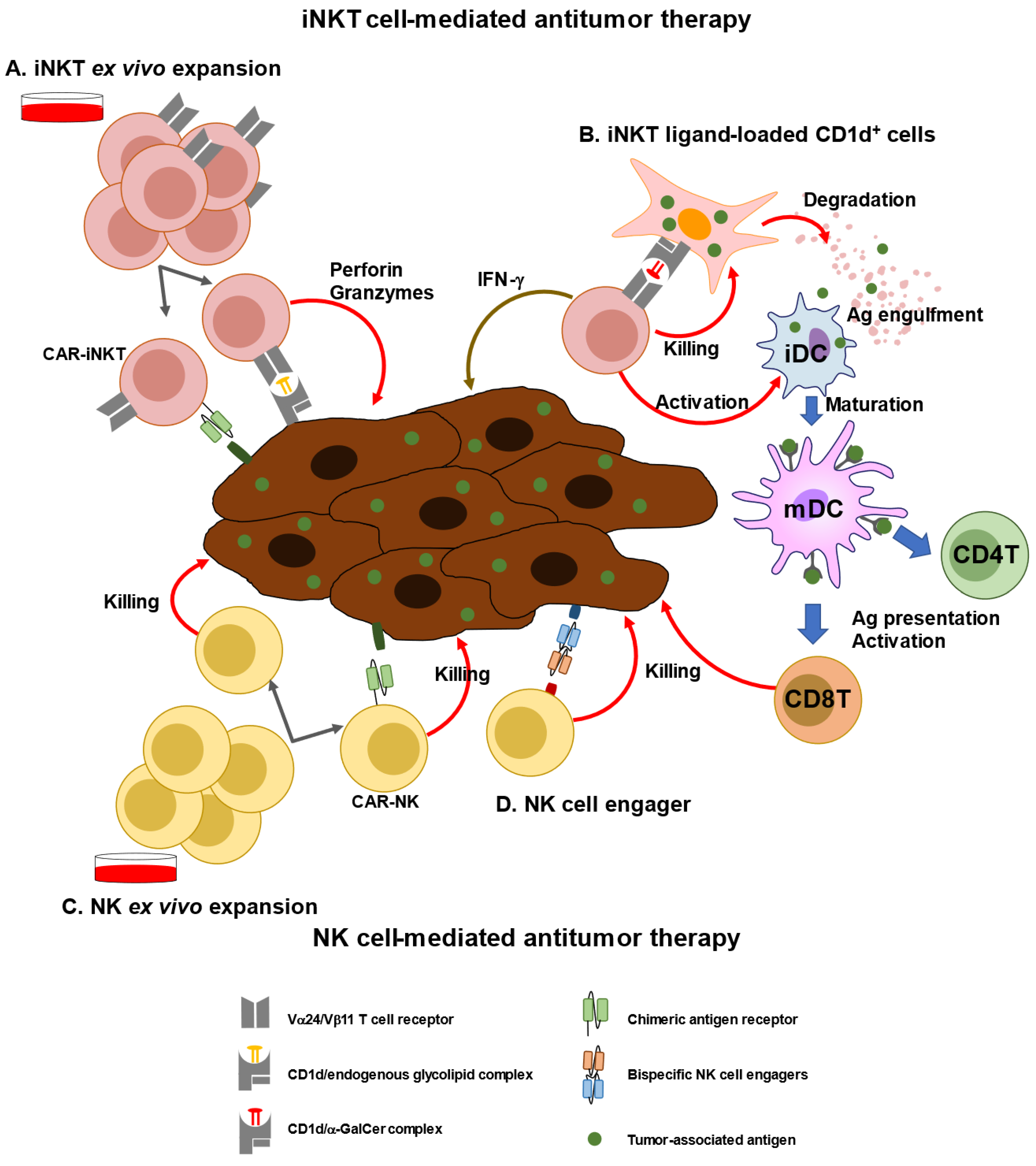
Both natural killer T (NKT) and natural killer (NK) cells are innate cytotoxic lymphoid cells that produce inflammatory cytokines and chemokines, and their role in the innate immune response to tumors and microorganisms has been investigated. Especially, emerging evidence has revealed their status and function in the tumor microenvironment (TME) of tumor cells. As a recent strategy in cancer immunotherapy, the mobilization or restoration of endogenous NKT or NK cells by novel vaccines or therapies has become a focus of research. Several new modalities based on the characteristics of NKT and NK cells, including artificial adjuvant vector cells, chimeric antigen receptor-expressing NK or NKT cell therapy, or their combination with immune checkpoint blockade have been developed. This research examines challenges and future directions for improving these therapies.

NKT cell NK cell dendritic cells antitumor effect cancer immunotherapy

1. Introduction

Unlike traditional cancer treatments, such as radiotherapy and chemotherapy, immunotherapy is an innovative treatment method that dynamically modulates the immune system to attack various target cancer cells at multiple locations. It is mainly used to strengthen the immune system by regulating the immune microenvironment, so that immune cells can attack and clear tumor cells at several important locations [1]. With the progress in research on antitumor immunotherapeutic drugs, including immune checkpoint inhibitors (ICIs) and chimeric antigen receptor (CAR)-engineered T cells, the potential market and application of new immunotherapy drugs will increase.
At present, in addition to conventional antitumor therapy and recently emerging ICIs, tumor immunotherapy is being developed from different aspects, such as innate immunity (natural killer T (NKT) and natural killer (NK) cell activation) and adaptive immunity (CD4+ and CD8+ T cell activation). After stimulating immune cells with a therapeutic vaccine, the body’s own tumor-specific immune response can block a tumor’s ability to escape immune surveillance. Immune cells can thus play a role in tumor surveillance and tumor cell clearance. In contrast, adoptive cell immunotherapy (ACI) is effective against hematologic tumors, but due to the heterogeneity within solid tumors and the local microenvironment, the efficacy of ACI against solid tumors may be limited. ACI treatment involves injecting immunologic effector cells expressing modified and amplified genes to kill tumor cells. ACI can be specific or nonspecific. Although nonspecific T cells, such as nonspecific ACI, are classified as activated lymphocytes, they exhibit only a weak tumor-killing activity. Specific ACI is induced by specific tumor–antigen stimulation and includes tumor-infiltrating lymphocyte therapy, T-cell receptor-T (TCR-T), and CAR T-cell immunotherapy (CAR-T) [2]. The main effector cells are CD8+ T and CD4+ T cells. This therapy can have strong specificity and targeting ability; hence, it can be used in patients with advanced cancers or patients showing no response to other therapies.
The innate immune system is the first line of defense against infection or cancer, and it takes time to induce an antigen-specific T cell response. When the body detects pathogens or cancer cells, it activates the innate immune system to attack and destroy them, while informing and modulating the adaptive immune response. As conserved innate effector lymphocytes, NK, NKT, γδ T, and mucosa-associated invariant (MAIT) cells are crucial for immune surveillance [3][4]. Notably, antigen recognition by the aforementioned unconventional T cells is not restricted to MHC class I and II molecules [3]. NKT cells are characterized as type I or type II [5][6]. Both types recognize glycolipids on CD1d molecules, but their functions in cancer defense are clearly distinct [7].

2. Characterization of iNKT Cells and Anti-Cancer Immunotherapy

iNKT cells from mice express an invariant TCR, which is formed by the rearrangement of the Vα14 and Jα18 TCRα gene segments. iNKT cells are preferentially paired with Vβ chains, including Vβ8.2, Vβ7, or Vβ2 TCR β gene segments [8][9][10]. In humans, iNKT cells express a rearranged Vα24-Jα18 TCRα-chain associated with a Vβ11 TCRβ-chain [11][12]. When iNKT cells are stimulated by a ligand, such as α-galactosylceramide (α-GalCer), they can produce large amounts of cytokines, such as IFN-γ, TNF-α, IL-2, and IL-4 [13][14]. Murine iNKT cells can be categorized into at least three distinct functional subsets, iNKT1, iNKT2, and iNKT17 cells, and this classification is regulated by the transcription factors T-bet, GATA3, and RORγ, respectively [15][16]. Each iNKT cell subset produces typical cytokines upon activation (IFN-γ, IL-4, or IL-17). A small population of iNKT17 cells is located in the lung and subcapsular regions of lymph nodes (LNs) [15]. Human iNKT cells develop within the thymus in a PLZF-dependent manner, similar to murine iNKT cells [17][18]. However, subsets of human iNKT cells are not as fully defined as those of mouse iNKT cells. Among CD4+ iNKT cells, CD8+ iNKT cells, and double-negative (DN) iNKT cells, DN iNKT and CD8+ iNKT cells are similar to mouse iNKT1 cells, with increased IFN-γ secretion and cytotoxic function when activated [19][20]. CD4+ iNKT cells produce a relatively higher amount of Th2-type cytokines, such as IL-4 and IL-13, than other subsets [19][20]. Human iNKT cells, particularly CD4- iNKT cells, predominantly express certain NK-related markers, such as 2B4, NKG2D, DNAM-1, CD94, and NKG2A [21]. The cytotoxicity of human iNKT cells against target cells may be mediated by either TCR or natural cytotoxicity receptor-mediated signaling.
iNKT cells recognize and kill tumor cells via NKG2D, CD161, NKp44, and NKp80 as well as via endogenous NKT ligands loaded on the CD1d molecule. Conversely, tumor cells suppress iNKT cell function by expressing PD-L1, FGL-1, galectin-9, and HVEM [22][23][24][25]. Further, the tumor microenvironment is inhibitory to immune cells, and lactic acid inhibits IFN-γ production by blocking mTORC1 in iNKT cells and suppressing PPARγ-mediated cholesterol synthesis [26] (Figure 1).
Figure 1. Stimulatory and inhibitory receptors on iNKT cells. (Left) Ligands and receptors for activating iNKT cells. (right) Ligands and receptors for suppressing iNKT cells. Lack of oxygen increases lactic acid in the tumor microenvironment. Lactic acid also inhibits iNKT cell activation via mTORC1 signaling.
Owing to their peripheral differentiation, iNKT cells can differentiate into memory-type iNKT cells. iNKT cells recognize a variety of lipid antigens, including ceramide-based glycolipids, such as glycosphingolipids, microbial lipids, and endogenous self-lipids, presented by MHC class I-like CD1d molecules on antigen-presenting cells (APCs). These lipid antigens are either intermediates of APCs’ intracellular metabolism or originate from the cell walls of bacteria, fungi, or protozoan parasites.
Various α-linked ceramide glycolipids are antigenic glycolipids produced only by microorganisms. α-glucuronosylceramides and α-galacturonosylceramides are produced by Sphingomonas spp., α-galactosyldiacylglycerols are produced by Borrelia burgdorferi, and α-glucosyldiacylglycerols are produced by Streptococcus pneumoniae [27]. α-GalCer, a prototypical iNKT cell ligand, is a sphingolipid that was initially isolated from the marine sponge, Agelas mauritiana, in 1994 [28]. Although some human cells produce β-GalCer via the glycosphingolipid metabolic pathway, there is no direct pathway in human cells to produce α-GalCer. Recently, it was reported that α-GalCer, rather than β-GlcCer or β-GalCer, in mouse gut was affected by the presence of the antibiotic vancomycin and not colistin [29]. Human gut bacteria, such as Bacteroides fragilis, Bacteroides vulgatus, and Prevotella copri, produce α-GalCer structures that can activate iNKT cells. α-GalCer can also be detected in certain tumor tissues, such as colon adenocarcinoma tissues. In fact, the presence of B. vulgatus in colon adenocarcinoma tissues correlates with better survival. Thus, α-GalCer-producing bacteria, members of the human gut microbiome, may infiltrate tumor tissues, resulting in antitumor activities [29]. Endogenous CD1d-binding iNKT cell ligand glycolipids, such as isoglobotrihexosylceramide (iGb3) [30][31], disialoganglioside GD3 [32], and α-glycosylceramides, have also been identified [33][34].
Tumor-derived lipids can help tumors evade immune surveillance using several mechanisms. An alteration in tumor lipid profile or the accumulation of specific lipids and fatty acids that favor tumor growth can limit antitumor immunity [35]. Sphingosine-1-phosphate (S1P) expression is upregulated in mantle cell lymphoma (MCL), an aggressive subtype of non-Hodgkin lymphoma. Knockdown of sphingosine kinase 1, the enzyme responsible for producing S1P, in human MCL cells induces upregulation of the expression of cardiolipin and glycerophospholipid, which are abundant in mitochondrial membranes and bind to CD1d [36], increasing iNKT cell activation [37]. Another report showed that iNKT cells from the infusion of donor lymphocytes efficiently lysed leukemia cell lines and primary acute myeloid leukemia (AML) blasts in a dose- and CD1d-dependent manner [38]. This implies that tumor metabolites present on tumor cell CD1d may be potential targets of iNKT-mediated immunotherapy against hematologic malignancies. Although CD1d expression varies among tumor types, in B cell chronic lymphocytic leukemia (CLL), the most common hematologic malignancy, the expression of CD1d is lower than that in normal B cells or is absent [39], which might allow the tumor to escape from iNKT-mediated surveillance.
iNKT cells are part of the innate immune system, and effector memory-like iNKT cells have also been shown to function as other innate immune cells, such as NK and γδT cells, which differentiate into memory cells after activation. Human iNKT cells may mediate immune response against Mycobacterium tuberculosis. In humans, CD3+TCR Vβ11+ NKT cells from pleural fluid mononuclear cells exhibit an effector memory phenotype. Increased levels of expression of cytolytic molecules and M. tuberculosis antigens stimulate the production of IFN-γ, indicating that iNKT cells participate in local immune responses against M. tuberculosis and protection against M. tuberculosis infection [40]. Killer cell lectin-like receptor subfamily G member 1-positive (KLRG1)-expressing iNKT cells were induced in the lungs after vaccination with α-GalCer-loaded CD1d+ cells, including α-GalCer-loaded DCs or α-GalCer-loaded CD1d-expressing NIH-3T3 cells. KLRG1+ iNKT cells co-expressing CD49d and granzyme A persisted for several months and retained a potent response to secondary stimulation [41]. Shimizu et al. also revealed that Eomes plays two roles in iNKT cells. First, during iNKT cell development in the thymus, Eomes regulates iNKT1 cell differentiation. Second, Eomes promotes differentiation into effector memory cells in peripheral organs and spleen. Eomes-conditional deficient iNKT cells have been shown to fail to differentiate into KLRG1+ granzyme A+ long-term effector memory cells, even after stimulation with α-GalCer-loaded DCs [42]. Furthermore, Prasit et al. reported that upon intratumoral administration of a TLR9 agonist, CpG and α-GalCer suppressed tumor growth in murine tumor models. This treatment also suppressed distant untreated tumors, known as the abscopal effect. They showed that sustained activity of iNKT cells was associated with the infiltration of KLRG1+ effector memory iNKT cells into both tumors and regional LNs [43].

3. Immunological and Clinical Findings of iNKT-Based Immunotherapy

Various therapies have been developed that are effective for treating hematopoietic tumors. Activating iNKT cells is an attractive strategy for anti-cancer therapy for several reasons. First, iNKT cells are available for treating many types of cancers because all human beings share the same invariant TCR and CD1d [5][44]. Second, iNKT cells can not only kill cancer cells directly but also induce activation of NK cells as an adjunct effect [45]. Third, iNKT cells exert an adjuvant effect on T cells via DCs. Fourth, iNKT cells can alter the tumor microenvironment by inhibiting immunosuppressive cells, such as tumor-associated macrophages and myeloid-derived suppressor cells [46][47]. Although iNKT cell level is often decreased in patients with certain cancers, whether iNKT cells can be expanded or not is of higher importance than their level in patients [48]. Reduced levels and functional impairment of iNKT cells have been reported in a wide range of solid and hematologic malignancies, including prostate [49], lung, esophageal, colorectal, gastric, gallbladder, uterine, bile duct, and pancreatic cancers [50] and multiple myeloma [51] as well as in patients with late stage head and neck squamous cell carcinoma (HNSCC) [52], neuroblastomas [53], AML, and CLL [54]. Conversely, increased level of intratumoral iNKT cells is correlated with good clinical outcomes and improved survival in colorectal cancer, neuroblastoma, periampullary adenocarcinoma, and hematologic malignancies [55]. In prostate cancer and oral cell squamous carcinoma, iNKT cells exhibit defective IFN-γ production but acquire IL-4 production ability [49]. Studies on cancer patients also showed that iNKT cells respond to chemotactic signals released by tumor cells, particularly in a range of primary and metastatic solid tumors. iNKT cell infiltration is associated with CCL2 expression in neuroblastoma cells [56] and CCL20-producing tumor-associated macrophages [57]. Patient-derived iNKT cells can be restored in vitro by culturing them in the presence of IL-2 or IL-12 or in vivo upon therapeutic administration of APCs pre-loaded with α-GalCer. Therefore, several clinical trials using autologous α-GalCer-pulsed DCs or adoptive transfer of ex vivo expanded iNKT cells have been conducted. α-GarCer-pulsed DC injections were well tolerated in all patients, with no major toxicity or altered immune response. The best clinical responses included decreased urine or serum M protein level in three cases of myeloma, one stable disease in a patient with renal cell cancer, and a reduction in tumor-associated monoclonal immunoglobulin level by administering α-GarCer-pulsed DC in combination with low-dose lenalidomide in three of four patients with measurable disease [58]. In patients with advanced lung cancer, IFN-γ-producing iNKT cell therapy, including Vα24+ iNKT cell and α-GalCer-pulsed APC (APC/Gal) therapy, correlated with clinical responses in 17 patients with non-small cell lung cancer [59]. Researchers established an mRNA-transfected cell-based vaccine (the artificial adjuvant vector cell (aAVC) system [60][61][62]) that was co-transfected with an antigen-derived mRNA and CD1d mRNA, and subsequently loaded with iNKT cell ligand. When administered intravenously, aAVC activated iNKT and NK cells, and the activated iNKT and NK cells rejected aAVC. Subsequently, the killed aAVCs were taken up by DCs in situ, thereby activating several DC-specific immunogenic features. Previous studies on tumor therapeutic models reported that administration of tumor antigen (e.g., melanoma-associated antigen recognized by T cells-1 and Wilms’ tumor 1 (WT1))-expressing aAVCs induced potent CD4+ and CD8+ T cell responses [61][62][63].
Next generation engineered iNKT cell transfer approaches have already been introduced (Figure 2). Engineered human hematopoietic stem cells (HSCs) expressing a human iNKT TCR gene were examined for their ability to produce human iNKT cells that can target cancer. These HSC-derived iNKT cells effectively suppressed multiple myeloma cells in a xenograft model in a CD1d-dependent manner [64]. Using genetically engineered CD34+ HSCs, yield and purity of human allogeneic CD34+ HSC-engineered iNKT (AlloHSC-iNKT) cells have been increased to increase the number of endogenous iNKT cells. AlloHSC-iNKT cells exhibited enhanced tumor-killing efficacy against five tumor cell lines due to increased expression of NK-activating receptors and increased production of highly cytotoxic molecules. BCMA-targeting CAR-engineered AlloHSC-iNKT cells targeting MM tumors using the NK/TCR/CAR triple mechanism exhibited increased cell-killing activity [65]. Furthermore, third-party HSC-iNKT cells are useful as preclinical models of lymphoma and leukemia [66]. Moreover, engineered NKT cells co-expressing both anti-GD2 CAR and IL-15 were used in a phase I study for children with relapsed or resistant neuroblastoma. The results showed that CAR-NKT cells were expanded in vivo and migrated to the tumor site, resulting in the regression of bone metastatic lesions in one patient [67].
Figure 2. iNKT or NK cell-mediated immunotherapy. (A) iNKT cells can be expanded ex vivo using specific ligands (i.e., a-GalCer) and cytokines (e.g., IL-2, and IL-15). Expanded iNKT cells or chimeric antigen receptor (CAR)-transfected NKT cells can be adoptively transferred to patients. (B) As a new modality of vaccines, iNKT ligand-loaded CD1d+ cells with tumor-associated antigen (TAA), called artificial advent vector cells, can induce TAA-specific CD4+ and CD8+T cells via maturation of dendritic cells. (C) NK cells can be expanded ex vivo using cytokines, such as IL-15 and IL-2. CAR-transfected NK cells can be adoptively transferred to patients. (D) Bispecific NK cells exhibit cytotoxic activity against tumor cells by promoting cell–cell interaction.

References

  1. Chevolet, I.; Speeckaert, R.; Schreuer, M.; Neyns, B.; Krysko, O.; Bachert, C.; Hennart, B.; Allorge, D.; van Geel, N.; Van Gele, M.; et al. Characterization of the in vivo immune network of IDO, tryptophan metabolism, PD-L1, and CTLA-4 in circulating immune cells in melanoma. Oncoimmunology 2015, 4, e982382.
  2. Marei, H.E.; Althani, A.; Caceci, T.; Arriga, R.; Sconocchia, T.; Ottaviani, A.; Lanzilli, G.; Roselli, M.; Caratelli, S.; Cenciarelli, C.; et al. Recent perspective on CAR and Fcgamma-CR T cell immunotherapy for cancers: Preclinical evidence versus clinical outcomes. Biochem. Pharmacol. 2019, 166, 335–346.
  3. Godfrey, D.I.; Le Nours, J.; Andrews, D.M.; Uldrich, A.P.; Rossjohn, J. Unconventional T Cell Targets for Cancer Immunotherapy. Immunity 2018, 48, 453–473.
  4. Stolk, D.; van der Vliet, H.J.; de Gruijl, T.D.; van Kooyk, Y.; Exley, M.A. Positive & Negative Roles of Innate Effector Cells in Controlling Cancer Progression. Front. Immunol. 2018, 9, 1990.
  5. Fujii, S.; Shimizu, K. Immune Networks and Therapeutic Targeting of iNKT Cells in Cancer. Trends Immunol. 2019, 40, 984–997.
  6. Terabe, M.; Berzofsky, J.A. Tissue-Specific Roles of NKT Cells in Tumor Immunity. Front. Immunol. 2018, 9, 1838.
  7. Kato, S.; Berzofsky, J.A.; Terabe, M. Possible Therapeutic Application of Targeting Type II Natural Killer T Cell-Mediated Suppression of Tumor Immunity. Front. Immunol. 2018, 9, 314.
  8. Koseki, H.; Imai, K.; Nakayama, F.; Sado, T.; Moriwaki, K.; Taniguchi, M. Homogenous junctional sequence of the V14+ T-cell antigen receptor alpha chain expanded in unprimed mice. Proc. Natl. Acad. Sci. USA 1990, 87, 5248–5252.
  9. Lantz, O.; Bendelac, A. An invariant T cell receptor alpha chain is used by a unique subset of major histocompatibility complex class I-specific CD4+ and CD4-8- T cells in mice and humans. J. Exp. Med. 1994, 180, 1097–1106.
  10. Fujii, S.; Shimizu, K.; Hemmi, H.; Steinman, R.M. Innate Valpha14(+) natural killer T cells mature dendritic cells, leading to strong adaptive immunity. Immunol. Rev. 2007, 220, 183–198.
  11. Porcelli, S.; Yockey, C.E.; Brenner, M.B.; Balk, S.P. Analysis of T cell antigen receptor (TCR) expression by human peripheral blood CD4-8- alpha/beta T cells demonstrates preferential use of several V beta genes and an invariant TCR alpha chain. J. Exp. Med. 1993, 178, 1–16.
  12. Dellabona, P.; Padovan, E.; Casorati, G.; Brockhaus, M.; Lanzavecchia, A. An invariant Vα24-JαQ/Vβ11 T cell receptor is expressed in all individuals by clonally expanded CD4-8- T cells. J. Exp. Med. 1994, 180, 1171–1176.
  13. Rogers, P.R.; Matsumoto, A.; Naidenko, O.; Kronenberg, M.; Mikayama, T.; Kato, S. Expansion of human Vα24+ NKT cells by repeated stimulation with KRN7000. J. Immunol. Meth. 2004, 285, 197–214.
  14. Coquet, J.M.; Chakravarti, S.; Kyparissoudis, K.; McNab, F.W.; Pitt, L.A.; McKenzie, B.S.; Berzins, S.P.; Smyth, M.J.; Godfrey, D.I. Diverse cytokine production by NKT cell subsets and identification of an IL-17-producing CD4−NK1.1− NKT cell population. Proc. Natl. Acad. Sci. USA 2008, 105, 11287–11292.
  15. Crosby, C.M.; Kronenberg, M. Tissue-specific functions of invariant natural killer T cells. Nat. Rev. Immunol. 2018, 18, 559–574.
  16. Shissler, S.C.; Webb, T.J. The ins and outs of type I iNKT cell development. Mol. Immunol. 2019, 105, 116–130.
  17. Berzins, S.P.; Cochrane, A.D.; Pellicci, D.G.; Smyth, M.J.; Godfrey, D.I. Limited correlation between human thymus and blood NKT cell content revealed by an ontogeny study of paired tissue samples. Eur. J. Immunol. 2005, 35, 1399–1407.
  18. Savage, A.K.; Constantinides, M.G.; Han, J.; Picard, D.; Martin, E.; Li, B.; Lantz, O.; Bendelac, A. The transcription factor PLZF directs the effector program of the NKT cell lineage. Immunity 2008, 29, 391–403.
  19. Gumperz, J.E.; Miyake, S.; Yamamura, T.; Brenner, M.B. Functionally distinct subsets of CD1d-restricted natural killer T cells revealed by CD1d tetramer staining. J. Exp. Med. 2002, 195, 625–636.
  20. Lee, P.T.; Benlagha, K.; Teyton, L.; Bendelac, A. Distinct functional lineages of human valpha24 natural killer T cells. J. Exp. Med. 2002, 195, 637–641.
  21. Kuylenstierna, C.; Bjorkstrom, N.K.; Andersson, S.K.; Sahlstrom, P.; Bosnjak, L.; Paquin-Proulx, D.; Malmberg, K.J.; Ljunggren, H.G.; Moll, M.; Sandberg, J.K. NKG2D performs two functions in invariant NKT cells: Direct TCR-independent activation of NK-like cytolysis and co-stimulation of activation by CD1d. Eur. J. Immunol. 2011, 41, 1913–1923.
  22. Stojanovic, A.; Correia, M.P.; Cerwenka, A. The NKG2D/NKG2DL Axis in the Crosstalk Between Lymphoid and Myeloid Cells in Health and Disease. Front. Immunol. 2018, 9, 827.
  23. Winkler, I.; Wos, J.; Bojarska-Junak, A.; Semczuk, A.; Rechberger, T.; Baranowski, W.; Markut-Miotla, E.; Tabarkiewicz, J.; Wolinska, E.; Skrzypczak, M. An association of iNKT+/CD3+/CD161+ lymphocytes in ovarian cancer tissue with CA125 serum concentration. Immunobiology 2020, 225, 152010.
  24. Pisibon, C.; Ouertani, A.; Bertolotto, C.; Ballotti, R.; Cheli, Y. Immune Checkpoints in Cancers: From Signaling to the Clinic. Cancers 2021, 13, 4573.
  25. Li, Y.; Sharma, A.; Maciaczyk, J.; Schmidt-Wolf, I.G.H. Recent Development in NKT-Based Immunotherapy of Glioblastoma: From Bench to Bedside. Int. J. Mol. Sci. 2022, 23, 1311.
  26. Fu, S.; He, K.; Tian, C.; Sun, H.; Zhu, C.; Bai, S.; Liu, J.; Wu, Q.; Xie, D.; Yue, T.; et al. Impaired lipid biosynthesis hinders anti-tumor efficacy of intratumoral iNKT cells. Nat. Commun. 2020, 11, 438.
  27. Oh, S.F.; Jung, D.J.; Choi, E. Gut Microbiota-Derived Unconventional T Cell Ligands: Contribution to Host Immune Modulation. Immunohorizons 2022, 6, 476–487.
  28. Natori, T.; Morita, M.; Akimoto, K.; Koezuka, Y. Agelasphins, novel antitumor and immunostimulatory cerebrosides from the sponge Agelas mauritanus. Tetrahedron 1994, 50, 2771–2784.
  29. Ustjanzew, A.; Sencio, V.; Trottein, F.; Faber, J.; Sandhoff, R.; Paret, C. Interaction between Bacteria and the Immune System for Cancer Immunotherapy: The alpha-GalCer Alliance. Int. J. Mol. Sci. 2022, 23, 5896.
  30. Dias, B.R.; Rodrigues, E.G.; Nimrichter, L.; Nakayasu, E.S.; Almeida, I.C.; Travassos, L.R. Identification of iGb3 and iGb4 in melanoma B16F10-Nex2 cells and the iNKT cell-mediated antitumor effect of dendritic cells primed with iGb3. Mol. Cancer 2009, 8, 116.
  31. Cameron, G.; Cheng, J.M.H.; Godfrey, D.I.; Timmer, M.S.M.; Stocker, B.L.; Dangerfield, E.M. The NKT cell TCR repertoire can accommodate structural modifications to the lipid and orientation of the terminal carbohydrate of iGb3. RSC Adv. 2022, 12, 18493–18500.
  32. Wu, D.Y.; Segal, N.H.; Sidobre, S.; Kronenberg, M.; Chapman, P.B. Cross-presentation of disialoganglioside GD3 to natural killer T cells. J. Exp. Med. 2003, 198, 173–181.
  33. Tsuji, M. Glycolipids and phospholipids as natural CD1d-binding NKT cell ligands. Cell. Mol. Life Sci. 2006, 63, 1889–1898.
  34. Kain, L.; Costanzo, A.; Webb, B.; Holt, M.; Bendelac, A.; Savage, P.B.; Teyton, L. Endogenous ligands of natural killer T cells are alpha-linked glycosylceramides. Mol. Immunol. 2015, 68, 94–97.
  35. Tiwary, S.; Berzofsky, J.A.; Terabe, M. Altered Lipid Tumor Environment and Its Potential Effects on NKT Cell Function in Tumor Immunity. Front. Immunol. 2019, 10, 2187.
  36. Cox, D.; Fox, L.; Tian, R.; Bardet, W.; Skaley, M.; Mojsilovic, D.; Gumperz, J.; Hildebrand, W. Determination of cellular lipids bound to human CD1d molecules. PLoS ONE 2009, 4, e5325.
  37. Lee, M.S.; Sun, W.; Webb, T.J. Sphingosine Kinase Blockade Leads to Increased Natural Killer T Cell Responses to Mantle Cell Lymphoma. Cells 2020, 9, 1030.
  38. Jahnke, S.; Schmid, H.; Secker, K.A.; Einhaus, J.; Duerr-Stoerzer, S.; Keppeler, H.; Schober-Melms, I.; Baur, R.; Schumm, M.; Handgretinger, R.; et al. Invariant NKT Cells From Donor Lymphocyte Infusions (DLI-iNKTs) Promote ex vivo Lysis of Leukemic Blasts in a CD1d-Dependent Manner. Front. Immunol. 2019, 10, 1542.
  39. Gorini, F.; Azzimonti, L.; Delfanti, G.; Scarfo, L.; Scielzo, C.; Bertilaccio, M.T.; Ranghetti, P.; Gulino, A.; Doglioni, C.; Di Napoli, A.; et al. Invariant NKT cells contribute to chronic lymphocytic leukemia surveillance and prognosis. Blood 2017, 129, 3440–3451.
  40. Li, Z.; Yang, B.; Zhang, Y.; Ma, J.; Chen, X.; Lao, S.; Li, B.; Wu, C. Mycobacterium tuberculosis-specific memory NKT cells in patients with tuberculous pleurisy. J. Clin. Immunol. 2014, 34, 979–990.
  41. Shimizu, K.; Sato, Y.; Shinga, J.; Watanabe, T.; Endo, T.; Asakura, M.; Yamasaki, S.; Kawahara, K.; Kinjo, Y.; Kitamura, H.; et al. KLRG+ invariant natural killer T cells are long-lived effectors. Proc. Natl. Acad. Sci. USA 2014, 111, 12474–12479.
  42. Shimizu, K.; Sato, Y.; Kawamura, M.; Nakazato, H.; Watanabe, T.; Ohara, O.; Fujii, S. Eomes transcription factor is required for the development and differentiation of invariant NKT cells. Commun. Biol. 2019, 2, 150.
  43. Prasit, K.K.; Ferrer-Font, L.; Burn, O.K.; Anderson, R.J.; Compton, B.J.; Schmidt, A.J.; Mayer, J.U.; Chen, C.J.; Dasyam, N.; Ritchie, D.S.; et al. Intratumoural administration of an NKT cell agonist with CpG promotes NKT cell infiltration associated with an enhanced antitumour response and abscopal effect. Oncoimmunology 2022, 11, 2081009.
  44. Fujii, S.; Shimizu, K. Exploiting Antitumor Immunotherapeutic Novel Strategies by Deciphering the Cross Talk between Invariant NKT Cells and Dendritic Cells. Front. Immunol. 2017, 8, 886.
  45. Krijgsman, D.; Hokland, M.; Kuppen, P.J.K. The Role of Natural Killer T Cells in Cancer-A Phenotypical and Functional Approach. Front. Immunol. 2018, 9, 367.
  46. Cortesi, F.; Delfanti, G.; Grilli, A.; Calcinotto, A.; Gorini, F.; Pucci, F.; Luciano, R.; Grioni, M.; Recchia, A.; Benigni, F.; et al. Bimodal CD40/Fas-Dependent Crosstalk between iNKT Cells and Tumor-Associated Macrophages Impairs Prostate Cancer Progression. Cell Rep. 2018, 22, 3006–3020.
  47. Mussai, F.; De Santo, C.; Cerundolo, V. Interaction between invariant NKT cells and myeloid-derived suppressor cells in cancer patients: Evidence and therapeutic opportunities. J. Immunother. 2012, 35, 449–459.
  48. Lam, P.Y.; Nissen, M.D.; Mattarollo, S.R. Invariant Natural Killer T Cells in Immune Regulation of Blood Cancers: Harnessing Their Potential in Immunotherapies. Front. Immunol. 2017, 8, 1355.
  49. Tahir, S.M.; Cheng, O.; Shaulov, A.; Koezuka, Y.; Bubley, G.J.; Wilson, S.B.; Balk, S.P.; Exley, M.A. Loss of IFN-γ production by invariant NK T cells in advanced cancer. J. Immunol. 2001, 167, 4046–4050.
  50. Yanagisawa, K.; Seino, K.; Ishikawa, Y.; Nozue, M.; Todoroki, T.; Fukao, K. Impaired Proliferative Response of Vα24 NKT Cells from Cancer Patients Against α-galactosylceramide. J. Immunol. 2002, 168, 6494–6499.
  51. Dhodapkar, M.V.; Geller, M.D.; Chang, D.H.; Shimizu, K.; Fujii, S.; Dhodapkar, K.M.; Krasovsky, J. A reversible defect in natural killer T cell function characterizes the progression of premalignant to malignant multiple myeloma. J. Exp. Med. 2003, 197, 1667–1676.
  52. Motohashi, S.; Okamoto, Y.; Yoshino, I.; Nakayama, T. Anti-tumor immune responses induced by iNKT cell-based immunotherapy for lung cancer and head and neck cancer. Clin. Immunol. 2011, 140, 167–176.
  53. Wienke, J.; Dierselhuis, M.P.; Tytgat, G.A.M.; Kunkele, A.; Nierkens, S.; Molenaar, J.J. The immune landscape of neuroblastoma: Challenges and opportunities for novel therapeutic strategies in pediatric oncology. Eur. J. Cancer 2021, 144, 123–150.
  54. Boeck, C.L.; Amberger, D.C.; Doraneh-Gard, F.; Sutanto, W.; Guenther, T.; Schmohl, J.; Schuster, F.; Salih, H.; Babor, F.; Borkhardt, A.; et al. Significance of Frequencies, Compositions, and/or Antileukemic Activity of (DC-stimulated) Invariant NKT, NK and CIK Cells on the Outcome of Patients With AML, ALL and CLL. J. Immunother. 2017, 40, 224–248.
  55. Nelson, A.; Lukacs, J.D.; Johnston, B. The Current Landscape of NKT Cell Immunotherapy and the Hills Ahead. Cancers 2021, 13, 5174.
  56. Metelitsa, L.S.; Wu, H.W.; Wang, H.; Yang, Y.; Warsi, Z.; Asgharzadeh, S.; Groshen, S.; Wilson, S.B.; Seeger, R.C. Natural killer T cells infiltrate neuroblastomas expressing the chemokine CCL2. J. Exp. Med. 2004, 199, 1213–1221.
  57. Liu, D.; Song, L.; Wei, J.; Courtney, A.N.; Gao, X.; Marinova, E.; Guo, L.; Heczey, A.; Asgharzadeh, S.; Kim, E.; et al. IL-15 protects NKT cells from inhibition by tumor-associated macrophages and enhances antimetastatic activity. J. Clin. Investig. 2012, 122, 2221–2233.
  58. Richter, J.; Neparidze, N.; Zhang, L.; Nair, S.; Monesmith, T.; Sundaram, R.; Miesowicz, F.; Dhodapkar, K.M.; Dhodapkar, M.V. Clinical regressions and broad immune activation following combination therapy targeting human NKT cells in myeloma. Blood 2013, 121, 423–430.
  59. Ishibashi, F.; Sakairi, Y.; Iwata, T.; Moriya, Y.; Mizobuchi, T.; Hoshino, H.; Yoshida, S.; Hanaoka, H.; Yoshino, I.; Motohashi, S. A phase I study of loco-regional immunotherapy by transbronchial injection of alpha-galactosylceramide-pulsed antigen presenting cells in patients with lung cancer. Clin. Immunol. 2020, 215, 108457.
  60. Fujii, S.; Goto, A.; Shimizu, K. Antigen mRNA-transfected, allogeneic fibroblasts loaded with NKT-cell ligand confer antitumor immunity. Blood 2009, 113, 4262–4272.
  61. Shimizu, K.; Mizuno, T.; Shinga, J.; Asakura, M.; Kakimi, K.; Ishii, Y.; Masuda, K.; Maeda, T.; Sugahara, H.; Sato, Y.; et al. Vaccination with antigen-transfected, NKT cell ligand-loaded, human cells elicits robust in situ immune responses by dendritic cells. Cancer Res. 2013, 73, 62–73.
  62. Shimizu, K.; Yamasaki, S.; Shinga, J.; Sato, Y.; Watanabe, T.; Ohara, O.; Kuzushima, K.; Yagita, H.; Komuro, Y.; Asakura, M.; et al. Systemic DC Activation Modulates the Tumor Microenvironment and Shapes the Long-Lived Tumor-Specific Memory Mediated by CD8+ T Cells. Cancer Res. 2016, 76, 3756–3766.
  63. Yamasaki, S.; Shimizu, K.; Kometani, K.; Sakurai, M.; Kawamura, M.; Fujii, S. In vivo dendritic cell targeting cellular vaccine induces CD4(+) Tfh cell-dependent antibody against influenza virus. Sci Rep. 2016, 6, 35173.
  64. Zhu, Y.; Smith, D.J.; Zhou, Y.; Li, Y.R.; Yu, J.; Lee, D.; Wang, Y.C.; Di Biase, S.; Wang, X.; Hardoy, C.; et al. Development of Hematopoietic Stem Cell-Engineered Invariant Natural Killer T Cell Therapy for Cancer. Cell Stem Cell 2019, 25, 542–557.e9.
  65. Li, Y.R.; Zhou, Y.; Kim, Y.J.; Zhu, Y.; Ma, F.; Yu, J.; Wang, Y.C.; Chen, X.; Li, Z.; Zeng, S.; et al. Development of allogeneic HSC-engineered iNKT cells for off-the-shelf cancer immunotherapy. Cell Rep. Med. 2021, 2, 100449.
  66. Li, Y.R.; Zeng, S.; Dunn, Z.S.; Zhou, Y.; Li, Z.; Yu, J.; Wang, Y.C.; Ku, J.; Cook, N.; Kramer, A.; et al. Off-the-shelf third-party HSC-engineered iNKT cells for ameliorating GvHD while preserving GvL effect in the treatment of blood cancers. iScience 2022, 25, 104859.
  67. Heczey, A.; Courtney, A.N.; Montalbano, A.; Robinson, S.; Liu, K.; Li, M.; Ghatwai, N.; Dakhova, O.; Liu, B.; Raveh-Sadka, T.; et al. Anti-GD2 CAR-NKT cells in patients with relapsed or refractory neuroblastoma: An interim analysis. Nat. Med. 2020, 26, 1686–1690.
More
Information
Subjects: Immunology
Contributors MDPI registered users' name will be linked to their SciProfiles pages. To register with us, please refer to https://encyclopedia.pub/register : , , , ,
View Times: 475
Revisions: 2 times (View History)
Update Date: 21 Feb 2023
1000/1000
Video Production Service