Submitted Successfully!
To reward your contribution, here is a gift for you: A free trial for our video production service.
Thank you for your contribution! You can also upload a video entry or images related to this topic.
Version Summary Created by Modification Content Size Created at Operation
1 -- 2477 2023-01-31 20:05:04 |
2 Reference format revised. Meta information modification 2477 2023-02-01 03:02:08 |

Video Upload Options

We provide professional Video Production Services to translate complex research into visually appealing presentations. Would you like to try it?

Confirm

Are you sure to Delete?
Cite
If you have any further questions, please contact Encyclopedia Editorial Office.
Kruszewski, M.;  Aksenov, M.O. Myostatin Gene Polymorphisms with Strength Phenotype of Athletes. Encyclopedia. Available online: https://encyclopedia.pub/entry/40688 (accessed on 16 November 2024).
Kruszewski M,  Aksenov MO. Myostatin Gene Polymorphisms with Strength Phenotype of Athletes. Encyclopedia. Available at: https://encyclopedia.pub/entry/40688. Accessed November 16, 2024.
Kruszewski, Marek, Maksim Olegovich Aksenov. "Myostatin Gene Polymorphisms with Strength Phenotype of Athletes" Encyclopedia, https://encyclopedia.pub/entry/40688 (accessed November 16, 2024).
Kruszewski, M., & Aksenov, M.O. (2023, January 31). Myostatin Gene Polymorphisms with Strength Phenotype of Athletes. In Encyclopedia. https://encyclopedia.pub/entry/40688
Kruszewski, Marek and Maksim Olegovich Aksenov. "Myostatin Gene Polymorphisms with Strength Phenotype of Athletes." Encyclopedia. Web. 31 January, 2023.
Myostatin Gene Polymorphisms with Strength Phenotype of Athletes
Edit

Polymorphism (rs1805086), c.458A>G, p.Lys(K)153Arg(R), (K153R) of the myostatin gene (MSTN) has been associated with a skeletal muscle phenotype (hypertrophic response in muscles due to strength training). The K153R polymorphism is significant in the development of muscle mass and strength. The rare R variant increases the inhibition of MSTN synthesis, thereby leading to an increase in skeletal muscle mass and muscle strength. The R variant is favorable for sports in which muscle strength and mass are important, such as bodybuilding, powerlifting, weightlifting, arm wrestling, kettlebell lifting, shot put, and bobsleigh. It can be assumed that the strong effect of this allele on the ability to become a successful athlete in weightlifting and speed-power sports is based on the inhibition of MSTN synthesis. 

myostatin MSTN muscle strength

1. History

In addition to sports training, environmental exposure, nutrition, and professional activity of a person, genetic factors also have a great influence on the strength indicators of an athlete’s skeletal muscles [1]. The study of genetic foundations, including gene polymorphisms and their connections with a body’s resistance to physical load as a whole, and their contribution to an athlete’s strength abilities and development should reasonably be considered as one of the most important and significant areas of modern sports science [2].
Myostatin (MSTN) protein was discovered in 1997 and was encoded by the MSTN gene, located on chromosome 2 2q32.2; it encodes 375 amino acids in three exons and occupies a site of approximately 8 kb [3]. This discovery was considered a significant success in the study of genetic factors for increasing muscle mass and developing strength abilities.
This gene was named MSTN because of its ability to inhibit muscle differentiation and growth [4], whereas the overexpression of MSTN is associated with muscle atrophy [5]. However, these studies have confirmed the central and critical role of MSTN in suppressing muscle growth [6][7].
Special attention is paid to MSTN because the very first publications on this factor concluded its absence to affect an increase in muscle mass due to hypertrophy and hyperplasia of the muscle fibers [8]. The increase in the detailed scientific studies of MSTN and the possibilities of using the published data for various biomedical and sports purposes, including gene doping [9], has increased interest in the subject.
The ability of MSTN to limit the growth of muscle mass immediately attracted the attention of researchers, as it can be used in sports and sports medicine.
MSTN, also known as the growth differentiation factor-8 (GDF-8), is a protein-based hormone that acts as a negative regulator of muscle growth. This was first mentioned by McPherron et al. [10]. The authors found that a mutation in MSTN leads to an increase in the size of muscle tissue. In the initial stages, these were primarily conducted on animals followed by those on humans. MSTN is particularly of interest in sports, wherein one can monitor its correlation with performance, especially in sports that require muscle strength and mass [10].
Mutations in MSTN lead to a significant increase in muscle mass [11]. It is an important gene that affects myogenesis as its role is to regulate the growth and differentiation of muscle cells [12]. In particular, the genetic predisposition to gain muscle mass is due to the low expression of MSTN, which is advantageous in the improvement of strength [13].
As MSTN is the most common type of skeletal muscle protein, it is of interest in studies related to sports science [14]. However, its expression is also noted in the heart and adipose tissues [15][16].

1.1. MSTN Inhibitors

There are a number of factors that act as inhibitors of MSTN synthesis, including the myocyte 2 enhancing factor (MEF2); γ-receptors activated by peroxisome proliferator (PPARγ); MyoD; and hormones, such as insulin-like growth factor (IGF-1), angiotensin II, thyroid hormone, erythropoietin [17], sex steroids, follistatin, and estradiol [18].
One of the main factors in sports that significantly affects the level of MSTN secreted is power-oriented physical activity, hypoxia, and dietary supplements. Moreover, the production of MSTN is influenced by essential amino acids, which are often consumed by athletes after intensive training [19].
Currently, the study of antibodies against MSTN, e.g., MYO—029 and BYM338, are attracting much attention, but their effectiveness is still poorly studied [20][21]. In addition to antibodies, other MSTN inhibitors, such as hormone follistatin, can also suppress its activity [22][23][24].
Recent studies have shown that essential amino acids suppress MSTN expression in human skeletal muscles [25][26].
In high-performance sports, MSTN inhibition is prohibited by WADA (https://www.wada-ama.org/en/resources/world-anti-doping-program/2023-prohibited-list, accessed on 15 October 2022; page 11 class S4).
MSTN is a potential genetic marker of the athletic abilities in strength sports because of the involvement of a large number of skeletal muscles and the functions of myokines. Some research related to the study of MSTN and its role in hypertrophy and skeletal muscle strength seemed contradictory [27][28][29].

1.2. Mechanism of Effect of MSTN on Skeletal Muscle Mass and Strength

Physical activity causes muscle hypertrophy and performing physical power-oriented exercises clearly demonstrated this. This type of exercise causes mechanical damage to sarcomeres and sarcolemmas. After a certain period of time, the balance shifts toward protein synthesis and, as a result, phenotypic changes increase the volume and strength of skeletal muscles. These processes release active MSTN, which affects satellite cells and fibroblasts located near the damaged area. MSTN can cause protein degradation in myofibrils, which are important for the normal functioning of muscle fibers as they remove unnecessary, wasted proteins from the muscle cells [30].
MSTN is one of the main factors associated with muscle atrophy. In studies involving humans, it was found that by the 25th day of the sedentary regime, the level of MSTN increased by 12% [9]. MSTN can regulate the function of muscle fibers and nearby cells, which include fibroblasts and satellite cells or satellites. Mature muscle fibers are the products of final differentiation [23].
An increase in muscle size is achieved by the fusion of satellite-proliferating cells with fibers. Primarily, microtrauma in a single muscle fiber acts as a stimulus for the proliferation of satellite cells in adult organisms. When these cells are activated and emerge from a dormant state, genes characteristic of myoblasts are also activated. Therefore, satellite cells become myoblasts that migrate to the damaged areas of muscle tissue and depending on the degree of damage, either merge with the damaged muscle fiber (hypertrophy) or merge with each other, thus creating new fibers (hyperplasia). Therefore, satellite cells regulate the functional state of skeletal muscles in the adult body (Figure 1). They are necessary for the restoration of damaged muscle fibers and are a source of additional nuclei in case of muscle hypertrophy after training sessions. MSTN negatively affects the proliferation of satellite cells [31]. Power-oriented training sessions result in mechanical stretching of the muscle and lead to microdamage. There is also evidence that MSTN negatively regulates the activation of resting satellite cells, hindering their development. Such inhibitory effects are necessary for normal muscle regeneration as a premature fusion of satellite cells with myofibrils can impair muscle fiber functions.
Figure 1. Molecular mechanisms of athletes’ strength abilities.
In general, the mechanism by which MSTN controls the number of muscle fibers is not well studied. It is synthesized as an inactive protein and undergoes changes to turn into a mature active form in two stages [32]. It enters the bloodstream as a latent precursor protein and undergoes a proteolytic process, turning into a mature peptide that binds to the extracellular type II receptor (ActRIIB) activin. The binding of MSTN to ActRIIB induces the intracellular activation of proteins, by which MSTN modulates the proliferation and differentiation of myoblasts, and ultimately, the muscle mass [25][33][34].

1.3. Effect of MSTN on Tendons and Bones

Tendons are an important component in the manifestation of the maximum strength of the skeletal muscle. Weightlifting and speed-strength sports athletes with high indicators of skeletal muscle strength often have tendon injuries as their muscle strength exceeds their endurance. During strength training, fibroblasts proliferate, collagen synthesis increases and the cross-sectional area of the tendons increases to make them stiffer. This allows the tendons to withstand high-intensity physical loads and reduce the risk of damage to them [35].
MSTN can change the mechanical properties of tendons by impairing their ability to stretch, increasing the risk of damage. Such data cast doubt on the feasibility of inhibiting MSTN expression for sports purposes [31]. The exact mechanisms of the effect of MSTN on tendons and ligaments are still unknown, and further studies are needed to assess its regulatory role in these processes [36]. When studying the regeneration of muscles and tendon fibroblasts, it is assumed that MSTN affects the expression of type 1 collagen. Recent studies have reported that local injections of exogenous MSTN during tendon healing increase the cross-sectional area of the tendon [37].
In both human and animal studies, there is evidence that MSTN is an important regulator of muscle mass as well as bone density. The mechanisms by which MSTN regulates bone formation are not completely understood, but it is clear that it has a direct effect on the proliferation and differentiation of stem cells [38][39]. Since MSTN and its receptor are expressed during bone regeneration, it affects bone density [39]. It is likely that MSTN directly affects bones, increasing bone mineral density. Some features of different phenotypes may be associated with increased biomechanical load, e.g., in weightlifters or under the influence of other factors, such as mechanical growth factors or growth hormones. These issues have yet to be studied in more detail, but if a number of studies prove that MSTN does have an effect on bones, then it can be assumed that MSTN inhibitors will be useful not only for increasing muscle mass but also for bone density. This assumption is supported by recent data showing that MSTN significantly increases bone volume during fibular healing [40].

2. Myostatin Mutations

2.1. MSTN Mutation (rs397515373, c.373 + 5 G>A)

This mutation is very rare, with an average prevalence of 0.0004% in the population. It was necessary to obtain 500,000 samples to detect a mutation once. In 2004, a paper describing a case of MSTN mutation in a child was published. In both the allelic copies of MSTN, the newborn boy had mutations that suppressed the synthesis of the functioning MSTN protein. The child was observed to have enlarged muscles of the thighs and upper extremities at birth. Ultrasonography of this child showed that the cross-section of the quadriceps femoris muscle was 7.2 SD, which was higher than the average (± standard deviation) value for 10 persons matched for age and gender. Moreover, the thickness of his subcutaneous fat was 2.88 SD below the average value of that of his peers. All reflexes of the child were normal, except for those associated with tendons. Interestingly, this mutation was also present in other members of this family. One of the relatives was extraordinarily strong, and the 24-year-old mother of the child was a professional athlete and had developed muscles, although to a lesser extent than her son. It showed for the first time that the MSTN rs397515373 mutation (c.373 + 5 G>A) leads to an increase in muscle mass and strength [41].

2.2. MSTN A55T Mutation (rs180565, 163 G>A)

A55T is important for the stability of the inhibitory activity of MSTN and affects the mature MSTN [42].
A study devoted to physical exercise reported that after 8 weeks of exercise with weights the subjects with A55T polymorphism AT and TT genotypes had greater muscle hypertrophy than those with AA genotype [43]. Studies have shown that MSTN polymorphisms can affect the skeletal muscle phenotype after exercise with weights. However, previous studies of MSTN SNPs associated with muscle hypertrophy in response to prolonged power-oriented strength exercises have not confirmed pronounced muscle hypertrophy after strength physical load [44].
Chinese scientists found that people with the MSTN A55T AT + TT genotype showed a significant increase in the thickness of biceps (0.292 ± 0.210 cm, P = 0.03) but not of quadriceps (0.254 ± 0.198 cm, P = 0.07) compared to those of AA genotype carriers. Thus, the obtained results suggest a possible association between A55T polymorphism and muscle hypertrophy caused by strength training in Chinese individuals [43].
Korean researchers have found that the A55T polymorphism is associated with skeletal muscle recovery after strength training. The study sample included 48 young, healthy college students (age 24.8 ± 2.2 years, height 176.7 ± 5.3 cm, weight 73.7 ± 8.3 kg) who performed 50 repetitions in strength exercises. After strength exercises subjects with heterozygous AT showed significantly faster muscle recovery than those in the AA group (P = 0.042). These results prove that the A55T polymorphism AT genotype is associated with a faster recovery of skeletal muscle strength after intense strength exercise [45].

2.3. Mutation of MSTN E164K rs35781413 (c.490G>A, p.Glu164Lus)

In a number of studies related to the influence of this genotype on the phenotype of athletes and people not engaged in sports, the results of experiments showed no statistically significant differences [46]. According to the website http://www.ensembl.org (accessed on 15 October 2022), the average frequency of a rare allele was 1%.
There are only indirect assumptions that this mutation can affect the manifestation of muscle mass and strength in humans. These assumptions are based on the fact that this polymorphism can make a significant contribution to the biochemical variability of mature MSTN, and accordingly, affect the state of the vertebrate muscular system.

2.4. Mutation of MSTN K153R (rs1805086, p.Lys153Arg, c.458A>G)

The MSTN rs1805086 RR genotype gene is more common in top-class weightlifting athletes [47]. Some researchers found a positive association between the K153R rs1805086 polymorphisms and the manifestation of strength abilities and muscle hypertrophy [13][43][48][49], whereas other researchers did not find any significant connection [29][48][50]. Some studies have proven a connection with high performance in high jumps (P < 0.05) [48]. Studies on the relationship between K153R and skeletal muscle phenotypes in elderly Caucasian women have shown that the heterozygote MSTN rs1805086 KR is a favorable polymorphism for the increased muscle mass in the biceps of the shoulder [51].

2.5. MSTN K153R (rs1805086) Polymorphism Frequency
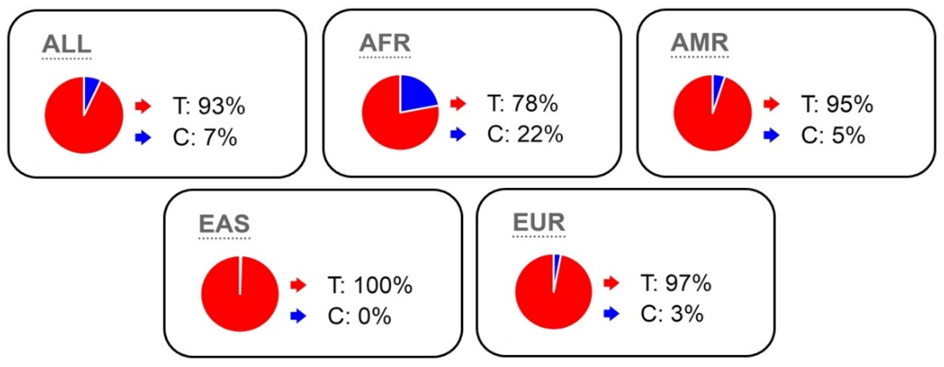
According to the Ensembl database, the frequency of the rare variant K153R is on an average 7% (3% in Caucasians and 22% in Africans), and large sample sizes are necessary to reliably identify the association of this polymorphism with strength abilities and muscle hypertrophy (Figure 2).
Figure 2. T and C allele frequencies of the MSTN K153R polymorphism according to the Ensembl database (All-General picture; AFR- African; AMR- American; EAS- Asian; EUR- Caucasian).
The conducted studies could not always prove a connection between the athletes’ skeletal muscle strength, muscle mass, and competitive performance [28][50]. Due to the low frequency of K153R polymorphism in Caucasian athletes of cyclic sports, the scholars point out the possibility of evaluating MSTN K153R polymorphism during sports selection and that this mutation needs further study.
The problem with studying mutations in MSTN is the low frequency of some alleles. Obtaining the required number of subjects and statistically significant data would need very specific subjects, for example highly qualified athletes of weightlifting sports or people with an exceptional proportion of skeletal muscles [52]. Such subjects, for instance, can also include some ethnicities, considering their residence [29].

References

  1. Beunen, G.; Thomis, M. Gene powered? Where to go from heritability (H-2) in muscle strength and power? Exerc. Sport Sci. Rev. 2004, 32, 148–154.
  2. Mangine, G.T.; Hoffman, J.R.; Gonzalez, A.M.; Townsend, J.R.; Wells, A.J.; Jajtner, A.R.; Beyer, K.S.; Boone, C.H.; Miramonti, A.A.; Wang, R.; et al. The effect of training volume and intensity on improvements in muscular strength and size in resistance-trained men. Physiol. Rep. 2015, 3, 17.
  3. Rodriguez, J.; Vernus, B.; Chelh, I.; Cassar-Malek, I.; Gabillard, J.C.; Sassi, A.H.; Seiliez, I.; Picard, B.; Bonnieu, A. Myostatin and the skeletal muscle atrophy and hypertrophy signaling pathways. Cell. Mol. Life Sci. 2014, 71, 4361–4371.
  4. Yamada, A.K.; Verlengia, R.; Bueno, C.R. Myostatin: Genetic variants, therapy and gene doping. Braz. J. Pharm. Sci. 2012, 48, 369–377.
  5. Dalbo, V.J.; Roberts, M.D.; Sunderland, K.L.; Poole, C.N.; Stout, J.R.; Beck, T.W.; Bemben, M.; Kerksick, C.M. Acute Loading and Aging Effects on Myostatin Pathway Biomarkers in Human Skeletal Muscle After Three Sequential Bouts of Resistance Exercise. J. Gerontol. Ser. A-Biol. Sci. Med. Sci. 2011, 66, 855–865.
  6. Allen, D.L.; Hittel, D.S.; McPherron, A.C. Expression and Function of Myostatin in Obesity, Diabetes, and Exercise Adaptation. Med. Sci. Sport. Exerc. 2011, 43, 1828–1835.
  7. Zheng, L.-F.; Chen, P.-J.; Xiao, W.-H. Signaling pathways controlling skeletal muscle mass. Acta Physiol. Sin. 2019, 49, 671–679.
  8. Roth, S.M.; Martel, G.F.; Ferrell, R.E.; Metter, E.J.; Hurley, B.F.; Rogers, M.A. Myostatin gene expression is reduced in humans with heavy resistance strength training: A brief communication. Exp. Biol. Med. 2003, 228, 706–709.
  9. Shishkin, S.S. Miostatin i nekotorye drugie biohimicheskie faktory, reguliruyushchie rost myshechnyh tkanej u cheloveka i ryada vysshih pozvonochnyh. Uspekhi Biol. Him. 2004, 44, 209–262.
  10. McPherron, A.C.; Lawler, A.M.; Lee, S.J. Regulation of skeletal muscle mass in mice by a new TGF-β superfamily member. Nature 1997, 387, 83–90.
  11. Kollias, H.D.; McDermott, J.C. Transforming growth factor-β and myostatin signaling in skeletal muscle. J. Appl. Physiol. 2008, 104, 579–587.
  12. McFarlane, C.; Hui, G.Z.; Amanda, W.Z.W.; Lau, H.Y.; Lokireddy, S.; Ge, X.J.; Mouly, V.; Butler-Browne, G.; Gluckman, P.D.; Sharma, M.; et al. Human myostatin negatively regulates human myoblast growth and differentiation. Am. J. Physiol.-Cell Physiol. 2011, 301, C195–C203.
  13. Ferrell, R.E.; Conte, V.; Lawrence, E.C.; Roth, S.M.; Hagberg, J.M.; Hurley, B.F. Frequent sequence variation in the human myostatin (GDF8) gene as a marker for analysis of muscle-related phenotypes. Genomics 1999, 62, 203–207.
  14. Sergeeva, K.V.; Miroshnikov, A.B.; Smolensky, A.V. Effect of Growth Hormone Administration on the Mass And Strength of Muscles in Healthy Young Adults: A Systematic Review and Meta-Analysis. Hum. Physiol. 2019, 4, 452–460.
  15. Pan, H.; Ping, X.C.; Zhu, H.J.; Gong, F.Y.; Dong, C.X.; Li, N.S.; Wang, L.J.; Yang, H.B. Association of myostatin gene polymorphisms with obesity in Chinese north Han human subjects. Gene 2012, 494, 237–241.
  16. Thomis, M.A.; Huygens, W.; Peeters, M.; Vlietinck, R.; Beunen, G.P. Linkage analysis of myostatin-pathway genes in human adiposity: The Leuven Genes for Muscular Strength Project. Med. Sci. Sport. Exerc. 2004, 36, S99.
  17. Feder, D.; Rugollini, M.; Santomauro, A.; Oliveira, L.P.; Lioi, V.P.; dos Santos, R.; Ferreira, L.G.; Nunes, M.T.; Carvalho, M.H.; Delgado, P.O.; et al. Erythropoietin reduces the expression of myostatin in mdx dystrophic mice. Braz. J. Med. Biol. Res. 2014, 47, 966–971.
  18. Gentile, M.A.; Nantermet, P.V.; Vogel, R.L.; Phillips, R.; Holder, D.; Hodor, P.; Cheng, C.; Dai, H.Y.; Freedman, L.P.; Ray, W.J. Androgen-mediated improvement of body composition and muscle function involves a novel early transcriptional program including IGF1, mechano growth factor, and induction of β-catenin. J. Mol. Endocrinol. 2010, 44, 55–73.
  19. Kim, J.S.; Cross, J.M.; Bamman, M.M. Impact of resistance loading on myostatin expression and cell cycle regulation in young and older men and women. Am. J. Physiol.-Endocrinol. Metab. 2005, 288, E1110–E1119.
  20. Lach-Trifilieff, E.; Minetti, G.C.; Sheppard, K.; Ibebunjo, C.; Feige, J.N.; Hartmann, S.; Brachat, S.; Rivet, H.; Koelbing, C.; Morvan, F.; et al. An Antibody Blocking Activin Type II Receptors Induces Strong Skeletal Muscle Hypertrophy and Protects from Atrophy. Mol. Cell. Biol. 2014, 34, 606–618.
  21. Jespersen, J.G.; Nedergaard, A.; Andersen, L.L.; Schjerling, P.; Andersen, J.L. Myostatin expression during human muscle hypertrophy and subsequent atrophy: Increased myostatin with detraining. Scand. J. Med. Sci. Sport. 2011, 21, 215–223.
  22. Lee, S.J.; McPherron, A.C. Regulation of myostatin activity and muscle growth. Proc. Natl. Acad. Sci. USA 2001, 98, 9306–9311.
  23. Walker, R.G.; Poggioli, T.; Katsimpardi, L.; Buchanan, S.M.; Oh, J.; Wattrus, S.; Heidecker, B.; Fong, Y.W.; Rubin, L.L.; Ganz, P.; et al. Biochemistry and Biology of GDF11 and Myostatin Similarities, Differences, and Questions for Future Investigation. Circ. Res. 2016, 118, 1125–1141.
  24. Hill, J.J.; Qiu, Y.C.; Hewick, R.M.; Wolfman, N.M. Regulation of myostatin in vivo by growth and differentiation factor-associated serum protein-1: A novel protein with protease inhibitor and follistatin domains. Mol. Endocrinol. 2003, 17, 1144–1154.
  25. Huang, Z.Q.; Chen, X.L.; Chen, D.W. Myostatin: A novel insight into its role in metabolism, signal pathways, and expression regulation. Cell. Signal. 2011, 23, 1441–1446.
  26. Drummond, M.J.; Glynn, E.L.; Fry, C.S.; Dhanani, S.; Volpi, E.; Rasmussen, B.B. Essential Amino Acids Increase MicroRNA-499,-208b, and-23a and Downregulate Myostatin and Myocyte Enhancer Factor 2C mRNA Expression in Human Skeletal Muscle. J. Nutr. 2009, 139, 2279–2284.
  27. Gonzalez-Freire, M.; Rodriguez-Romo, G.; Santiago, C.; Bustamante-Ara, N.; Yvert, T.; Gomez-Gallego, F.; Rexach, J.A.S.; Ruiz, J.R.; Lucia, A. The K153R variant in the myostatin gene and sarcopenia at the end of the human lifespan. Age 2010, 32, 405–409.
  28. Ben-Zaken, S.; Meckel, Y.; Nemet, D.; Rabinovich, M.; Kassem, E.; Eliakim, A. Frequency of the MSTN Lys(K)-153Arg(R) polymorphism among track & field athletes and swimmers. Growth Horm. IGF Res. 2015, 25, 196–200.
  29. Fuku, N.; Alis, R.; Yvert, T.; Zempo, H.; Naito, H.; Abe, Y.; Arai, Y.; Murakami, H.; Miyachi, M.; Pareja-Galeano, H.; et al. Muscle-Related Polymorphisms (MSTN rs1805086 and ACTN3 rs1815739) Are Not Associated with Exceptional Longevity in Japanese Centenarians. PLoS ONE 2016, 11, 0166605.
  30. Joulia-Ekaza, D.; Cabello, G. The myostatin gene: Physiology and pharmacological relevance. Curr. Opin. Pharmacol. 2007, 7, 310–315.
  31. Kostyunina, D.S.; Ivanova, A.D.; Smirnova, O.V. Myostatin: 20 years later. Hum. Physiol. 2018, 1, 99–114.
  32. Baczek, J.; Silkiewicz, M.; Wojszel, Z.B. Myostatin as a Biomarker of Muscle Wasting and other Pathologies-State of the Art and Knowledge Gaps. Nutrients 2020, 12, 2401.
  33. Szlama, G.; Trexler, M.; Buday, L.; Patthy, L. K153R polymorphism in myostatin gene increases the rate of promyostatin activation by furin. FEBS Lett. 2015, 589, 295–301.
  34. Walsh, F.S.; Celeste, A.J. Myostatin: A modulator of skeletal-muscle stem cells. Biochem. Soc. Trans. 2005, 33, 1513–1517.
  35. Zhang, Z.L.; He, J.W.; Qin, Y.J.; Hu, Y.Q.; Li, M.; Zhang, H.; Hu, W.W.; Liu, Y.J.; Gu, J.M. Association between myostatin gene polymorphisms and peak BMD variation in Chinese nuclear families. Osteoporos. Int. 2008, 19, 39–47.
  36. Elkasrawy, M.N.; Hamrick, M.W. Myostatin (GDF-8) as a key factor linking muscle mass and bone structure. J. Musculoskelet. Neuronal Interact. 2010, 10, 56–63.
  37. Zhu, J.; Li, Y.; Shen, W.; Qiao, C.; Ambrosio, F.; Lavasani, M.; Nozaki, M.; Branca, M.F.; Huard, J. Relationships between transforming growth factor-β 1, myostatin, and decorin—Implications for skeletal muscle fibrosis. J. Biol. Chem. 2007, 282, 25852–25863.
  38. Guo, W.; Flanagan, J.; Jasuja, R.; Kirkland, J.; Jiang, L.; Bhasin, S. The effects of myostatin on adipogenic differentiation of human bone marrow-derived mesenchymal stem cells are mediated through cross-communication between Smad3 and Wnt/β-catenin signaling pathways. J. Biol. Chem. 2008, 283, 9136–9145.
  39. Artaza, J.N.; Bhasin, S.; Magee, T.R.; Reisz-Porszasz, S.; Shen, R.Q.; Groome, N.P.; Fareez, M.M.; Gonzalez-Cadavid, N.F. Myostatin inhibits myogenesis and promotes adipogenesis in C3H 10T(1/2) mesenchymal multipotent cells. Endocrinology 2005, 146, 3547–3557.
  40. Hamrick, M.W.; Arounleut, P.; Kellum, E.; Cain, M.; Immel, D.; Liang, L.F. Recombinant Myostatin (GDF-8) Propeptide Enhances the Repair and Regeneration of Both Muscle and Bone in a Model of Deep Penetrant Musculoskeletal Injury. J. Trauma-Inj. Infect. Crit. Care 2010, 69, 579–583.
  41. Schuelke, M.; Wagner, K.R.; Stolz, L.E.; Hübner, C.; Riebel, T.; Kömen, W.; Braun, T.; Tobin, J.F.; Lee, S.-J. 2004. Myostatin Mutation Associated with Gross Muscle Hypertrophy in a Child. N. Engl. J. Med. 2004, 350, 2682–2688.
  42. Corsi, A.M.; Ferrucci, L.; Gozzini, A.; Tanini, A.; Brandi, M.L. Myostatin polymorphisms and age-related sarcopenia in the Italian population. J. Am. Geriatr. Soc. 2002, 50, 1463.
  43. Li, X.; Wang, S.J.; Tan, S.C.; Chew, P.L.; Liu, L.H.; Wang, L.; Wen, L.; Ma, L.H. The A55T and K153R polymorphisms of MSTN gene are associated with the strength training-induced muscle hypertrophy among Han Chinese men. J. Sport Sci. 2014, 32, 883–891.
  44. Kostek, M.A.; Angelopoulos, T.J.; Clarkson, P.M.; Gordon, P.M.; Moyna, N.M.; Visich, P.S.; Zoeller, R.F.; Price, T.B.; Seip, R.L.; Thompson, P.D.; et al. Myostatin and Follistatin Polymorphisms Interact with Muscle Phenotypes and Ethnicity. Med. Sci. Sport Exerc. 2009, 41, 1063–1071.
  45. Kim, J.; Park, K.; Lee, J. Myostatin A55T Genotype is Associated with Strength Recovery Following Exercise-Induced Muscle Damage. Int. J. Environ. Res. Public Health 2020, 17, 4900.
  46. Juffer, P.; Furrer, R.; Gonzalez-Freire, M.; Santiago, C.; Verde, Z.; Serratosa, L.; Morate, F.J.; Rubio, J.C.; Martin, M.A.; Ruiz, J.R.; et al. Genotype Distributions in Top-level Soccer Players: A Role for ACE? Int. J. Sport Med. 2009, 30, 387–392.
  47. Aksenov, M.O.; Andryushchenko, L.B. Myostatin gene role in strength building process. Teor. Prakt. Fiz. Kult. 2018, 4, 71–73.
  48. Santiago, C.; Ruiz, J.R.; Rodriguez-Romo, G.; Fiuza-Luces, C.; Yvert, T.; Gonzalez-Freire, M.; Gomez-Gallego, F.; Moran, M.; Lucia, A. The K153R Polymorphism in the Myostatin Gene and Muscle Power Phenotypes in Young, Non-Athletic Men. PLoS ONE 2011, 6, e16323.
  49. Filonzi, L.; Franchini, N.; Vaghi, M.; Chiesa, S.; Nonnis Marzano, F. The potential role of myostatin and neurotransmission genes in elite sport performances. J. Biosci. 2015, 40, 531–537.
  50. Ben-Zaken, S.; Meckel, Y.; Nemet, D.; Eliakim, A. The combined frequency of IGF and myostatin polymorphism among track & field athletes and swimmers. Growth Horm. IGF Res. 2017, 32, 29–32.
  51. Khanal, P.; He, L.X.; Herbert, A.J.; Stebbings, G.K.; Onambele-Pearson, G.L.; Degens, H.; Morse, C.I.; Thomis, M.; Williams, A.G. The Association of Multiple Gene Variants with Ageing Skeletal Muscle Phenotypes in Elderly Women. Genes 2020, 11, 1459.
  52. Grealy, R.; Herruer, J.; Smith, C.L.E.; Hiller, D.; Haseler, L.J.; Griffiths, L.R. Evaluation of a 7-Gene Genetic Profile for Athletic Endurance Phenotype in Ironman Championship Triathletes. PLoS ONE 2015, 10, e0145171.
More
Information
Contributors MDPI registered users' name will be linked to their SciProfiles pages. To register with us, please refer to https://encyclopedia.pub/register : ,
View Times: 1.0K
Revisions: 2 times (View History)
Update Date: 01 Feb 2023
1000/1000
ScholarVision Creations