Submitted Successfully!
To reward your contribution, here is a gift for you: A free trial for our video production service.
Thank you for your contribution! You can also upload a video entry or images related to this topic.
Version Summary Created by Modification Content Size Created at Operation
1 -- 2598 2023-01-12 14:55:49 |
2 format change Meta information modification 2598 2023-01-13 02:36:24 |

Video Upload Options

Do you have a full video?

Confirm

Are you sure to Delete?
Cite
If you have any further questions, please contact Encyclopedia Editorial Office.
Pocino, K.;  Stefanile, A.;  Basile, V.;  Napodano, C.;  D’ambrosio, F.;  Santo, R.D.;  Callà, C.A.M.;  Gulli, F.;  Saporito, R.;  Ciasca, G.; et al. Cytokines and Hepatocellular Carcinoma. Encyclopedia. Available online: https://encyclopedia.pub/entry/40120 (accessed on 30 July 2024).
Pocino K,  Stefanile A,  Basile V,  Napodano C,  D’ambrosio F,  Santo RD, et al. Cytokines and Hepatocellular Carcinoma. Encyclopedia. Available at: https://encyclopedia.pub/entry/40120. Accessed July 30, 2024.
Pocino, Krizia, Annunziata Stefanile, Valerio Basile, Cecilia Napodano, Francesca D’ambrosio, Riccardo Di Santo, Cinzia Anna Maria Callà, Francesca Gulli, Raffaele Saporito, Gabriele Ciasca, et al. "Cytokines and Hepatocellular Carcinoma" Encyclopedia, https://encyclopedia.pub/entry/40120 (accessed July 30, 2024).
Pocino, K.,  Stefanile, A.,  Basile, V.,  Napodano, C.,  D’ambrosio, F.,  Santo, R.D.,  Callà, C.A.M.,  Gulli, F.,  Saporito, R.,  Ciasca, G.,  Equitani, F.,  Basile, U., & Marino, M. (2023, January 12). Cytokines and Hepatocellular Carcinoma. In Encyclopedia. https://encyclopedia.pub/entry/40120
Pocino, Krizia, et al. "Cytokines and Hepatocellular Carcinoma." Encyclopedia. Web. 12 January, 2023.
Cytokines and Hepatocellular Carcinoma
Edit

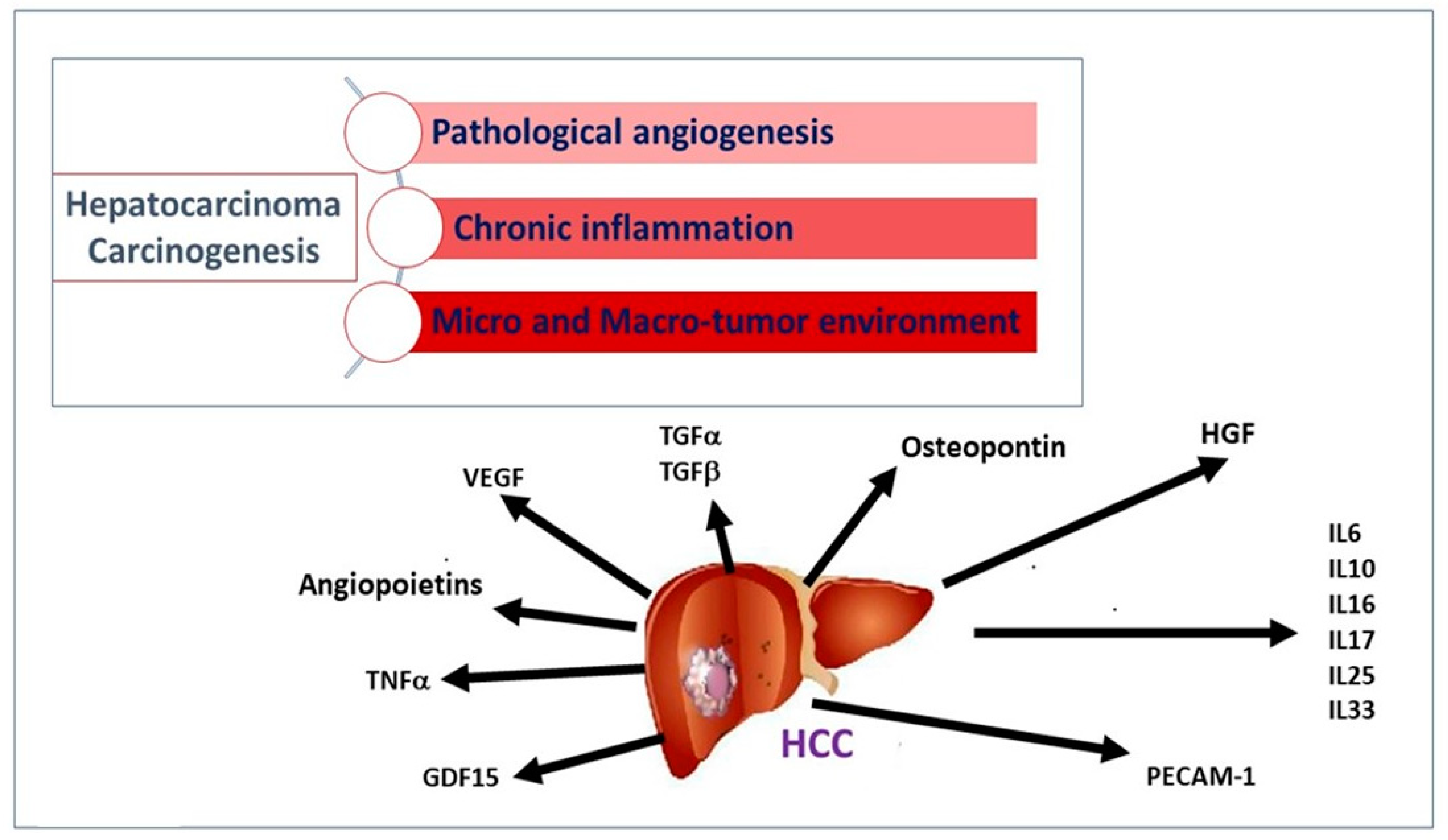
Hepatocellular carcinoma (HCC) represents a worldwide health matter with a major care burden, high prevalence, and poor prognosis. Its pathogenesis mainly varies depending on the underlying etiological factors, although it develops from liver cirrhosis in the majority of cases. In the premalignant environment, inflammatory cells release a wide range of cytokines, chemokines, growth factors, prostaglandins, and proangiogenic factors, making the liver environment more suitable for hepatocyte tumor progression that starts from acquired genetic mutations. A complex interaction of pro-inflammatory (IL-6, TNF-α) and anti-inflammatory cytokines (TGF-α and -β), pro-angiogenic molecules (including the Angiopoietins, HGF, PECAM-1, HIF-1α, VEGF), different transcription factors (NF-kB, STAT-3), and their signaling pathways are involved in the development of HCC. Since cytokines are expressed and released during the different stages of HCC progression, their measurement, by different available methods, can provide in-depth information on the identification and management of HCC. 

biomarkers cytokines hepatocellular carcinoma personalized medicine

1. Introduction

Hepatocellular carcinoma (HCC) is the most frequent type of cancer affecting the liver, and its incidence almost exceeds mortality. All those risk factors (chronic HBV or HCV infection, alcohol, aflatoxin B1, NAFLD/NASH) that concur with liver cirrhosis may be involved in HCC pathogenesis [1].
Cirrhotic liver tissue is characterized by low levels of hepatocyte cell proliferation in favor of a greater abundance of inflammatory mediators, fibrosis, and activation of the extracellular matrix environment. Therefore, a hepatocyte clone with a deregulated proliferative rate finds more suitable conditions for expansion, unlike in a normal and proliferating liver [2].
Following a viral infection or toxic tissue damage, a tightly regulated and coordinated multistep process may start in the liver, characterized by activation and local infiltration of immune cells and subsequent engagement in tissue repair. In this refined orchestration of events, the release of a wide range of soluble factors takes place [3].
In liver cirrhosis, a wide proliferation of stellate cells has been described, generating an abundance of extracellular matrix proteins, cytokines, growth factors, and oxidative stress products. The unbalanced expression of these factors and the initial unresolved inflammatory response produce a suitable microenvironment for developing neoplasm. Cytokines released by the tumor, neighboring non-tumor cells, and immune cells can act as a promoter of tumor survival [4] (Figure 1). Carcinogenetic events of HCC involve angiogenesis, chronic inflammation, and tumor micro and macro-environment (Figure 1).
Figure 1. Major pathogenetic events and cytokine network involved in hepatocellular carcinogenesis. Following persistent liver damage, locally activated chronic inflammation favors the release of soluble factors sustaining the proliferation and survival of tumor cells. Angiogenic factors (angiopoietins, VEGF), growth factors for normal and transformed hepatocytes (HGF), adhesion molecules and cytokines for recruitment and activation of leukocytes (PECAM, IL-6), and stellate cells upregulates TGF in the liver and, consequently, regeneration, proliferation, and hepatocyte dysplasia, and, ultimately, the development of HCC.

2. Cytokines and Growth Factors

2.1. Stimulators of Angiogenesis and Tumor Invasiveness

The progression of liver disease takes into account pathological angiogenesis, a prerequisite that facilitates the development of HCC. Angiogenesis is the result of a multiphase process and is the limiting step of tumor growth. In normal conditions, there is a balance between angiogenic inducers and inhibitors that keeps the angiogenic process under control and prevents inappropriate tissue vascularization. Angiogenesis inhibitors often derive from circulating extracellular matrix proteins (because of injury to the matrix), e.g., fibronectin, prolactin, collagen XVIII (endostatin), Hepatocyte Growth Factor fragment NK1, and angiostatin. Although tumors initially engage the pre-existing vascularity, an angiogenetic “switch” consisting of the production of factors inducing angiogenesis crucially modifies the tumor phenotype [5].
Vascular endothelial growth factor (VEGF) is the most powerful stimulator of normal and pathological angiogenesis. Circulating VEGF may be derived mainly from the large burden of tumor cells released under hypoxic conditions. Its expression is regulated by the hypoxia-inducible factor 1α (HIF-1α), which, induced during the hypoxic conditions, triggers the transcription of VEGF that stimulates the formation of new vessels (Figure 2A) [6]. This indicates that VEGF participates in the initial phase of angiogenesis. As a result, the transition of endothelial cells from an inactive to an active state can occur along with their proliferation, migration, and formation of new vessels, which can act as new gates for the recruitment of inflammatory cells, releasing cytokines and inducing further inflammation (Figure 2B). Different reports analyzed serum levels of VEGF in HCC patients in comparison to patients with or without HCV-related cirrhosis, often with opposite results [7][8]. Mukozu et al. showed that VEGF was higher in HCC patients compared to controls [7], while the results from Abden–Ramahal et al. displayed significantly higher serum levels of VEGF in HCC in comparison to cirrhotic patients, but no significant differences in healthy controls [8]. However, altogether these data highlight the important role of VEGF as a biomarker of vascular invasion in disease progression from liver cirrhosis to HCC.
Figure 2. (A) Hypoxic environment stimulates neoangiogenesis, thanks to a circuit involving HIF-1α -induced VEGF release by tumor cells. (B) A wide range of cellular released growth factors, derived from a tumor, stroma, and leukocytes, ensures the formation of new vascularity and the sustainment of cell growth, allowing the worsening progression of the initial tumor burden.
Patients with HCC showed a significant increase in VEGF after anticancer therapy compared to the values reported at the time of diagnosis, as well as to the levels of lymphocytes [9]. This may be partly explained by the rebound effect of VEGF, induced by hypoxia following locoregional treatments, often associated with treatment failure and low survival rates in patients [10]. Levels of VEGF are increased in patients who later experienced progression of HCC compared to those who remained stable [11]. Additionally, higher VEGF levels prior to sorafenib treatment (a multikinase inhibitor employed in several locally recurrent or metastatic solid tumors, including HCC) are associated with shorter survival [12][13].
The Tyrosine kinase proteins with Ig and EGF-homology domains 1 and 2 (Tie1 and Tie2) and their angiopoietin ligands 1–4 (Ang1, 2, 3, and 4) play a key role during the late phase of angiogenesis and are responsible for the maturation of newly established vascular structures. Ang1 and Ang2 have been deeply studied and characterized. The activity of the Angiopoietin/Tie system determines the stabilization of new vessels [14]. There is growing evidence that the angiopoietin/Tie signal can modify ongoing inflammation [15]. Ang1 appears to be a powerful activator of Tie2, as well as a regulator of blood vessel formation and development. Experimental studies showed that Ang1 acts as an anti-inflammatory molecule [16] but can induce pulmonary hypertension as a complication [17]. Ang1 also neutralizes tissue factor activity that is relevant for the induction of coagulation, thrombosis, and inflammatory response. Furthermore, Ang1 reduces the adhesion of VEGF-related leukocytes to the endothelium [18][19]. Conversely, Ang2 acts as a competitive antagonist of Ang1, deregulates the signal pathway of Tie2 [14], and plays a pro-inflammatory role [20][21]. Additionally, significantly high Ang2 serum levels have been observed in patients during liver carcinogenesis [22]. An increase in Ang2 levels has been observed in correlation with the liver disease progression [9][23], while Ang1 negatively correlates with the model for end-stage liver disease (MELD) and the hepatic fibrosis index. All these data confirm the potential diagnostic utility of Ang1 and Ang2 levels as developing new quantitative biomarkers for staging cirrhosis. Serum Ang2 concentrations decrease significantly after treatment with Direct Antiviral Drugs (DAAs), and, consequently, the Ang2/Ang1 ratio also drops. Ang2 is potentially useful in monitoring antiviral therapy [9].
It has been shown that the Hepatocyte growth factor (HGF) is over-expressed in HCC compared to the normal and cirrhotic liver without signs of neoplasia [9][24][25]. Expression of HGF and its receptor supports the existence of both autocrine and paracrine mechanisms of HGF action in HCC if compared to the unique paracrine mechanism found in normal liver tissue (in the absence of cancer), suggesting that it also plays a role in tumor development and progression [24][25][26]. Stellate cells and myofibroblasts are induced to secrete HGF from tumor cell products, and HGF, in turn, stimulates tumor cell invasiveness. Recent reports show that higher serum HGF levels negatively correlate with patient survival time [27] and positively with tumor size [28].
Furthermore, the comparison of cirrhotic patients with and without HCC suggests that HGF levels are potentially useful for monitoring the onset of HCC after a diagnosis of cirrhosis [9]. Interestingly, patients with lower HGF levels prior to treatment display major benefits from sorafenib therapy in terms of overall survival and time to progression [12].
Platelet endothelial cell adhesion molecule-1 (PECAM-1), also known as CD31, is normally found on the surface of endothelial cells, platelets, leukocyte subpopulations, and Kupffer cells [29]. This molecule is highly expressed within the vascular compartment but largely concentrated at junctions between adjacent cells, and its receptors mediate these interactions that play a crucial role during angiogenesis. In this context, PECAM-1 can mediate both homophilic and heterophilic adhesion [30]. PECAM-1 has been found to positively correlate with MELD, and its identification may aid in assessing the degree of tumor angiogenesis, which may indicate a rapidly growing tumor [31]. Furthermore, PECAM-1 promotes the formation of metastases by inducing the epithelium-mesenchymal transition in HCC by increasing the regulation of β1 integrin through the FAK (focal adhesion kinase) /Akt signaling pathway [32].

2.2. Stimulators of Chronic Inflammation, Liver Fibrosis, and Proliferation

Interleukin (IL)-6 acts as an important inducer of the acute phase response and infection defense in the liver [33]. IL-6 binds to the signal-transducing subunit gp130 on target cells either in complex with the membrane-bound or with the soluble IL-6 receptor to activate intracellular signaling. By the latter ‘trans-signaling’ mechanism, IL-6 can target monocyte chemotaxis and maintain sustained chronic inflammation towards any injured tissue [34][35]. Increased serum levels of IL-6 have been found in patients with advanced HCC compared to those with the early stage [36]. Additionally, elevated serum IL-6 levels in HCC patients undergoing hepatectomy are associated with lower overall survival and are prone to early relapses [37]. Research results from mouse models of HCC have shown that isolated HCC progenitor cells can give rise to cancer in the presence of ongoing liver damage and that these cells promote their own growth and progress to malignancy via autocrine IL-6 signaling [38]. A clinical study analyzing 128 HCC patients treated with sorafenib evaluated the prognostic value of serum IL-6 levels before treatment. Elevated pretreatment IL-6 concentrations have been found to be an independent predictor of poor overall survival, although there is no association with the efficacy of sorafenib [39].
Transforming growth factors (TGF)-α and TGF-β are closely related to the hepatocarcinogenesis process. Normal hepatocytes show low TGF-α expression compared to tumor cells. In fact, following chronic inflammation due to persistent liver damage, the secreted cytokine pool upregulates TGF-α in the liver and, consequently, regeneration, proliferation, hepatocyte dysplasia, and, ultimately, the development of HCC [40]. TGF-β is a key regulator of the late phase of inflammatory processes, not only promoting tissue repair but also inhibiting leukocyte activation and infiltration, acting at least in part using control of adhesion molecules on parenchymal cells [41]. In this way, TGF-β counteracts the effects of proinflammatory cytokines, leading to the inhibition of cellular processes, such as proliferation, differentiation, and survival. Paradoxically, cancer cells may exploit these microenvironment modifications to their advantage [42]. During carcinogenesis, malignant cells can often attenuate the suppressive TGF-β signaling by altering the expression of its receptors but also hijacking the signaling cascade. HCC cell lines with metastatic potential have been described to downregulate TGF-βR2. Interestingly, a reduced expression of TGF-βR2 in HCC correlates with larger tumor size and various metastatic features, such as poor differentiation, portal vein invasion, and intrahepatic metastases [43][44]. In the early stages of cancer, TGF-β acts as a tumor suppressor by inducing cytostasis and apoptosis, while in the later stages, it promotes pro-tumorigenic events, such as the transition of epithelial cells to mesenchymal, invasion, metastasis, and angiogenesis [45]. The simultaneous exposure (or addition) of TGF-β and IL-6 to human HCC cell cultures (Huh) highlighted an attenuation of the pro-proliferative effects induced by IL-6 by TGF-β. This explains a decrease in the transcription levels of the IL-6 receptor (IL-6R), in the expression of STAT-3 (signal transducer and activator of transcription) induced by IL-6, its nuclear localization, and, finally, a reduced activation of p65 compared to the unperturbed activation of the pathway. SMAD (small mother against decapentaplegic)-dependent TGF-β, resulting in the transition from epithelial to mesenchymal cells and, thus, loss of cell polarity and cell adhesion, as well as the acquisition of invasive and migratory properties, coupled with cell growth arrest [46].
IL-10 is a potent anti-inflammatory cytokine. Its role in HCC is less documented than in viral infections. A recent meta-analysis showed that IL-10 levels in HCC patients increased compared to cirrhotic patients and healthy controls but not compared to viral hepatitis patients [47]. Individuals with resectable HCC and IL-10 levels > 12 pg/mL display worse postoperative outcomes [48]. A study showed that in unresectable HCC, the serum levels of IL-10 acted as a negative prognostic factor [49].
Tumor necrosis factor (TNF) is a cytokine produced by proteolytic cleavage from a transmembrane protein precursor (mTNF) into a soluble TNF (sTNF). sTNF binds TNF receptors (TNFR1) (constitutively expressed in most tissues) and TNFR2 (expressed only in hematopoietic and endothelial cells), while mTNF only the type 2 receptor. TNF induces numerous biological responses in the liver, such as apoptosis, necrosis hepatocytes, hepatic inflammation, and regeneration, as well as the progression of HCC [50].
Expression of IL-6 and TNF-α during chronic liver injury activates the transduction pathway downstream of the transcription factor STAT3, which drives neoplastic transformation in the hepatic microenvironment [51]. IL-6 mediates its pro-proliferative effects through activation and direct interaction with the p65 subunit of NF-kB, activation of which is associated with a frequent and early event in liver fibrosis and HCC, regardless of etiology [52][53].

2.3. Liver Tumor Inducers

IL-16 is a pleiotropic cytokine whose activity influences both the chemical attraction and the modulation of the activation of T lymphocytes [54]. It has been identified as an important over-expressed cytokine in human liver tissue of HCC in both non-tumor and tumor regions compared to benign tumors and non-cancerous liver levels. Furthermore, IL-16 production can activate the ERK (extracellular signal-regulated kinase)/cyclin D1 signaling pathway, leading to tumor growth [55].
Different research groups evaluate osteopontin as an early marker of HCC. Produced by Kupffer cells, stellate cells, and hepatocytes, this cytokine is highly expressed at sites of inflammation and tissue remodeling [56]. Osteopontin mediates a wide range of biological functions in the immune and vascular systems and has been extensively studied in numerous cancers [57]. An increase in serum osteopontin levels was found in individuals with HCC compared to liver cirrhosis alone or chronic liver disease. The specific diagnostic efficacy of osteopontin in detecting early-stage HCC by differentiating them from non-HCC patients varies considerably among studies. Two studies report that osteopontin levels within two years of diagnosis have a reasonable predictive value of HCC with an AUC (area under the curve) of 0.82 [58][59].
After HCV eradication, patients undergo follow-up for the risk of developing HCC. Growth differentiation factor 15 (GDF15) is a cytokine, induced by mitochondrial dysfunction or oxidative stress. In one study, serum levels of GDF15 were measured from patients with chronic HCV infection without a history of HCC who had achieved a sustained virological response with DAAs. Serum levels of GDF15 were higher in patients with HCC onset after treatment with DAAs than in untreated patients. Furthermore, the score obtained using an algorithm composed of the GDF15, AFP (alpha-fetoprotein), and the FIB-4 index stratifies the risk of developing HCC de novo after the elimination of HCV [60].

3. Detection and Measurement of Cytokines

The demand for increased testing, particularly for early events of hepatocellular carcinogenesis, for its recurrence or detection of minimal residual disease in those asymptomatic patients requiring alternative approaches. Different methods for measurement of cytokines are currently available, including immunoassays for the detection of single molecules (ELISA, western blot), multiplex assays (chemiluminescent, bead-based (Luminex), and planar antibody arrays), and mass spectrometry [61].

References

  1. AIOM; AIRTUM. I Numeri Del Cancro in Italia, XI Edizione 2021. Available online: https://www.aiom.it/i-numeri-del-cancro-in-italia/ (accessed on 1 January 2021).
  2. Yang, Y.; Kim, S.; Seki, E. Inflammation and Liver Cancer: Molecular Mechanisms and Therapeutic Targets. Semin. Liver Dis. 2019, 39, 026–042.
  3. Turner, M.D.; Nedjai, B.; Hurst, T.; Pennington, D.J. Cytokines and Chemokines: At the Crossroads of Cell Signalling and Inflammatory Disease. Biochim. Biophys. Acta 2014, 1843, 2563–2582.
  4. Cabillic, F.; Corlu, A. Regulation of Transdifferentiation and Retrodifferentiation by Inflammatory Cytokines in Hepatocellular Carcinoma. Gastroenterology 2016, 151, 607–615.
  5. Medina, J.; Arroyo, A.G.; Sánchez-Madrid, F.; Moreno-Otero, R. Angiogenesis in Chronic Inflammatory Liver Disease. Hepatology 2004, 39, 1185–1195.
  6. Tammela, T.; Enholm, B.; Alitalo, K.; Paavonen, K. The Biology of Vascular Endothelial Growth Factors. Cardiovasc. Res. 2005, 65, 550–563.
  7. Mukozu, T.; Nagai, H.; Matsui, D.; Kanekawa, T.; Sumino, Y. Serum VEGF as a Tumor Marker in Patients with HCV-Related Liver Cirrhosis and Hepatocellular Carcinoma. Anticancer. Res. 2013, 33, 1013–1021.
  8. Zekri, A.-R.N.; Bahnassy, A.A.; Alam El-Din, H.M.; Morsy, H.M.; Shaarawy, S.; Moharram, N.Z.; Daoud, S.S. Serum Levels of β-Catenin as a Potential Marker for Genotype 4/Hepatitis C-Associated Hepatocellular Carcinoma. Oncol. Rep. 2011, 26, 825–831.
  9. Pocino, K.; Napodano, C.; Marino, M.; Di Santo, R.; Miele, L.; De Matthaeis, N.; Gulli, F.; Saporito, R.; Rapaccini, G.L.; Ciasca, G.; et al. A Comparative Study of Serum Angiogenic Biomarkers in Cirrhosis and Hepatocellular Carcinoma. Cancers 2021, 14, 11.
  10. Dong, G.; Lin, X.-H.; Liu, H.-H.; Gao, D.-M.; Cui, J.-F.; Ren, Z.-G.; Chen, R.-X. Intermittent Hypoxia Alleviates Increased VEGF and Pro-Angiogenic Potential in Liver Cancer Cells. Oncol. Lett. 2019, 18, 1831–1839.
  11. Miyahara, K.; Nouso, K.; Morimoto, Y.; Takeuchi, Y.; Hagihara, H.; Kuwaki, K.; Onishi, H.; Ikeda, F.; Miyake, Y.; Nakamura, S.; et al. Pro-Angiogenic Cytokines for Prediction of Outcomes in Patients with Advanced Hepatocellular Carcinoma. Br. J. Cancer 2013, 109, 2072–2078.
  12. Llovet, J.M.; Peña, C.E.A.; Lathia, C.D.; Shan, M.; Meinhardt, G.; Bruix, J. SHARP Investigators Study Group Plasma Biomarkers as Predictors of Outcome in Patients with Advanced Hepatocellular Carcinoma. Clin. Cancer Res. 2012, 18, 2290–2300.
  13. Gong, L.; Giacomini, M.M.; Giacomini, C.; Maitland, M.L.; Altman, R.B.; Klein, T.E. PharmGKB Summary: Sorafenib Pathways. Pharm. Genom. 2017, 27, 240–246.
  14. Augustin, H.G.; Koh, G.Y.; Thurston, G.; Alitalo, K. Control of Vascular Morphogenesis and Homeostasis through the Angiopoietin-Tie System. Nat. Rev. Mol. Cell Biol. 2009, 10, 165–177.
  15. Naldini, A.; Carraro, F. Role of Inflammatory Mediators in Angiogenesis. Curr. Drug Targets. Inflamm. Allergy 2005, 4, 3–8.
  16. Jeon, B.H.; Khanday, F.; Deshpande, S.; Haile, A.; Ozaki, M.; Irani, K. Tie-Ing the Antiinflammatory Effect of Angiopoietin-1 to Inhibition of NF-KappaB. Circ. Res. 2003, 92, 586–588.
  17. Sullivan, C.C.; Du, L.; Chu, D.; Cho, A.J.; Kido, M.; Wolf, P.L.; Jamieson, S.W.; Thistlethwaite, P.A. Induction of Pulmonary Hypertension by an Angiopoietin 1/TIE2/Serotonin Pathway. Proc. Natl. Acad. Sci. USA 2003, 100, 12331–12336.
  18. Koh, G.Y. Orchestral Actions of Angiopoietin-1 in Vascular Regeneration. Trends Mol. Med. 2013, 19, 31–39.
  19. Fiedler, U.; Augustin, H.G. Angiopoietins: A Link between Angiogenesis and Inflammation. Trends Immunol. 2006, 27, 552–558.
  20. Fiedler, U.; Reiss, Y.; Scharpfenecker, M.; Grunow, V.; Koidl, S.; Thurston, G.; Gale, N.W.; Witzenrath, M.; Rosseau, S.; Suttorp, N.; et al. Angiopoietin-2 Sensitizes Endothelial Cells to TNF-Alpha and Has a Crucial Role in the Induction of Inflammation. Nat. Med. 2006, 12, 235–239.
  21. Scharpfenecker, M.; Fiedler, U.; Reiss, Y.; Augustin, H.G. The Tie-2 Ligand Angiopoietin-2 Destabilizes Quiescent Endothelium through an Internal Autocrine Loop Mechanism. J. Cell Sci. 2005, 118, 771–780.
  22. Sugimachi, K.; Tanaka, S.; Taguchi, K.; Aishima, S.; Shimada, M.; Tsuneyoshi, M. Angiopoietin Switching Regulates Angiogenesis and Progression of Human Hepatocellular Carcinoma. J. Clin. Pathol. 2003, 56, 854–860.
  23. Scholz, A.; Rehm, V.A.; Rieke, S.; Derkow, K.; Schulz, P.; Neumann, K.; Koch, I.; Pascu, M.; Wiedenmann, B.; Berg, T.; et al. Angiopoietin-2 Serum Levels Are Elevated in Patients with Liver Cirrhosis and Hepatocellular Carcinoma. Am. J. Gastroenterol. 2007, 102, 2471–2481.
  24. García-Vilas, J.A.; Medina, M.Á. Updates on the hepatocyte growth factor/c-Met axis in hepatocellular carcinoma and its therapeutic implications. World J. Gastroenterol. 2018, 24, 3695–3708.
  25. Noguchi, O.; Enomoto, N.; Ikeda, T.; Kobayashi, F.; Marumo, F.; Sato, C. Gene Expressions of C-Met and Hepatocyte Growth Factor in Chronic Liver Disease and Hepatocellular Carcinoma. J. Hepatol. 1996, 24, 286–292.
  26. Ljubimova, J.Y.; Petrovic, L.M.; Wilson, S.E.; Geller, S.A.; Demetriou, A.A. Expression of HGF, Its Receptor c-Met, c-Myc, and Albumin in Cirrhotic and Neoplastic Human Liver Tissue. J. Histochem. Cytochem. 1997, 45, 79–87.
  27. Vejchapipat, P.; Tangkijvanich, P.; Theamboonlers, A.; Chongsrisawat, V.; Chittmittrapap, S.; Poovorawan, Y. Association between Serum Hepatocyte Growth Factor and Survival in Untreated Hepatocellular Carcinoma. J. Gastroenterol. 2004, 39, 1182–1188.
  28. Breuhahn, K.; Longerich, T.; Schirmacher, P. Dysregulation of Growth Factor Signaling in Human Hepatocellular Carcinoma. Oncogene 2006, 25, 3787–3800.
  29. Fodor, D.; Jung, I.; Turdean, S.; Satala, C.; Gurzu, S. Angiogenesis of hepatocellular carcinoma: An immunohistochemistry study. World J. Hepatol. 2019, 11, 294–304.
  30. DeLisser, H.M.; Christofidou-Solomidou, M.; Strieter, R.M.; Burdick, M.D.; Robinson, C.S.; Wexler, R.S.; Kerr, J.S.; Garlanda, C.; Merwin, J.R.; Madri, J.A.; et al. Involvement of Endothelial PECAM-1/CD31 in Angiogenesis. Am. J. Pathol. 1997, 151, 671–677.
  31. McCormick, B.A.; Zetter, B.R. Adhesive Interactions in Angiogenesis and Metastasis. Pharmacol. Ther. 1992, 53, 239–260.
  32. Zhang, Y.-Y.; Kong, L.-Q.; Zhu, X.-D.; Cai, H.; Wang, C.-H.; Shi, W.-K.; Cao, M.-Q.; Li, X.-L.; Li, K.-S.; Zhang, S.-Z.; et al. CD31 Regulates Metastasis by Inducing Epithelial-Mesenchymal Transition in Hepatocellular Carcinoma via the ITGB1-FAK-Akt Signaling Pathway. Cancer Lett. 2018, 429, 29–40.
  33. Schmidt-Arras, D.; Rose-John, S. IL-6 Pathway in the Liver: From Physiopathology to Therapy. J. Hepatol. 2016, 64, 1403–1415.
  34. Bartoccioni, E.; Scuderi, F.; Marino, M.; Provenzano, C. IL-6, Monocyte Infiltration and Parenchymal Cells. Trends Immunol. 2003, 24, 299–300, author reply 300–301.
  35. Marino, M.; Scuderi, F.; Ponte, E.; Maiuri, M.T.; De Cristofaro, R.; Provenzano, C.; Rose-John, S.; Cittadini, A.; Bartoccioni, E. Novel Path to IL-6 Trans-Signaling through Thrombin-Induced Soluble IL-6 Receptor Release by Platelets. J. Biol. Regul. Homeost. Agents 2013, 27, 841–852.
  36. Kao, J.-T.; Lai, H.-C.; Tsai, S.-M.; Lin, P.-C.; Chuang, P.-H.; Yu, C.-J.; Cheng, K.-S.; Su, W.-P.; Hsu, P.-N.; Peng, C.-Y.; et al. Rather than Interleukin-27, Interleukin-6 Expresses Positive Correlation with Liver Severity in Naïve Hepatitis B Infection Patients. Liver Int. 2012, 32, 928–936.
  37. Lai, S.-C.; Su, Y.-T.; Chi, C.-C.; Kuo, Y.-C.; Lee, K.-F.; Wu, Y.-C.; Lan, P.-C.; Yang, M.-H.; Chang, T.-S.; Huang, Y.-H. DNMT3b/OCT4 Expression Confers Sorafenib Resistance and Poor Prognosis of Hepatocellular Carcinoma through IL-6/STAT3 Regulation. J. Exp. Clin. Cancer Res. 2019, 38, 474.
  38. He, G.; Dhar, D.; Nakagawa, H.; Font-Burgada, J.; Ogata, H.; Jiang, Y.; Shalapour, S.; Seki, E.; Yost, S.E.; Jepsen, K.; et al. Identification of Liver Cancer Progenitors Whose Malignant Progression Depends on Autocrine IL-6 Signaling. Cell 2013, 155, 384–396.
  39. Shao, Y.-Y.; Lin, H.; Li, Y.-S.; Lee, Y.-H.; Chen, H.-M.; Cheng, A.-L.; Hsu, C.-H. High Plasma Interleukin-6 Levels Associated with Poor Prognosis of Patients with Advanced Hepatocellular Carcinoma. Jpn. J. Clin. Oncol. 2017, 47, 949–953.
  40. Zhang, J.; Wang, W.-L.; Li, Q.; Qiao, Q. Expression of Transforming Growth Factor-Alpha and Hepatitis B Surface Antigen in Human Hepatocellular Carcinoma Tissues and Its Significance. World J. Gastroenterol. 2004, 10, 830–833.
  41. Marino, M.; Scuderi, F.; Mannella, F.; Bartoccioni, E. TGF-Β1 and IL-10 Modulate IL-1β-Induced Membrane and Soluble ICAM-1 in Human Myoblasts. J. Neuroimmunol. 2003, 134, 151–157.
  42. Massagué, J. TGFbeta in Cancer. Cell 2008, 134, 215–230.
  43. Furuta, K.; Misao, S.; Takahashi, K.; Tagaya, T.; Fukuzawa, Y.; Ishikawa, T.; Yoshioka, K.; Kakumu, S. Gene Mutation of Transforming Growth Factor Beta1 Type II Receptor in Hepatocellular Carcinoma. Int. J. Cancer 1999, 81, 851–853.
  44. Alqahtani, A.; Khan, Z.; Alloghbi, A.; Said Ahmed, T.S.; Ashraf, M.; Hammouda, D.M. Hepatocellular Carcinoma: Molecular Mechanisms and Targeted Therapies. Medicina 2019, 55, E526.
  45. Gonzalez-Sanchez, E.; Vaquero, J.; Férnandez-Barrena, M.G.; Lasarte, J.J.; Avila, M.A.; Sarobe, P.; Reig, M.; Calvo, M.; Fabregat, I. The TGF-β Pathway: A Pharmacological Target in Hepatocellular Carcinoma? Cancers 2021, 13, 3248.
  46. Srivastava, A.; Sharma, H.; Khanna, S.; Sadhu Balasundaram, T.; Chowdhury, S.; Chowdhury, R.; Mukherjee, S. Interleukin-6 Induced Proliferation Is Attenuated by Transforming Growth Factor-β-Induced Signaling in Human Hepatocellular Carcinoma Cells. Front. Oncol. 2021, 11, 811941.
  47. Shakiba, E.; Ramezani, M.; Sadeghi, M. Evaluation of Serum Interleukin-6 Levels in Hepatocellular Carcinoma Patients: A Systematic Review and Meta-Analysis. Clin. Exp. Hepatol. 2018, 4, 182–190.
  48. Chau, G.Y.; Wu, C.W.; Lui, W.Y.; Chang, T.J.; Kao, H.L.; Wu, L.H.; King, K.L.; Loong, C.C.; Hsia, C.Y.; Chi, C.W. Serum Interleukin-10 but Not Interleukin-6 Is Related to Clinical Outcome in Patients with Resectable Hepatocellular Carcinoma. Ann. Surg. 2000, 231, 552–558.
  49. Shakiba, E.; Ramezani, M.; Sadeghi, M. Evaluation of serum interleukin-10 levels in hepatocellular carcinoma patients: A systematic review and meta-analysis. Clin. Exp. Hepatol. 2018, 4, 35–40.
  50. Tiegs, G.; Horst, A.K. TNF in the Liver: Targeting a Central Player in Inflammation. In Seminars in Immunopathology; Springer: Berlin/Heidelberg, Germany, 2022.
  51. Villanueva, A.; Luedde, T. The Transition from Inflammation to Cancer in the Liver. Clin. Liver Dis. 2016, 8, 89–93.
  52. Moran, D.M.; Mattocks, M.A.; Cahill, P.A.; Koniaris, L.G.; McKillop, I.H. Interleukin-6 Mediates G(0)/G(1) Growth Arrest in Hepatocellular Carcinoma through a STAT 3-Dependent Pathway. J. Surg. Res. 2008, 147, 23–33.
  53. Xu, J.; Lin, H.; Wu, G.; Zhu, M.; Li, M. IL-6/STAT3 Is a Promising Therapeutic Target for Hepatocellular Carcinoma. Front. Oncol. 2021, 11, 760971.
  54. Cruikshank, W.W.; Kornfeld, H.; Center, D.M. Interleukin-16. J. Leukoc. Biol. 2000, 67, 757–766.
  55. Takeba, Y.; Ohta, Y.; Ootaki, M.; Kobayashi, T.; Kida, K.; Watanabe, M.; Koizumi, S.; Otsubo, T.; Iiri, T.; Matsumoto, N. Identification of Interleukin-16 Production on Tumor Aggravation in Hepatocellular Carcinoma by a Proteomics Approach. Tumour Biol. 2021, 43, 309–325.
  56. Brown, L.F.; Berse, B.; Van de Water, L.; Papadopoulos-Sergiou, A.; Perruzzi, C.A.; Manseau, E.J.; Dvorak, H.F.; Senger, D.R. Expression and Distribution of Osteopontin in Human Tissues: Widespread Association with Luminal Epithelial Surfaces. Mol. Biol. Cell 1992, 3, 1169–1180.
  57. Zhao, H.; Chen, Q.; Alam, A.; Cui, J.; Suen, K.C.; Soo, A.P.; Eguchi, S.; Gu, J.; Ma, D. The Role of Osteopontin in the Progression of Solid Organ Tumour. Cell Death Dis. 2018, 9, 356.
  58. Da Costa, A.N.; Plymoth, A.; Santos-Silva, D.; Ortiz-Cuaran, S.; Camey, S.; Guilloreau, P.; Sangrajrang, S.; Khuhaprema, T.; Mendy, M.; Lesi, O.A.; et al. Osteopontin and Latent-TGF β Binding-Protein 2 as Potential Diagnostic Markers for HBV-Related Hepatocellular Carcinoma. Int. J. Cancer 2015, 136, 172–181.
  59. Duarte-Salles, T.; Misra, S.; Stepien, M.; Plymoth, A.; Muller, D.; Overvad, K.; Olsen, A.; Tjønneland, A.; Baglietto, L.; Severi, G.; et al. Circulating Osteopontin and Prediction of Hepatocellular Carcinoma Development in a Large European Population. Cancer Prev. Res. 2016, 9, 758–765.
  60. Myojin, Y.; Hikita, H.; Tahata, Y.; Doi, A.; Kato, S.; Sasaki, Y.; Shirai, K.; Sakane, S.; Yamada, R.; Kodama, T.; et al. Serum Growth Differentiation Factor 15 Predicts Hepatocellular Carcinoma Occurrence after Hepatitis C Virus Elimination. Aliment. Pharmacol. Ther. 2022, 55, 422–433.
  61. Kupcova Skalnikova, H.; Cizkova, J.; Cervenka, J.; Vodicka, P. Advances in Proteomic Techniques for Cytokine Analysis: Focus on Melanoma Research. Int. J. Mol. Sci. 2017, 18, E2697.
More
Information
Contributors MDPI registered users' name will be linked to their SciProfiles pages. To register with us, please refer to https://encyclopedia.pub/register : , , , , , , , , , , , ,
View Times: 443
Revisions: 2 times (View History)
Update Date: 13 Jan 2023
1000/1000
Video Production Service