 +1 credit
| Version | Summary | Created by | Modification | Content Size | Created at | Operation |
|---|---|---|---|---|---|---|
| 1 | Magdalena Zemelka-Wiacek | -- | 2810 | 2023-01-07 19:31:33 | | | |
| 2 | Peter Tang | -1 word(s) | 2809 | 2023-01-08 10:27:54 | | |
Video Upload Options
Metal allergy is mainly an environmental disorder which can cause allergic contact dermatitis. Environmental metal exposures include jewelry, everyday metal items, mobile phones, leather, metal-rich food and implants, including stents or anchors. While consumer exposure is liable for the majority of metal hypersensitivity cases, the significance of occupational exposure to metals remains relevant. The most common metal allergens are nickel, chromium, and cobalt.
1. Introduction
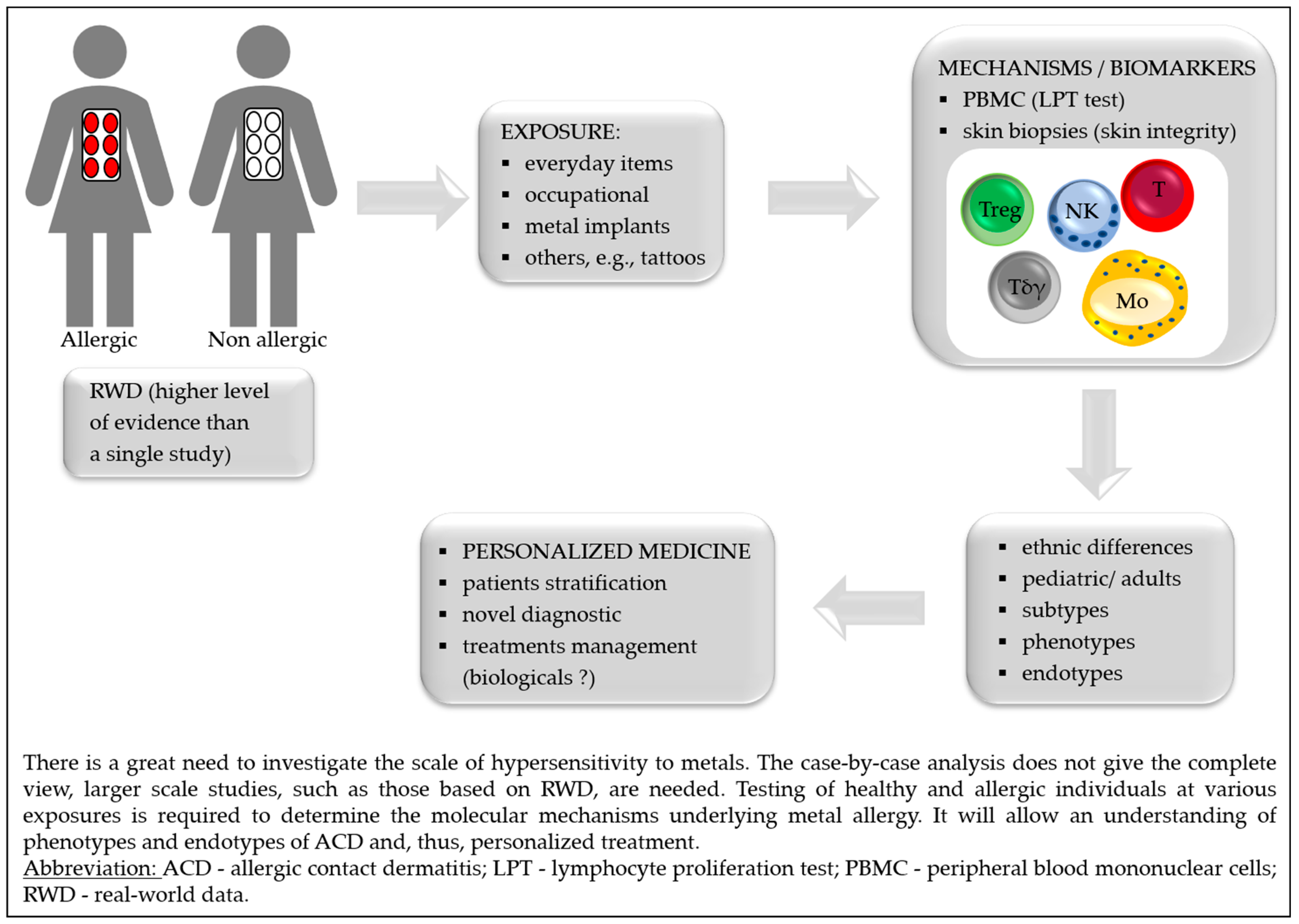
2. Exposure
3. Diagnostics
4. Mechanisms and Biomarkers
|
Outcomes |
Exposition to Metal |
Ref. |
|---|---|---|
|
↑ metal-specific CD154+ CD4+ Tmem (overexpression of TRAV9-2 and CDR3 histidine) |
Allergic and non-allergic subjects stimulated with Ni, Co or Pd (PBMC) |
|
|
↑ IL-5 |
Ni-allergic patients (PBMC) |
[69] |
|
Ni-allergy patients, differentiation between independent Ni or cross-reactivity of Ni/Pd allergy (PBMC) |
[70] |
|
|
Cross reactivity between Ni/Cr and Pd |
Sensitization with Ni or Cr, challenge with Pd (mice model) |
[71] |
|
Skin barrier defects: ↓ terminal differentiation—FLG, FLG2, LOR, LCEs, tight junction—CLDN1/CLDN8, lipid metabolism—FA2H, FABP7 |
Biopsies from healthy subjects after Ni-topical application |
[72] |
|
Cellular infiltrates: ↑ CD3+ T, CD11c+ myeloid DC, DC-LAMP+ mature DC, MBP+ eosinophils, FOXP3+ Treg |
||
|
↑ M1, mast cells, neutrophils, NK, CD4+ Tmem, CD8+ T |
Biopsies from Ni-allergic patients |
[73] |
|
↓ M2, resting mast cells, Tγδ, Treg |
||
|
↓ SBSN |
Ni-allergic patients (serum) |
[74] |
|
↑ Sema3A (activates MAPK and TNF-α) |
Ni-induced allergy (mouse ear tissue) |
[75] |
|
↑ TSLP in keratinocytes and TNF-α in epithelium |
OLPs metal-allergy patients |
[76] |
|
↑ IL-6, CXCL8, CCL2, CCL5, and CCL20 |
RHS exposure to Ni and Streptococcus mitis exposure |
[77] |
|
Lipid profile: ↑ cholesterol, DAG, MAG |
Non-allergic skin exposed to Co |
[78] |
|
Lipid profile: ↑ DAG |
Non-allergic skin exposed to Cr |
|
|
Lipid profile: ↓ DAG and MAG |
Non-allergic skin exposed to Ni |
|
|
LC emigration of the epidermis (in an IL-10 but not IL-1B dependent way) |
RHS exposure to Ti |
[79] |
Abbreviations: ↑ = increase; ↓ = decrease; + = positive; CCL = C-C motif ligand; CD = cluster of differentiation; CDR3 = complementarity determining region 3; CLDN = claudin; Co = cobalt; Cr = chromium; CXCL = C-X-C motif chemokine ligand; DC = dendritic cell; FA2H = fatty acid 2-hydroxylase; FABP7 = fatty acid binding protein 7, brain; FLG = filaggrin; FOXP3 = forkhead box P3; IL = interleukin; LAMP = lysosomal associated membrane glycoprotein; LCEs = late cornified envelope proteins; LOR = loricrin; M1/M2 = proinflammatory/anti-inflammatory macrophages; MAG/DAG = mono/diacylglycerols; MAPK = mitogen-activated protein kinases; MBP = myelin basic protein; Ni = nickel; NK = natural killer cells; OLP = oral lichen planus; PBMC = peripheral blood mononuclear cells; Pd = palladium; RHS = reconstructed human skin; SBNS = suprabasin; Sema3A = semaphorin; Tmem = memory lymphocytes T; TNF-α = tumor necrosis factor-alpha; TRAV9-2 = α-chain V-segment; TSLP = thymic stromal lymphopoietin; Tγδ = T gamma-delta cells.
5. Implants
|
Metal that Caused Hypersensitivity/Implant Type |
Symptoms |
Ref. |
|---|---|---|
|
Multiple metals/orthopedic implants: hip, knee shoulder joint |
Different; the most common—delayed wound healing and/or recurrent wound issues after the implantation, joint failure or loosening |
|
|
Ni/endovascular implants (stents) |
In-stent restenosis or prominent eczematous reaction overlying the endovascular implant, eosinophilia |
|
|
Co, Ni/drug-eluting stents |
Pruritic rash with hypereosinophilia |
[89] |
|
Au and Pd/dental implant |
Oral lichenoid contact lesion (OLCLs) |
[90] |
|
Ti/dental implants |
Rash, urticaria, pruritus, redness, dermatitis and facial eczema, pain, hyperaemia of soft tissues, swelling in submental and labial sulcus, gingival hyperplasia acne-like facial inflammation |
[91] |
|
Ni/metal anchors |
Erythematous and vesicular lesions around the grafted tattoo skin, but the tattoo was not affected (placed with clips or anchors) |
[59] |
|
Ni/stainless-steel skull pins |
Erythema on sites of the head where the skull pins inserted |
[92] |
|
Ti/cervical implant |
Persistent refractory neck pain; subsequently, after eight years, a planter rush |
[93] |
|
Ti/metal clips for cholecystectomy |
Right upper quadrant pain, diarrhea, and nausea |
[94] |
|
Low-grade fever, nausea, vomiting, joint pain, bloody diarrhea |
[95] |
|
|
Co, Ni, Hg/metal clips for cholecystectomy |
Myalgia, joint pain and tenderness, mental fogginess, mild forgetfulness, irritable bowel syndrome, stomach cramps, dry skin and hair, hair loss |
[96] |
|
Cu/intrauterine device |
Cutaneous eruption |
[97] |
|
Multiple-metals/dental implant |
Palmoplantar pustulosis (PPP) and periodontitis |
|
|
Au/dental implants |
Oral lesions with characteristic Wickham’s striae |
[100] |
|
Ti/temporary tissue expander |
Well-demarcated, erythematous plaque over the left breast reconstructive breast surgery |
[101] |
Abbreviations: Au = gold; Co = cobalt; Cu = copper, Hg = mercury; Ni = nickel; Pd = palladium; Ti = titanium.
References
- Johansen, J.D.; Bonefeld, C.M.; Schwensen, J.F.B.; Thyssen, J.P.; Uter, W. Novel insights into contact dermatitis. J. Allergy Clin. Immunol. 2022, 149, 1162–1171.
- Luger, T.; Amagai, M.; Dreno, B.; Dagnelie, M.-A.; Liao, W.; Kabashima, K.; Schikowski, T.; Proksch, E.; Elias, P.M.; Simon, M.; et al. Atopic dermatitis: Role of the skin barrier, environment, microbiome, and therapeutic agents. J. Dermatol. Sci. 2021, 102, 142–157.
- Fonacier, L.; Frankel, D.; Mawhirt, S. Contact allergens for the allergist. Ann. Allergy Asthma Immunol. 2022, 128, 629–644.
- Agache, I.; Akdis, C.A. Precision medicine and phenotypes, endotypes, genotypes, regiotypes, and theratypes of allergic diseases. J. Clin. Investig. 2019, 129, 1493–1503.
- Tokura, Y.; Hayano, S. Subtypes of atopic dermatitis: From phenotype to endotype. Allergol. Int. 2022, 71, 14–24.
- Wang, T.; Yin, L.; Ma, Z.; Zhang, Y. Chlorogenic Acid-Loaded Mesoporous Silica Nanoparticles Modified with Hexa-Histidine Peptides Reduce Skin Allergies by Capturing Nickel. Molecules 2022, 27, 1430.
- Quintana, B.R.; Hernández, A.F.; Guedes, A.S.; Borrego, L. Contact Dermatitis to Allergens in the Spanish Standard Series: PT Findings in the South of Gran Canaria. Actas Dermosifiliogr. 2022, 113, T555–T562.
- Linauskienė, K.; Malinauskienė, L.; Blažienė, A. Metals Are Important Contact Sensitizers: An Experience from Lithuania. BioMed Res. Int. 2017, 2017, 3964041.
- Silverberg, J.I.; Patel, N.; Warshaw, E.M.; Maibach, H.I.; Belsito, D.V.; DeKoven, J.G.; Zug, K.A.; Taylor, J.S.; Sasseville, D.; DeLeo, V.A.; et al. Patch testing with cobalt in children and adolescents: North American contact dermatitis group experience, 2001–2018. Contact Dermat. 2022, 87, 420–429.
- Mercan, S.; Vehid, H.; Semen, S.; Celik, U.; Yayla, M.; Engin, B. An ICP-MS Study for Quantitation of Nickel and Other Inorganic Elements in Urine Samples: Correlation of Patch Test Results with Lifestyle Habits. Biol. Trace Element Res. 2021, 200, 49–58.
- Linauskiene, K.; Dahlin, J.; Ezerinskis, Z.; Isaksson, M.; Sapolaite, J.; Malinauskiene, L. The Penetration of Chromium: An Up-To-Date 0.5% Potassium Dichromate Vehicle Comparison. Dermatitis 2022, 33, 368–372.
- Kim, D.; Kim, A.R.; Kim, H.; Lee, S.; Seo, B.; Suh, H.S.; Sim, C.S.; Lee, H.; Yoo, C. Nickel dust-induced occupational contact dermatitis by welding and grinding work in shipyard workers: A report of nine cases. Ann. Occup. Environ. Med. 2022, 34, e7.
- Özkaya, E.; Aslan, M.S.E. Occupational allergic contact dermatitis: A 24-year, retrospective cohort study from Turkey. Contact Dermat. 2021, 85, 503–513.
- Linde, S.J.L.; Franken, A.; du Plessis, J.L. Skin and respiratory exposure to soluble lead, cobalt, nickel, copper, arsenic and silver at two South African precious metals refineries. Int. Arch. Occup. Environ. Health 2022, 1–12.
- Babaahmadifooladi, M.; Jacxsens, L.; De Meulenaer, B.; Du Laing, G. Nickel in foods sampled on the Belgian market: Identification of potential contamination sources. Food Addit. Contam. Part A Chem. Anal. Control. Expo. Risk Assess. 2020, 37, 607–621.
- Babaahmadifooladi, M.; Jacxsens, L. Chronic dietary exposure to nickel from selected foods consumed in Belgium. Food Addit. Contam. Part A Chem. Anal. Control. Expo. Risk Assess. 2021, 38, 95–112.
- Cubadda, F.; Iacoponi, F.; Ferraris, F.; D’Amato, M.; Aureli, F.; Raggi, A.; Sette, S.; Turrini, A.; Mantovani, A. Dietary exposure of the Italian population to nickel: The national Total Diet Study. Food Chem. Toxicol. 2020, 146, 111813.
- Mania, M.; Rebeniak, M.; Orshulyak, O.; Postupolski, J. Assessment of exposure to nickel intake with selected cereal grains and cereal-based products. Rocz. Panstw. Zakl. Hig. 2020, 71, 371–376.
- EFSA Panel on Contaminants in the Food Chain (CONTAM); Schrenk, D.; Bignami, M.; Bodin, L.; Chipman, J.K.; Del Mazo, J.; Grasl-Kraupp, B.; Hogstrand, C.; Hoogenboom, L.; Leblanc, J.; et al. Update of the risk assessment of nickel in food and drinking water. EFSA J. 2020, 18, e06268.
- Pearson, A.J.; Ashmore, E. Risk assessment of antimony, barium, beryllium, boron, bromine, lithium, nickel, strontium, thallium and uranium concentrations in the New Zealand diet. Food Addit. Contam. Part A Chem. Anal. Control. Expo. Risk Assess. 2020, 37, 451–464.
- Picarelli, A.; Greco, N.; Sciuttini, F.; Marini, C.; Meacci, A. High consumption of Nickel-containing foods and IBS-like disorders: Late events in a gluten-free diet. Ecotoxicol. Environ. Saf. 2021, 222, 112492.
- Borghini, R.; De Amicis, N.; Bella, A.; Greco, N.; Donato, G.; Picarelli, A. Beneficial Effects of a Low-Nickel Diet on Relapsing IBS-Like and Extraintestinal Symptoms of Celiac Patients during a Proper Gluten-Free Diet: Nickel Allergic Contact Mucositis in Suspected Non-Responsive Celiac Disease. Nutrients 2020, 12, 2277.
- Borghini, R.; Porpora, M.G.; Casale, R.; Marino, M.; Palmieri, E.; Greco, N.; Donato, G.; Picarelli, A. Irritable Bowel Syndrome-Like Disorders in Endometriosis: Prevalence of Nickel Sensitivity and Effects of a Low-Nickel Diet. An Open-Label Pilot Study. Nutrients 2020, 12, 341.
- Yousaf, A.; Hagen, R.; Mitchell, M.; Ghareeb, E.; Fang, W.; Correa, R.; Zinn, Z.; Gayam, S. The effect of a low-nickel diet and nickel sensitization on gastroesophageal reflux disease: A pilot study. Indian J. Gastroenterol. 2021, 40, 137–143.
- Kageyama, Y.; Shimokawa, Y.; Kawauchi, K.; Morimoto, M.; Aida, K.; Akiyama, T.; Nakamura, T. Higher Prevalence of Nickel and Palladium Hypersensitivity in Patients with Ulcerative Colitis. Int. Arch. Allergy Immunol. 2020, 181, 456–461.
- Rizzi, A.; Chini, R.; Inchingolo, R.; Carusi, V.; Pandolfi, F.; Gasbarrini, A.; Nucera, E. Nickel allergy in lipid transfer protein sensitized patients: Prevalence and clinical features. Int. J. Immunopathol. Pharmacol. 2020, 34, 2058738420974895.
- Risi, R.; Masieri, S.; Poggiogalle, E.; Watanabe, M.; Caputi, A.; Tozzi, R.; Gangitano, E.; Masi, D.; Mariani, S.; Gnessi, L.; et al. Nickel Sensitivity Is Associated with GH-IGF1 Axis Impairment and Pituitary Abnormalities on MRI in Overweight and Obese Subjects. Int. J. Mol. Sci. 2020, 21, 9733.
- Kluger, N. Nickel and tattoos: Where are we? Contact Dermat. 2021, 85, 136–140.
- Blaser, P.; Rothmund, B.; Schmid, P.; Stadler, R.; Gemperle, C.; McCombie, G. Nickel release from metal items in contact with skin: A comparison of methods and practical implications for regulation in Europe. J. Environ. Sci. Health Part A Tox/Hazard. Subst. Environ. Eng. 2022, 57, 45–51.
- Wennervaldt, M.; Ahlström, M.G.; Menné, T.; Thyssen, J.P.; Johansen, J.D. Copper release from metals may mask positive nickel spot test results. Contact Dermat. 2022, 86, 431–433.
- Pavesi, T.; Moreira, J.C. A comprehensive study of nickel levels in everyday items in Brazil. Contact Dermat. 2020, 83, 88–93.
- Nayak, S.U.K.; Amala, D.; Shenoi, S.D. Nickel release from laptop detected by dimethylglyoxime (DMG) test. Indian J. Dermatol. 2021, 66, 696–697.
- Margulies, S.; Samia, A.M.; Montañez-Wiscovich, M.; Saikaly, S.K. Microneedling in the nickel-allergic patient. JAAD Int. 2022, 9, 48–49.
- Symanzik, C.; Uter, W.; Becker, S.; Skudlik, C.; John, S.M. Nickel and cobalt release from beauty tools: A field study in the German cosmetics trade. Contact Dermat. 2022, 87, 162–169.
- Symanzik, C.; Skudlik, C.; John, S.M. Nickel and cobalt: Underestimated contact allergens in hairdressers? Allergol. Sel. 2022, 6, 98–103.
- Symanzik, C.; Skudlik, C.; John, S.M. Experimental evaluation of nickel and cobalt release from tools and self-reported prevalence of nickel and cobalt allergy in the German hairdressing trade. J. Eur. Acad. Dermatol. Venereol. 2021, 35, 965–972.
- Available online: https://eur-lex.europa.eu/LexUriServ/LexUriServ.do?uri=OJ:L:2004:301:0051:0052:EN:PDF (accessed on 14 October 2022).
- Wennervaldt, M.; Ahlström, M.G.; Menné, T.; Thyssen, J.P.; Johansen, J.D. Nickel release from metallic earrings: A survey of the Danish market and validation of the nickel spot test. Contact Dermat. 2021, 85, 178–185.
- Mercan, S. A Comprehensive Artificial Sweat Study for Quantitation of Nickel and Other Inorganic Elements Released from Imitation Earrings Purchased in Istanbul Market. Biol. Trace Element Res. 2020, 194, 303–312.
- Wennervaldt, M.; Ahlström, M.G.; Menné, T.; Haulrig, M.B.; Alinaghi, F.; Thyssen, J.P.; Johansen, J.D. Chromium and cobalt release from metallic earrings from the Danish market. Contact Dermat. 2021, 85, 523–530.
- Saxena, S.; Tiwari, S. Effects of pH and time on Nickel Ion release from pediatric stainless-steel crowns: An In-Vitro Comparative Study. J. Pharm. Bioallied Sci. 2022, 14 (Suppl. S1), S545–S549.
- Hayakawa, M.; Suzuki, C.; Zhu, Y.; Anzai, H. Allergic contact dermatitis to gold in the parts of in-ear headphones. Contact Dermat. 2022, 86, 328–330.
- Oppel, E.; Kapp, F.; Böhm, A.S.; Pohl, R.; Thomas, P.; Summer, B. Contact sensitization to iron: A potentially underestimated metal allergen and elicitor of complications in patients with metal implants. Contact Dermat. 2022, 86, 531–538.
- Mistry, B.D.; DeKoven, J.G. Widespread cutaneous eruption after aluminum-containing vaccination: A case report and review of current literature. Pediatr. Dermatol. 2021, 38, 872–874.
- Aquino, M.R.; Bingemann, T.A.; Nanda, A.; Maples, K.M. Delayed allergic skin reactions to vaccines. Allergy Asthma Proc. 2022, 43, 20–29.
- Hoffmann, S.S.; Wennervaldt, M.; Alinaghi, F.; Simonsen, A.B.; Johansen, J.D. Aluminium contact allergy without vaccination granulomas: A systematic review and meta-analysis. Contact Dermat. 2021.
- Novack, D.E.; Yu, J.; Adler, B.L. Aluminum: The 2022 American Contact Dermatitis Society Allergen of the Year. Cutis 2022, 110, 21–24.
- Hoffmann, S.S.; Elberling, J.; Thyssen, J.P.; Hansen, K.S.; Johansen, J.D. Does aluminium in sunscreens cause dermatitis in children with aluminium contact allergy: A repeated open application test study. Contact Dermat. 2021, 86, 9–14.
- Bruze, M.; Netterlid, E.; Siemund, I. Aluminum—Allergen of the Year 2022. Dermatitis 2022, 33, 10–15.
- Fonacier, L.; Noor, I. Contact dermatitis and patch testing for the allergist. Ann. Allergy Asthma Immunol. 2018, 120, 592–598.
- Tupker, R.A.; Stapper, W.G.C.; Kelder, J.C. Predictive factors for Day 7 positive patch test readings at a secondary referral centre. Ski. Health Dis. 2021, 2, e79.
- Forkel, S.; Schubert, S.; Dickel, H.; Gina, M.; Schröder-Kraft, C.; Vieluf, D.; Brans, R.; Kreft, B.; Wurpts, G.; Geier, J.; et al. The benefit of late readings in patch testing depends both on allergen and patient characteristics. Allergy 2022, 77, 1477–1485.
- Kato, M.; Oiso, N.; Yanagihara, S.; Kawada, A. Long-lasting allergic patch test reactions to dental metal allergens in a patient with palmoplantar pustulosis and pustulotic arthro-osteitis. J. Dermatol. 2020, 47, e324–e325.
- Mufti, A.; Lu, J.D.; Sachdeva, M.; Zaaroura, H.; Kashetsky, N.; Yeung, J.; Maibach, H.I.; DeKoven, J. Patch Testing During Immunosuppressive Therapy: A Systematic Review. Dermatitis 2021, 32, 365–374.
- de Wijs, L.E.M.; van der Waa, J.D.; Nijsten, T.; Silverberg, J.I.; Kunkeler, A.C.M.; Hijnen, D.J. Effects of dupilumab treatment on patch test reactions: A retrospective evaluation. Clin. Exp. Allergy 2021, 51, 959–967.
- Raffi, J.; Suresh, R.; Botto, N.; Murase, J.E. The impact of dupilumab on patch testing and the prevalence of comorbid allergic contact dermatitis in recalcitrant atopic dermatitis: A retrospective chart review. J. Am. Acad. Dermatol. 2020, 82, 132–138.
- Johnson, H.; Adler, B.L.; Yu, J. Dupilumab for Allergic Contact Dermatitis: An Overview of Its Use and Impact on patch testing. Cutis 2022, 109, 265–267.
- Todberg, T.; Zachariae, C.; Krustrup, D.; Skov, L. The effect of anti-IL-17 treatment on the reaction to a nickel patch test in patients with allergic contact dermatitis. Int. J. Dermatol. 2019, 58, e58–e61.
- Marcant, P.; Alcaraz, I.; Beauval, N.; de Lassalle, E.M.; Chantelot, C.; Staumont-Sallé, D. Metal implant allergy: A diagnostic challenge illustrating the limits of the nickel spot test. Contact Dermat. 2021, 85, 251–253.
- Rodriguez, R.L.; Bujan, J.G. Silver: An underdiagnosed allergen? Contact Dermat. 2021, 84, 464–466.
- Bach, R.O.; Svendsen, M.T.; Mose, K.F.; Bruze, M.; Svedman, C.; Andersen, K.E. A comparison of patch testing with nickel sulfate in TRUE Test and in petrolatum at 2.5% and 5% concentrations. Contact Dermat. 2022, 86, 233–234.
- De Graaf, N.P.J.; Bontkes, H.J.; Roffel, S.; Kleverlaan, C.J.; Rustemeyer, T.; Gibbs, S.; Feilzer, A.J. Non–heat inactivated autologous serum increases accuracy of in vitro CFSE lymphocyte proliferation test (LPT) for nickel. Clin. Exp. Allergy 2020, 50, 722–732.
- Sachs, B.; Fatangare, A.; Sickmann, A.; Glässner, A. Lymphocyte transformation test: History and current approaches. J. Immunol. Methods 2021, 493, 113036.
- Glässner, A.; Dubrall, D.; Weinhold, L.; Schmid, M.; Sachs, B. Lymphocyte transformation test for drug allergy detection: When does it work? Ann. Allergy Asthma Immunol. 2022, 129, 497–506.e3.
- Blom, L.H.; Elrefaii, S.A.; Zachariae, C.; Thyssen, J.P.; Poulsen, L.K.; Johansen, J.D. Memory T helper cells identify patients with nickel, cobalt, and chromium metal allergy. Contact Dermat. 2021, 85, 7–16.
- Renert-Yuval, Y.; Thyssen, J.P.; Bissonnette, R.; Bieber, T.; Kabashima, K.; Hijnen, D.; Guttman-Yassky, E. Biomarkers in atopic dermatitis—A review on behalf of the International Eczema Council. J. Allergy Clin. Immunol. 2021, 147, 1174–1190.e1.
- Aparicio-Soto, M.; Riedel, F.; Leddermann, M.; Bacher, P.; Scheffold, A.; Kuhl, H.; Timmermann, B.; Chudakov, D.M.; Molin, S.; Worm, M.; et al. TCRs with segment TRAV9-2 or a CDR3 histidine are overrepresented among nickel-specific CD4+ T cells. Allergy 2020, 75, 2574–2586.
- Riedel, F.; Aparicio-Soto, M.; Curato, C.; Münch, L.; Abbas, A.; Thierse, H.; Peitsch, W.K.; Luch, A.; Siewert, K. Unique and common TCR repertoire features of Ni2+-, Co2+-, and Pd2+-specific human CD154+ CD4+ T cells. Allergy 2022.
- De Graaf, N.P.J.; Roffel, S.; Gibbs, S.; Kleverlaan, C.J.; Gonzalez, M.L.; Rustemeyer, T.; Feilzer, A.J.; Bontkes, H.J. Nickel allergy is associated with a broad spectrum cytokine response. Contact Dermat. 2022.
- Kapp, F.; Summer, B.; Thomas, P. Usefulness of lymphocyte transformation test and in vitro cytokine release in differentiating between independent and cross-reacting nickel/palladium allergy. Immun. Inflamm. Dis. 2020, 8, 483–492.
- Shigematsu, H.; Kumagai, K.; Suzuki, M.; Eguchi, T.; Matsubara, R.; Nakasone, Y.; Nasu, K.; Yoshizawa, T.; Ichikawa, H.; Mori, T.; et al. Cross-Reactivity of Palladium in a Murine Model of Metal-induced Allergic Contact Dermatitis. Int. J. Mol. Sci. 2020, 21, 4061.
- Pavel, A.B.; Del Duca, E.; Cheng, J.; Wu, J.; Ungar, B.; Estrada, Y.D.; Jack, C.; Maari, C.; Proulx, E.S.-C.; Ramirez-Valle, F.; et al. Delayed type hypersensitivity reactions to various allergens may differently model inflammatory skin diseases. Allergy 2022.
- Wisgrill, L.; Werner, P.; Jalonen, E.; Berger, A.; Lauerma, A.; Alenius, H.; Fyhrquist, N. Integrative transcriptome analysis deciphers mechanisms of nickel contact dermatitis. Allergy 2021, 76, 804–815.
- Nakazawa, S.; Phadungsakswasdi, P.; Kageyama, H.; Fukuchi, K.; Shimauchi, T.; Fujiyama, T.; Ito, T.; Honda, T. Decreased serum level of suprabasin in patients with nickel allergy. J. Dermatol. 2022, 49, e189–e190.
- Liu, L.; Watanabe, M.; Minami, N.; Yunizar, M.F.; Ichikawa, T. Semaphorin 3A: A potential target for prevention and treatment of nickel allergy. Commun. Biol. 2022, 5, 671.
- Yunizar, M.F.; Watanabe, M.; Liu, L.; Minami, N.; Ichikawa, T. Metal Allergy Mediates the Development of Oral Lichen Planus via TSLP-TSLPR Signaling. J. Clin. Med. 2022, 11, 519.
- Shang, L.; Deng, D.; Roffel, S.; Gibbs, S. Differential influence of Streptococcus mitis on host response to metals in reconstructed human skin and oral mucosa. Contact Dermat. 2020, 83, 347–360.
- Knox, S.; Hagvall, L.; Malmberg, P.; O’Boyle, N.M. Topical Application of Metal Allergens Induces Changes to Lipid Composition of Human Skin. Front. Toxicol. 2022, 4, 867163.
- Neves, C.T.R.; Spiekstra, S.W.; de Graaf, N.P.; Rustemeyer, T.; Feilzer, A.J.; Kleverlaan, C.J.; Gibbs, S. Titanium salts tested in reconstructed human skin with integrated MUTZ-3-derived Langerhans cells show an irritant rather than a sensitizing potential. Contact Dermat. 2020, 83, 337–346.
- Schalock, P.C.; Thyssen, J.P. Patch Testers’ Opinions Regarding Diagnostic Criteria for Metal Hypersensitivity Reactions to Metallic Implants. Dermatitis 2013, 24, 183–185.
- Shahid, M.; Vassileva, S.; Drenovska, K. Nickel and Skin: From Allergy to Autoimmunity. Endocr. Metab. Immune Disord.-Drug Targets 2020, 20, 1032–1040.
- Peacock, C.J.H.; Fu, H.; Asopa, V.; Clement, N.D.; Kader, D.; Sochart, D.H. The effect of Nickel hypersensitivity on the outcome of total knee arthroplasty and the value of skin patch testing: A systematic review. Arthroplasty 2022, 4, 40.
- Baumann, C.A.; Crist, B.D. Nickel allergy to orthopaedic implants: A review and case series. J. Clin. Orthop. Trauma 2020, 11 (Suppl. S4), S596–S603.
- Yang, S.; Choi, E.; Ng, Y.H. Cutaneous Metal Hypersensitivity Reaction. Case Rep. Dermatol. 2022, 14, 61–65.
- Matar, H.E.; Porter, P.J.; Porter, M.L. Metal allergy in primary and revision total knee arthroplasty. Bone Jt. Open 2021, 2, 785–795.
- Lamb, L.; Dass, R.; Dass, K. M311 An unusual presentation of metal hypersensitivity symptom recurrence during omalizumab treatment successfully treated with dupilumab. Ann. Allergy Asthma Immunol. 2021, 127, S128–S129.
- Guéroult, A.M.; Al-Balah, A.; Davies, A.H.; Shalhoub, J. Nickel hypersensitivity and endovascular devices: A systematic review and meta-analysis. Heart 2021, 108, 1707–1715.
- Joshi, S.R.; Khan, D.A. Effective Use of Dupilumab in Managing Systemic Allergic Contact Dermatitis. Dermatitis 2018, 29, 282–284.
- Faybusovich, P.; Lim, J.; Ioffreda, M.D.; Al-Shaikhly, T. Mepolizumab for treating systemic allergic dermatitis with hypereosinophilia likely secondary to a nickel/cobalt-containing coronary artery stent. Contact Dermat. 2022, 86, 123–125.
- Tsushima, F.; Sakurai, J.; Shimizu, R.; Kayamori, K.; Harada, H. Oral lichenoid contact lesions related to dental metal allergy may resolve after allergen removal. J. Dent. Sci. 2022, 17, 1300–1306.
- Comino-Garayoa, R.; Brinkmann, J.C.-B.; Peláez, J.; López-Suárez, C.; Martínez-González, J.M.; Suárez, M.J. Allergies to Titanium Dental Implants: What Do We Really Know about Them? A Scoping Review. Biology 2020, 9, 404.
- Matsudate, Y. Case of allergic contact dermatitis due to nickel contained in stainless steel skull pins. J. Dermatol. 2022, 49, e307–e308.
- Aoyama, R.; Anazawa, U.; Hotta, H.; Watanabe, I.; Takahashi, Y.; Matsumoto, S. Cervical Implant Allergy With Chronic Neck Pain: A Case Report. Cureus 2022, 14, e28293.
- Yabit, F.; Hughes, L.; Sylvester, B.; Tiesenga, F. Hypersensitivity Reaction Post Laparoscopic Cholecystectomy Due to Retained Titanium Clips. Cureus 2022, 14, e26167.
- Jain, M.S.; Lingarajah, S.; Luvsannyam, E.; Somagutta, M.R.; Jagani, R.P.; Sanni, J.; Ebose, E.; Tiesenga, F.M.; Jorge, J.M. Delayed Titanium Hypersensitivity and Retained Foreign Body Causing Late Abdominal Complications. Case Rep. Surg. 2021, 2021, 5515401.
- Shah, R.N.; Tiesenga, F.; Jorge, J.; Chaudhry, A.F. Surgical clips metal allergy postlaparoscopic cholecystectomy. Int. J. Surgery Glob. Health 2020, 4, e48.
- Gara, S.; Litaiem, N.; Bacha, T.; Jones, M.; Houas, A.; Zeglaoui, F. Systemic allergic dermatitis caused by a copper-containing intra-uterine device. Contact Dermat. 2021, 84, 132–134.
- Takaoka, Y.; Akiba, Y.; Nagasawa, M.; Ito, A.; Masui, Y.; Akiba, N.; Eguchi, K.; Miyazawa, H.; Tabeta, K.; Uoshima, K. The relationship between dental metal allergy, periodontitis, and palmoplantar pustulosis: An observational study. J. Prosthodont. Res. 2022, 66, 438–444.
- Nagura, S.; Sakai, M.; Obi, H.; Fukahara, K. Aortic valve replacement in a patient with self-reported systemic multiple metal allergy. Gen. Thorac. Cardiovasc. Surg. 2021, 70, 79–82.
- Rasul, T.F.; Anderson, J.; Bergholz, D.R.; Faiz, A.; Prasad, R.R. Gold Dental Implant-Induced Oral Lichen Planus. Cureus 2022, 14, e21852.
- Buonomo, M.; Ruggiero, J.L.; Hylwa, S. Titanium allergy as a likely cause of post-reconstruction dermatitis of the breast. Contact Dermat. 2022, 86, 142–143.

