Submitted Successfully!
To reward your contribution, here is a gift for you: A free trial for our video production service.
Thank you for your contribution! You can also upload a video entry or images related to this topic.
Version Summary Created by Modification Content Size Created at Operation
1 -- 3499 2022-12-06 22:13:47 |
2 Reference format revised. + 3 word(s) 3502 2022-12-08 01:53:22 | |
3 Remove "(Section 4.1)” -1 word(s) 3501 2022-12-08 08:41:58 |

Video Upload Options

Do you have a full video?

Confirm

Are you sure to Delete?
Cite
If you have any further questions, please contact Encyclopedia Editorial Office.
Boccalatte, F.;  Mina, R.;  Aroldi, A.;  Leone, S.;  Suryadevara, C.M.;  Placantonakis, D.G.;  Bruno, B. CAR T Cell Immune Therapy for Solid Tumors. Encyclopedia. Available online: https://encyclopedia.pub/entry/38145 (accessed on 27 July 2024).
Boccalatte F,  Mina R,  Aroldi A,  Leone S,  Suryadevara CM,  Placantonakis DG, et al. CAR T Cell Immune Therapy for Solid Tumors. Encyclopedia. Available at: https://encyclopedia.pub/entry/38145. Accessed July 27, 2024.
Boccalatte, Francesco, Roberto Mina, Andrea Aroldi, Sarah Leone, Carter M. Suryadevara, Dimitris G. Placantonakis, Benedetto Bruno. "CAR T Cell Immune Therapy for Solid Tumors" Encyclopedia, https://encyclopedia.pub/entry/38145 (accessed July 27, 2024).
Boccalatte, F.,  Mina, R.,  Aroldi, A.,  Leone, S.,  Suryadevara, C.M.,  Placantonakis, D.G., & Bruno, B. (2022, December 06). CAR T Cell Immune Therapy for Solid Tumors. In Encyclopedia. https://encyclopedia.pub/entry/38145
Boccalatte, Francesco, et al. "CAR T Cell Immune Therapy for Solid Tumors." Encyclopedia. Web. 06 December, 2022.
CAR T Cell Immune Therapy for Solid Tumors
Edit

Chimeric antigen receptor (CAR) T cells are genetically engineered T cells that recognize markers present on tumor cells and drive the degradation of the tumor itself. CAR T immunotherapy has obtained remarkable success in targeting a number of blood malignancies; however, its outcome is typically modest when applied to solid tumors, because of specific structural, biological, and metabolic aspects of the solid tumor environment. 

solid tumors chimeric antigen receptor (CAR) T cell adoptive immunotherapy tumor microenvironment

1. The Solid Tumor Microenvironment and Its Impact on CAR T Therapy

The tumor microenvironment (TME) is a complex ecosystem where tumor cells interact with soluble factors (cytokines, chemokines), immune cells (e.g., lymphocytes, phagocytic cells, and antigen-presenting cells), and non-immune cells (such as endothelial and stromal cells). Moreover, physical, metabolic, and biochemical factors contribute to the TME, and have a significant impact on the natural immune responses and on immunotherapies (Figure 1). Though solid tumors represent the majority of cancers, adoptive immunotherapies, including the most innovative chimeric antigen receptor (CAR)  T cells, have been far less effective in solid tumors compared to hematological malignancies.
Figure 1. The tumor microenvironment (TME) in solid tumors. Numerous factors in a solid tumor mass inhibit the function of CAR T cells. Cancer cells downregulate their antigens and express immune checkpoint inhibitor molecules (PD1 and others). They also generate an unfavorable microenvironment by secreting metabolites that inhibit the function of CAR T, such as kynurenine, adenosine, and lactate (the latter contributing to lowering the pH). Moreover, tumor cells scavenge nutrients, such as glucose, which, when converted into phosphoenolpyruvate (PEP), are essential for CAR T cell function. Immunosuppressive leucocytes also participate in dampening the function of CAR T cells; tumor-associated macrophages (TAMs), myeloid-derived suppressor cells (MDSCs), and regulatory T cells (Treg) secrete inhibitory cytokines (TGFb, IL-4, IL-10) that inhibit CAR T cell activation and favor tumor escape. Another cytokine, VEGF, stimulates the formation of an aberrant vasculature, which contributes to the hypoxic environment hostile to CAR T cell function. The cancer stroma also contains cancer-associated fibroblasts (CAFs) that secrete inhibitory cytokines and the extracellular matrix (ECM), forming a physical barrier to CAR T cell infiltration.

1.1. CAR T Cell Trafficking and Infiltration

In the setting of solid tumors, immune cells should efficiently reach tumor sites and infiltrate masses that can be of considerable size. Successful immune cell trafficking depends on the concordant expression of chemokines secreted by the tumor, and the appropriate chemokine receptors on the T cells. Similarly, the infiltration process is driven by a matched expression of adhesion receptors/ligands by the T cells and the tumor endothelium. Unfortunately, tumors often downmodulate the expression of chemoattractant molecules, therefore escaping immune surveillance [1].

1.2. The Solid Tumor Microenvironment: Physical and Metabolic Barriers

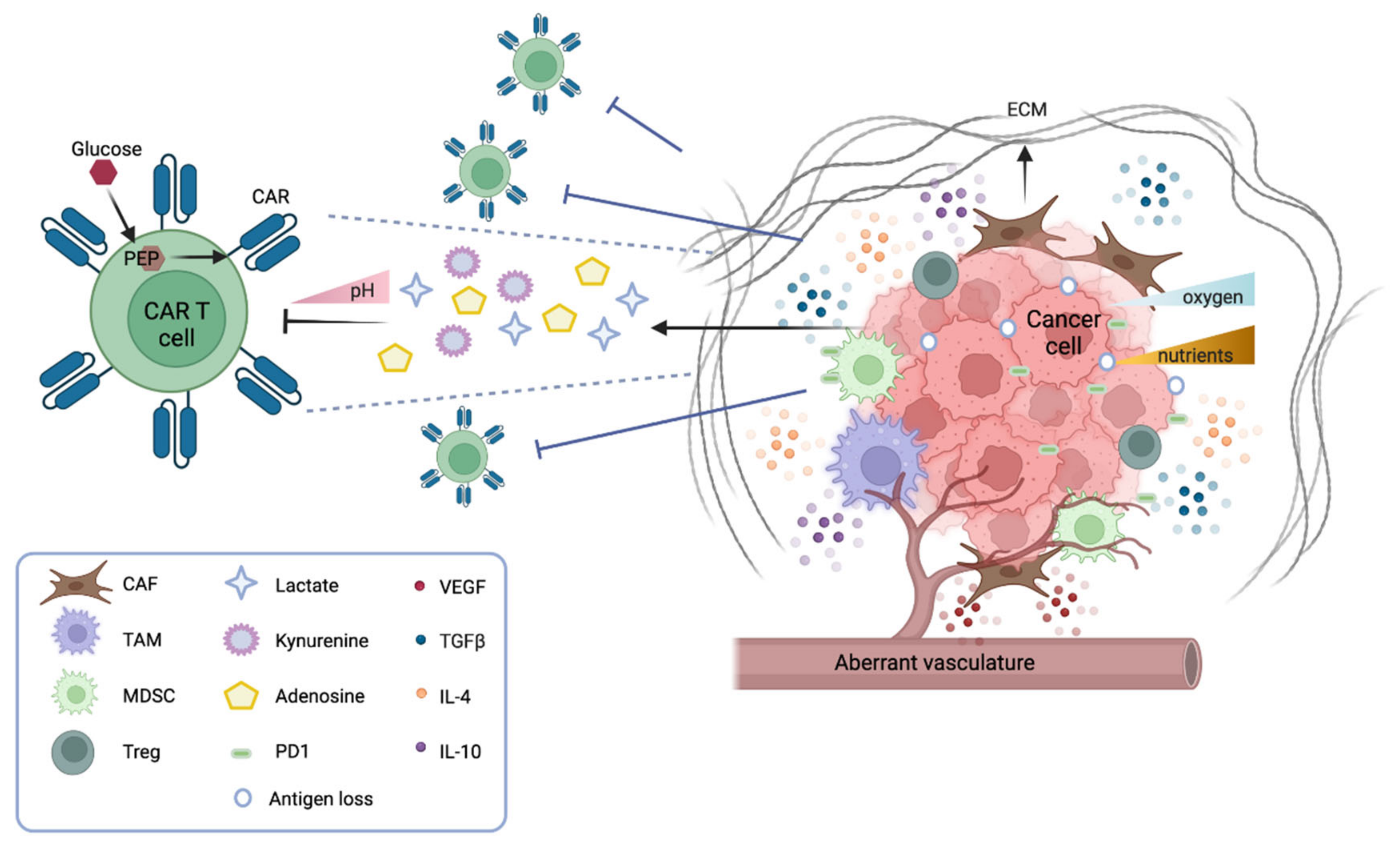
Several physical barriers hamper the accessibility of CAR T cells to a solid tumor mass, including thick surrounding tumor stroma, aberrant vasculature, and high interstitial pressure. The stroma is mostly constituted by cancer-associated fibroblasts (CAFs) that drive the deposition of the extracellular matrix (ECM), thus physically preventing the infiltration of immune cells. Several strategies are under investigation to overcome this obstacle. CAR T cells can be engineered to target fibroblast activation protein (FAP), therefore reducing the number of CAFs in the microenvironment [2]. Alternatively, CAR T cells can be armored with proteases that degrade the ECM. For instance, CAR T cells engineered to express heparanase, which degrades the heparan sulfate component of the ECM, have shown better infiltration and tumor clearance both in vitro and in animal models [3]. Moreover, the aberrant tumor vasculature causes interstitial hypertension that prevents extravasation and a hypoxic microenvironment, especially in the central part of the tumor. Thus, normalizing the tumor vasculature may be beneficial [4]. In this context, vascular endothelial growth factor (VEGF) signaling plays a pivotal role. Antiangiogenic therapy that blocks VEGF signaling improves immune cell infiltration [5], and anti-VEGFR CAR T cells can efficiently inhibit tumor growth—as shown in several syngeneic mouse models [6].

In terms of metabolic barriers, it is worth noticing that the particular anatomical structure of solid tumors generates hostile hypoxia and nutrient starvation for immune cells. The hypoxic environment caused by poor perfusion and abnormal vasculature hampers the expansion of CAR T cells and shifts their phenotype from effector to central memory [7]. Strategies to favor CAR T response in the hypoxic TME are under investigation by fusing an oxygen-sensitive domain of hypoxia-inducible factor 1 (HIF1a) to the CAR scaffold [8].
Of note, to mount an effective antitumor response, CAR T cells need to proliferate and produce cytokines and molecules that degrade tumor cells. Thus, CAR T cells must compete for nutrients and metabolites in a niche where tumor cells are scavenging most resources. CAR T cell effector functions rely on glucose and glycolytic metabolism [9], which becomes highly challenging in a nutrient-poor environment. In particular, it has been observed that the insufficient production of phosphoenolpyruvate (PEP) in T cells can dampen TCR signaling, and therefore limit the effector response, and that PEP supplementation can efficiently restore T cell responses [10]. It should also be noted that the addition of specific costimulatory molecules to the CAR structure has an impact on their glycolytic or fatty acid metabolism, and therefore their effectiveness in combating tumor cells [11].

1.3. The Solid Tumor Microenvironment: Soluble and Cellular Drivers of Immune Suppression

The TME is replete with soluble factors released by both tumor and immune cells. Most of these factors have a direct suppressive role on CAR T cell adoptive immunotherapy. For example, adenosine is an immunosuppressive metabolite secreted by tumor and immune cells in the TME. In melanoma models, antagonists of the adenosine 2a receptor strongly increase the efficacy of CAR T therapy, either alone or in combination with a PD-1 checkpoint blockade [12]. Another inhibitory factor is prostaglandin E2 (PGE2), an inflammatory molecule generated by tumor cells and macrophages that impairs CD4+ T cell proliferation and CD8+ T cell differentiation [13]. Both PGE2 and adenosine exert their immunosuppressive function by activating protein kinase A (PKA), which inhibits TCR signaling. Newick et al. engineered CAR T cells to express a PKA inhibitor peptide, and showed that these armored CAR T cells had improved TCR signaling, cytokine production, and enhanced tumor killing [14].
An important class of soluble factors in the TME is represented by cytokines and chemokines. These molecules can function either as boosters or inhibitors of antitumor responses. In the solid TME, cytokines act not only by impairing cytotoxic T cells, but also by recruiting immunosuppressor cells from peripheral sites, and by polarizing the resident immune cells towards an immunosuppressive phenotype. The most widely studied inhibitory cytokine in the context of the TME is tumor growth factor beta (TGFb). This factor acts both on the tumor stroma, where it enhances matrix deposition and shields tumor cells from immune surveillance [15], and on T cells, where it inhibits effector functions and skews their phenotype towards immune tolerance [16]. The systemic blockade of TGFb receptor signaling has been shown to enhance the efficacy of adoptive immunotherapy [17]. Other studies have generated synthetic receptors to target TGFb signaling, such as a TGFb dominant negative (DN) receptor and a TGFb CAR. The TGFb DN receptor is a truncated, non-functional form of TGFb receptor that cannot transduce the intracellular signal, and therefore competes with the natural TGFb ligand–receptor function [18]. The TGFb CAR has a double function, since it outcompetes the natural TGFb receptor for binding its ligand, and additionally stimulates the antitumor activity of neighboring cytotoxic T cells [19].
Other inhibitory cytokines belong to the family of interleukins, for example IL-10 and IL-4. To counteract the inhibitory effect of IL-4, two different groups have engineered chimeric IL-4 receptors by fusing its extracellular domain with either the intracellular domain of the IL-2 receptor [20] or the intracellular domain of the IL-7 receptor [21]. These chimeric receptors can be combined with other approaches, and have been efficiently used to boost adoptive immunotherapy in animal models [22]. Further studies have tried to increase the release of inflammatory cytokines, such as IL-12, in the TME to favor adoptive immunotherapy. CAR T cells that release IL-12 upon their activation can boost the natural immune cell response towards tumor cells that are escaping immunotherapy [23]. Although this approach has achieved promising results in animal models, the high toxicity of IL-12 has so far hampered its clinical application. 
Together with soluble factors, many different cell types harbor in the solid TME. Of note, suppressive cell populations are found both in the myeloid and in the lymphoid lineage. Regulatory T cells (Tregs), myeloid-derived suppressor cells (MDSCs), tumor-associated macrophages (TAMs), and tumor-associated neutrophils (TANs) have been extensively studied. Some of these cells, for instance TAMs, derive from an intrinsic proinflammatory and antitumorigenic macrophage phenotype, the so called M1 phenotype, characterized by Th1 cytokine secretion, but in the solid TME they convert into an anti-inflammatory and protumorigenic phenotype, the M2 phenotype, characterized by Th2 cytokine secretion [24][25].
The inhibitory effect of myeloid immunosuppressive cell populations on CAR T cells is currently a prominent area of research. As previously described, most immunosuppressive cells (TAMs, TANs, MDSCs) constantly release soluble factors, such as TGFb, PGE2, and IL-10, impairing CAR T cell functions [25]. Moreover, myeloid-derived suppressive cells express on their surface the programmed cell death ligand 1 (PDL-1), which acts as an inhibitory stimulus while binding to the PD1 receptor on T cells [26]. Based on this observation, several studies have shown that PD1 blockade improves the therapeutic efficacy of CAR T cells on solid tumors [27]. Other groups have instead focused on depleting or re-educating the suppressor cell types.
In the lymphoid lineage, CD4+/FOXP3+ T regs are known to inhibit T cell activity at multiple stages, either via the secretion of suppressive factors (TGFb, IL-10, IL-35, adenosine), by cell-to-cell contact, or through competition for activating cytokines [28]. Given their prominent role in generating immune tolerance and impairing T cell functions, several approaches have been attempted to deplete Tregs in the TME. However, since most Treg markers are shared with other cell populations, including CAR T cells, selective depletion of Tregs while sparing antitumor efficacy remains highly challenging [29]

2. CAR T Cell Immune Therapy for Solid Tumors

 In the last decade, the feasibility, safety, and preliminary efficacy of CAR T cells targeting a wide range of tumor antigens pertaining to solid tumors have been evaluated in early-phase trials. However, different from what has been reported for hematologic malignancies, there are several hurdles that currently limit the use of CAR T cells in the treatment of solid malignancies. When developing a CAR construct against neoplastic cells, a first key point concerns the specificity of tumor-associated antigens (TAAs), which should ideally be restricted to malignant cells and should be absent in normal cells, in order to mitigate the risk of on-target off-tumor toxicities. Another issue with TAA is their plasticity, which may lead to antigen loss or mutation, thus providing an antigen escape mechanism. The need for highly specific TAAs recognized by CAR T cells is substantiated by reports of severe toxicities caused by on-target off-tumor CAR T cells.

2.1. Toxicities

Importantly, CAR T may induce a number of potentially life-threatening side effects, such as Cytokine Release Syndrome (CRS), Immune effector Cell-Associated Neurotoxicity Syndrome (ICANS), hemophagocytosis, and prolonged cytopenias [30]. CRS consists of fever, hypotension, hypoxia, and organ toxicity, which can provoke organ failure in severe cases. ICANS includes several neurological symptoms such as reduced concentration, cognitive disorders, confusion, lethargy, aphasia, agitation, tremor, delirium, seizures, paresis, motor weakness, and/or signs of intracerebral pressure. ICANS commonly arises during or after CRS. A twice a day 10-point neurologic evaluation using the ICE screening tool is recommended for early detection [30].

2.2. Gastrointestinal Cancers

Gastric cancer represents approximately 9% of all cancers, and is the third cause of cancer deaths worldwide, with a 5–20% overall survival (OS) in advanced stages [31]. Several antigens expressed by gastric cancer cells have been identified: human epidermal growth factor receptor 2 (HER2), mucin 1 (MUC1), carcinoembryonic antigen (CEA), claudin 18.2 (CLDN18.2), and mesothelin (MSLN).
CLDN18.2 can be found in up to 70% of gastric adenocarcinomas, thus emerging as a promising target for CAR T cell therapy. A second-generation, autologous CAR T cell targeting CLDN18.2 was investigated in a first-in-human study on patients with advanced, CLDN18.2-positive gastrointestinal cancers [32], including seven patients with gastric and five with pancreatic metastatic adenocarcinoma. Among 11 evaluable patients, one complete response (CR) and two partial responses (PR) were reported, while five patients achieved a stable disease (SD). The median progression-free survival (PFS) was 133 days.
HER2 is a transmembrane glycoprotein that mediates cell proliferation and whose overexpression plays a central role in tumorigenesis [33][34]. A phase I trial published in 2018 by Feng et al. showed the feasibility of delivering anti-HER2 CAR T cells in patients with advanced, HER2-positive (>50% of cells) biliary tract and pancreatic cancer [99].
Another potential target for CAR T cells is CEA, a glycoprotein that can be found on epithelial cells of the gastrointestinal tract and lungs that is highly expressed on the surface of cancer cells of the gastrointestinal tract [35]. A phase I, dose-escalating study enrolled 14 patients with various gastrointestinal, metastatic cancers (esophagus, gastric, colorectal, pancreatic cancer) to study feasibility, safety, and early efficacy of a first-generation CAR T cell product targeting CEA. Despite some evidence of CAR T cells trafficking to tumor sites, no objective response was observed, the best response being an SD in seven out of 14 patients. 
An ideal CAR T cell target for the treatment of pancreatic ductal adenocarcinomas is mesothelin, a glycoprotein highly expressed by pancreatic ductal adenocarcinomas, ovarian cancers, and malignant pleural mesothelioma, with low levels of expression on mesothelial cells in the peritoneum, pleura, and pericardium [36]. In the phase I study published by Beatty et al., six patients with pancreatic ductal adenocarcinoma received second-generation CAR T cells, which were mRNA-engineered to transiently express anti-mesothelin CAR [37]. The scholars reported neither CRS nor neurological toxicity. Unfortunately, no objective response was observed, with the best response being an SD reported in two out of six treated patients.

2.3. Genitourinary Cancers and Beyond

Prostate cancer is the second most common cancer in developed countries. Prostate-specific membrane antigen (PSMA) is expressed by almost all prostate cancers, with its expression increasing in poorly differentiated and metastatic cancer cells. A phase I study investigating PSMA-targeting CAR T cells with concurrent infusion of IL-2 in patients with prostate cancer [110] . Out of 6 patients enrolled, no relevant anti- PSMA toxicities were reported, and preliminary activity was observed in two patients attaining a PR, and in one patient achieving a minimal response.
A different approach to the systemic delivery of CAR T cells relies on the local administration of anti-mesothelin CAR T cells. Adusumilli et al. reported the results of a phase I trial investigating the intrapleural delivery of mesothelin-targeted CAR T cells in patients with pleural cancer from malignant pleural mesothelioma, metastatic lung cancer, or breast cancer [38]. Twenty-seven patients were treated with intrapleural mesothelin-targeted CAR T cells, and 18 of them also received the anti–PD-1 monoclonal antibody pembrolizumab. CRS and neurotoxicity were limited to grade 1–2. Thirteen out of 27 patients achieved at least an SD, with two patients attaining a PR. The median time to next treatment was 15.3 months, and median OS was 17.7 months. In patients who received mesothelin-targeted CAR T cells plus pembrolizumab, the median time to next treatment was not reached, and median OS was 23.9 months.
Despite the availability of several antigens to target solid tumors with CAR T cells, and despite the encouraging preclinical data, the clinical experience in early-phase studies enrolling a small number of patients with various solid malignancies is less promising than that observed in studies in hematologic malignancies. More specifically, data generated so far show limited efficacy of several CAR T constructs, possibly related to impaired T cells trafficking to tumor sites, limited persistence and highly immunosuppressive tumor microenvironment, and potential safety concerns due to on-target off-tumor toxicities. Altogether, these factors challenge the adoption of CAR T cells to treat solid malignancies, and should be addressed in future trials.

2.4. Brain Tumors

Trials in most brain malignancies have not been encouraging so far [39]. Glioblastoma (GBM), the most common form of glioma and also the most frequent brain malignancy in adults, carries a poor prognosis due to its robust resistance to conventional chemoradiotherapy [40]. The search for novel therapeutic approaches has included CAR T strategies [41], but efficacy has not yet been demonstrated except in anecdotal instances [42][43]. Reasons for the limited efficacy of CAR T cells in brain malignancies extend beyond the identification of targetable antigens with tumor-specific/enriched expression, and include tumor-cell-intrinsic and tumor microenvironment biological properties, as well as brain-specific immunologic variables. Additionally, systemic side effects hamper the efficacy of CAR T cells against brain tumors, as is observed in most solid tumors.
While liquid malignancies are often genetically clonal, GBM shows marked intratumoral heterogeneity at the genomic and transcriptional level [44][45][46][47][48]. This property may lead to heterogeneous expression of any antigen targeted by CAR T cells, thus limiting efficacy [49]. The transcriptional heterogeneity can certainly extend to stereotypical mutant proteins that represent neoantigens, and are therefore favorable CAR T targets, such as the mutant epidermal growth factor receptor EGFRvIII [49][50][51][52][53][54], by virtue of their lack of expression in healthy tissues.
An important biological property of GBM that limits efficacy of immunologic approaches, including CAR T cells, is its highly immunosuppressive microenvironment, characterized by numerous TAMs, MDSCs, and a few lymphocytes [55]. Additionally, Treg cells are abundant and drive CAR T exhaustion, senescence, and anergy [56][57] mediated by immunosuppressive cytokines abundantly present in the GBM environment (TGFb among others) [58][59][60][61][62][63][64][65][66]. Furthermore, this GBM-induced immunosuppression is not just local and confined within the tumor, but rather systemic [57][67]. This immunosuppressive milieu is therefore predicted to act as a hostile environment for CAR T cells, even if delivered to the tumor in sufficient amounts. One key advantage to the CAR paradigm, however, is its modular design, which is evolving to bypass specific immunosuppressive axes as they become apparent in preclinical and clinical studies. For example, CARs have been modified to confer intrinsic resistance to Treg cells [68], and have also been engineered to target the tumor microenvironment (including cancer-associated fibroblasts, tumor vasculature, extracellular matrix, and tumor-associated macrophages [3][6][69][70]) rather than tumor cells themselves, in order to provide an indirect approach to limit tumor progression and promote endogenous antitumor activity. In addition to the immunosuppressive properties of GBM, the fact that the brain is an immune privileged organ, further limits efficacy of immunologic therapies. The paucity of brain lymphatic circulation, which was not demonstrated until very recently, and relative lack of dendritic cells, are just two examples of brain properties that may hinder immunologic therapies [71][72][73][74].

2.5. Pediatric Sarcomas

Osteosarcoma (OS) is a rare pediatric primary bone tumor most commonly diagnosed during adolescence due to rapid bone growth. OS is commonly treated with surgery, and chemotherapy [75][76]; however, the development of immunotherapies and adoptive cell transfers, namely chimeric antigen receptor T cells (CAR T), has led to novel targeted therapies for OS [76].
One of the most promising targets is the Human epidermal growth factor 2 (HER2), a widely known tumor antigen commonly associated with breast cancer that is also found in pediatric sarcomas. In vivo studies regarding expression of HER2 in OS is highly debated based on immunohistochemistry and flow cytometry data [77][78]. In addition, its use as a prognostic indicator is also contentious, as some studies suggest it is associated with poor prognosis [78], while others claim a favorable prognosis, or no association at all [79][80]. In spite of some skepticism in the use of HER2 as a target for OS,  cell lines treated in vitro with HER2 CAR T cells are effectively killed [157]. A following phase I/II clinical trial using HER2 CAR T cells to treat 19 relapsed/refractory HER2-positive sarcoma patients, 16 of which had osteosarcoma showed no benefits in 10 out of 16 cases [177].
Other pediatric sarcoma tumor targets include GD2 and B7-H3. GD2 is a glycolipid found in neuronal stem cells, but is also upregulated in pediatric cancers, including neuroblastoma, Ewing’s sarcoma, rhabdomyosarcoma, and osteosarcoma [81]. GD2 CAR T cells showed therapeutic potential in an in vitro study using an osteosarcoma cell line. Interestingly, OS cell lines exposed to GD2 CAR T cells showed increased cell surface levels of PD-1, suggesting tumor adaptability and immune system escape [82]. In addition, B7-H3, a checkpoint molecule associated with tumor growth, has been identified as a potential CAR T cell target for pediatric solid tumors and sarcomas. In vivo, OS mouse models treated with B7-H3 CAR T cells had increased survival and decreased tumor growth compared to control mice [83].

3. Conclusions

Following the encouraging results obtained for lymphoproliferative disorders, CAR T cells have become an intensive area of research as a potential curative treatment in solid tumors. However, their real efficacy remains to be determined. A pivotal role is played by the tumor microenvironment, where physical and functional barriers significantly hamper the interactions between cancer cells and immune cells. CAR T cell infiltration into the tumor tissue, or their exhaustion by the immunosuppressive factors typical of the tumor microenvironment, make this form of cell therapy challenging. However, despite the hurdles experienced in both preclinical and clinical studies, one advantage of this cellular therapy is the intrinsic potential to improve its design. Translational research efforts may considerably improve CAR T efficacy and safety in the near future.

References

  1. Beatty, G.L.; Gladney, W.L. Immune escape mechanisms as a guide for cancer immunotherapy. Clin. Cancer Res. 2015, 21, 687–692.
  2. Wang, L.C.; Lo, A.; Scholler, J.; Sun, J.; Majumdar, R.S.; Kapoor, V.; Antzis, M.; Cotner, C.E.; Johnson, L.A.; Durham, A.C.; et al. Targeting fibroblast activation protein in tumor stroma with chimeric antigen receptor T cells can inhibit tumor growth and augment host immunity without severe toxicity. Cancer Immunol. Res. 2014, 2, 154–166.
  3. Caruana, I.; Savoldo, B.; Hoyos, V.; Weber, G.; Liu, H.; Kim, E.S.; Ittmann, M.M.; Marchetti, D.; Dotti, G. Heparanase promotes tumor infiltration and antitumor activity of CAR-redirected T lymphocytes. Nat. Med. 2015, 21, 524–529.
  4. Newport, E.L.; Pedrosa, A.R.; Njegic, A.; Hodivala-Dilke, K.M.; Munoz-Felix, J.M. Improved Immunotherapy Efficacy by Vascular Modulation. Cancers 2021, 13, 5207.
  5. Allen, E.; Jabouille, A.; Rivera, L.B.; Lodewijckx, I.; Missiaen, R.; Steri, V.; Feyen, K.; Tawney, J.; Hanahan, D.; Michael, I.P.; et al. Combined antiangiogenic and anti-PD-L1 therapy stimulates tumor immunity through HEV formation. Sci. Transl. Med. 2017, 9, 385.
  6. Chinnasamy, D.; Yu, Z.; Theoret, M.R.; Zhao, Y.; Shrimali, R.K.; Morgan, R.A.; Feldman, S.A.; Restifo, N.P.; Rosenberg, S.A. Gene therapy using genetically modified lymphocytes targeting VEGFR-2 inhibits the growth of vascularized syngenic tumors in mice. J. Clin. Investig. 2010, 120, 3953–3968.
  7. Berahovich, R.; Liu, X.; Zhou, H.; Tsadik, E.; Xu, S.; Golubovskaya, V.; Wu, L. Hypoxia Selectively Impairs CAR-T Cells In Vitro. Cancers 2019, 11, 602.
  8. Juillerat, A.; Marechal, A.; Filhol, J.M.; Valogne, Y.; Valton, J.; Duclert, A.; Duchateau, P.; Poirot, L. An oxygen sensitive self-decision making engineered CAR T-cell. Sci. Rep. 2017, 7, 39833.
  9. Chang, C.H.; Curtis, J.D.; Maggi, L.B.; Faubert, B., Jr.; Villarino, A.V.; O’Sullivan, D.; Huang, S.C.; van der Windt, G.J.; Blagih, J.; Qiu, J.; et al. Posttranscriptional control of T cell effector function by aerobic glycolysis. Cell 2013, 153, 1239–1251.
  10. Ho, P.C.; Bihuniak, J.D.; Macintyre, A.N.; Staron, M.; Liu, X.; Amezquita, R.; Tsui, Y.C.; Cui, G.; Micevic, G.; Perales, J.C.; et al. Phosphoenolpyruvate Is a Metabolic Checkpoint of Anti-tumor T Cell Responses. Cell 2015, 162, 1217–1228.
  11. Kawalekar, O.U.; O’Connor, R.S.; Fraietta, J.A.; Guo, L.; McGettigan, S.E.; Posey, A.D., Jr.; Patel, P.R.; Guedan, S.; Scholler, J.; Keith, B.; et al. Distinct Signaling of Coreceptors Regulates Specific Metabolism Pathways and Impacts Memory Development in CAR T Cells. Immunity 2016, 44, 712.
  12. Beavis, P.A.; Henderson, M.A.; Giuffrida, L.; Mills, J.K.; Sek, K.; Cross, R.S.; Davenport, A.J.; John, L.B.; Mardiana, S.; Slaney, C.Y.; et al. Targeting the adenosine 2A receptor enhances chimeric antigen receptor T cell efficacy. J. Clin. Investig. 2017, 127, 929–941.
  13. Goodwin, J.S.; Bankhurst, A.D.; Messner, R.P. Suppression of human T-cell mitogenesis by prostaglandin. Existence of a prostaglandin-producing suppressor cell. J. Exp. Med. 1977, 146, 1719–1734.
  14. Newick, K.; O’Brien, S.; Sun, J.; Kapoor, V.; Maceyko, S.; Lo, A.; Pure, E.; Moon, E.; Albelda, S.M. Augmentation of CAR T-cell Trafficking and Antitumor Efficacy by Blocking Protein Kinase A Localization. Cancer Immunol. Res. 2016, 4, 541–551.
  15. Chakravarthy, A.; Khan, L.; Bensler, N.P.; Bose, P.; De Carvalho, D.D. TGF-beta-associated extracellular matrix genes link cancer-associated fibroblasts to immune evasion and immunotherapy failure. Nat. Commun. 2018, 9, 4692.
  16. Oh, S.A.; Li, M.O. TGF-beta: Guardian of T cell function. J. Immunol. 2013, 191, 3973–3979.
  17. Wallace, A.; Kapoor, V.; Sun, J.; Mrass, P.; Weninger, W.; Heitjan, D.F.; June, C.; Kaiser, L.R.; Ling, L.E.; Albelda, S.M. Transforming growth factor-beta receptor blockade augments the effectiveness of adoptive T-cell therapy of established solid cancers. Clin. Cancer Res. 2008, 14, 3966–3974.
  18. Bollard, C.M.; Rossig, C.; Calonge, M.J.; Huls, M.H.; Wagner, H.J.; Massague, J.; Brenner, M.K.; Heslop, H.E.; Rooney, C.M. Adapting a transforming growth factor beta-related tumor protection strategy to enhance antitumor immunity. Blood 2002, 99, 3179–3187.
  19. Hou, A.J.; Chang, Z.L.; Lorenzini, M.H.; Zah, E.; Chen, Y.Y. TGF-beta-responsive CAR-T cells promote anti-tumor immune function. Bioeng. Transl. Med. 2018, 3, 75–86.
  20. Wilkie, S.; Burbridge, S.E.; Chiapero-Stanke, L.; Pereira, A.C.; Cleary, S.; van der Stegen, S.J.; Spicer, J.F.; Davies, D.M.; Maher, J. Selective expansion of chimeric antigen receptor-targeted T-cells with potent effector function using interleukin-4. J. Biol. Chem. 2010, 285, 25538–25544.
  21. Leen, A.M.; Sukumaran, S.; Watanabe, N.; Mohammed, S.; Keirnan, J.; Yanagisawa, R.; Anurathapan, U.; Rendon, D.; Heslop, H.E.; Rooney, C.M.; et al. Reversal of tumor immune inhibition using a chimeric cytokine receptor. Mol. Ther. 2014, 22, 1211–1220.
  22. Sukumaran, S.; Watanabe, N.; Bajgain, P.; Raja, K.; Mohammed, S.; Fisher, W.E.; Brenner, M.K.; Leen, A.M.; Vera, J.F. Enhancing the Potency and Specificity of Engineered T Cells for Cancer Treatment. Cancer Discov. 2018, 8, 972–987.
  23. Chmielewski, M.; Hombach, A.A.; Abken, H. Of CARs and TRUCKs: Chimeric antigen receptor (CAR) T cells engineered with an inducible cytokine to modulate the tumor stroma. Immunol. Rev. 2014, 257, 83–90.
  24. Quail, D.F.; Joyce, J.A. Microenvironmental regulation of tumor progression and metastasis. Nat. Med. 2013, 19, 1423–1437.
  25. Gabrilovich, D.I.; Ostrand-Rosenberg, S.; Bronte, V. Coordinated regulation of myeloid cells by tumours. Nat. Rev. Immunol. 2012, 12, 253–268.
  26. Hamanishi, J.; Mandai, M.; Matsumura, N.; Abiko, K.; Baba, T.; Konishi, I. PD-1/PD-L1 blockade in cancer treatment: Perspectives and issues. Int. J. Clin. Oncol. 2016, 21, 462–473.
  27. Cherkassky, L.; Morello, A.; Villena-Vargas, J.; Feng, Y.; Dimitrov, D.S.; Jones, D.R.; Sadelain, M.; Adusumilli, P.S. Human CAR T cells with cell-intrinsic PD-1 checkpoint blockade resist tumor-mediated inhibition. J. Clin. Investig. 2016, 126, 3130–3144.
  28. Sojka, D.K.; Huang, Y.H.; Fowell, D.J. Mechanisms of regulatory T-cell suppression—A diverse arsenal for a moving target. Immunology 2008, 124, 13–22.
  29. Kim, J.H.; Kim, B.S.; Lee, S.K. Regulatory T Cells in Tumor Microenvironment and Approach for Anticancer Immunotherapy. Immune. Netw. 2020, 20, e4.
  30. Lee, D.W.; Santomasso, B.D.; Locke, F.L.; Ghobadi, A.; Turtle, C.J.; Brudno, J.N.; Maus, M.V.; Park, J.H.; Mead, E.; Pavletic, S.; et al. ASTCT Consensus Grading for Cytokine Release Syndrome and Neurologic Toxicity Associated with Immune Effector Cells. Biol. Blood Marrow. Transpl. 2019, 25, 625–638.
  31. Ferlay, J.; Soerjomataram, I.; Dikshit, R.; Eser, S.; Mathers, C.; Rebelo, M.; Parkin, D.M.; Forman, D.; Bray, F. Cancer incidence and mortality worldwide: Sources, methods and major patterns in GLOBOCAN 2012. Int. J. Cancer 2015, 136, E359–E386.
  32. Zhang, J.; Dong, R.; Shen, L. Evaluation and reflection on claudin 18.2 targeting therapy in advanced gastric cancer. Chin. J. Cancer Res. 2020, 32, 263–270.
  33. Cho, H.S.; Mason, K.; Ramyar, K.X.; Stanley, A.M.; Gabelli, S.B.; Denney, D.W., Jr.; Leahy, D.J. Structure of the extracellular region of HER2 alone and in complex with the Herceptin Fab. Nature 2003, 421, 756–760.
  34. Boku, N. HER2-positive gastric cancer. Gastric. Cancer 2014, 17, 1–12.
  35. Holzinger, A.; Abken, H. CAR T cells targeting solid tumors: Carcinoembryonic antigen (CEA) proves to be a safe target. Cancer Immunol. Immunother. 2017, 66, 1505–1507.
  36. Beatty, G.L.; O’Hara, M. Chimeric antigen receptor-modified T cells for the treatment of solid tumors: Defining the challenges and next steps. Pharmacol. Ther. 2016, 166, 30–39.
  37. Beatty, G.L.; O’Hara, M.H.; Lacey, S.F.; Torigian, D.A.; Nazimuddin, F.; Chen, F.; Kulikovskaya, I.M.; Soulen, M.C.; McGarvey, M.; Nelson, A.M.; et al. Activity of Mesothelin-Specific Chimeric Antigen Receptor T Cells Against Pancreatic Carcinoma Metastases in a Phase 1 Trial. Gastroenterology 2018, 155, 29–32.
  38. Adusumilli, P.S.; Zauderer, M.G.; Riviere, I.; Solomon, S.B.; Rusch, V.W.; O’Cearbhaill, R.E.; Zhu, A.; Cheema, W.; Chintala, N.K.; Halton, E.; et al. A Phase I Trial of Regional Mesothelin-Targeted CAR T-cell Therapy in Patients with Malignant Pleural Disease, in Combination with the Anti-PD-1 Agent Pembrolizumab. Cancer Discov. 2021, 11, 2748–2763.
  39. The Lancet Oncology. CAR T-cell therapy for solid tumours. Lancet Oncol. 2021, 22, 893.
  40. Stupp, R.; Mason, W.P.; van den Bent, M.J.; Weller, M.; Fisher, B.; Taphoorn, M.J.; Belanger, K.; Brandes, A.A.; Marosi, C.; Bogdahn, U.; et al. Radiotherapy plus concomitant and adjuvant temozolomide for glioblastoma. N. Engl. J. Med. 2005, 352, 987–996.
  41. Soler, D.C.; Kerstetter-Fogle, A.; McCormick, T.S.; Sloan, A.E. Using chimeric antigen receptor T-cell therapy to fight glioblastoma multiforme: Past, present and future developments. J. Neurooncol. 2022, 156, 81–96.
  42. Brown, C.E.; Alizadeh, D.; Starr, R.; Weng, L.; Wagner, J.R.; Naranjo, A.; Ostberg, J.R.; Blanchard, M.S.; Kilpatrick, J.; Simpson, J.; et al. Regression of Glioblastoma after Chimeric Antigen Receptor T-Cell Therapy. N. Engl. J. Med. 2016, 375, 2561–2569.
  43. Majzner, R.G.; Ramakrishna, S.; Yeom, K.W.; Patel, S.; Chinnasamy, H.; Schultz, L.M.; Richards, R.M.; Jiang, L.; Barsan, V.; Mancusi, R.; et al. GD2-CAR T cell therapy for H3K27M-mutated diffuse midline gliomas. Nature 2022, 603, 934–941.
  44. Verhaak, R.G.; Hoadley, K.A.; Purdom, E.; Wang, V.; Qi, Y.; Wilkerson, M.D.; Miller, C.R.; Ding, L.; Golub, T.; Mesirov, J.P.; et al. Integrated genomic analysis identifies clinically relevant subtypes of glioblastoma characterized by abnormalities in PDGFRA, IDH1, EGFR, and NF1. Cancer Cell 2010, 17, 98–110.
  45. Brennan, C.W.; Verhaak, R.G.; McKenna, A.; Campos, B.; Noushmehr, H.; Salama, S.R.; Zheng, S.; Chakravarty, D.; Sanborn, J.Z.; Berman, S.H.; et al. The somatic genomic landscape of glioblastoma. Cell 2013, 155, 462–477.
  46. Neftel, C.; Laffy, J.; Filbin, M.G.; Hara, T.; Shore, M.E.; Rahme, G.J.; Richman, A.R.; Silverbush, D.; Shaw, M.L.; Hebert, C.M.; et al. An Integrative Model of Cellular States, Plasticity, and Genetics for Glioblastoma. Cell 2019, 178, 835–849.
  47. Patel, A.P.; Tirosh, I.; Trombetta, J.J.; Shalek, A.K.; Gillespie, S.M.; Wakimoto, H.; Cahill, D.P.; Nahed, B.V.; Curry, W.T.; Martuza, R.L.; et al. Single-cell RNA-seq highlights intratumoral heterogeneity in primary glioblastoma. Science 2014, 344, 1396–1401.
  48. Snuderl, M.; Fazlollahi, L.; Le, L.P.; Nitta, M.; Zhelyazkova, B.H.; Davidson, C.J.; Akhavanfard, S.; Cahill, D.P.; Aldape, K.D.; Betensky, R.A.; et al. Mosaic amplification of multiple receptor tyrosine kinase genes in glioblastoma. Cancer Cell 2011, 20, 810–817.
  49. Thokala, R.; Binder, Z.A.; Yin, Y.; Zhang, L.; Zhang, J.V.; Zhang, D.Y.; Milone, M.C.; Ming, G.L.; Song, H.; O’Rourke, D.M. High-Affinity Chimeric Antigen Receptor With Cross-Reactive scFv to Clinically Relevant EGFR Oncogenic Isoforms. Front. Oncol. 2021, 11, 664236.
  50. Heimberger, A.B.; Hlatky, R.; Suki, D.; Yang, D.; Weinberg, J.; Gilbert, M.; Sawaya, R.; Aldape, K. Prognostic effect of epidermal growth factor receptor and EGFRvIII in glioblastoma multiforme patients. Clin. Cancer Res. 2005, 11, 1462–1466.
  51. Sampson, J.H.; Choi, B.D.; Sanchez-Perez, L.; Suryadevara, C.M.; Snyder, D.J.; Flores, C.T.; Schmittling, R.J.; Nair, S.K.; Reap, E.A.; Norberg, P.K.; et al. EGFRvIII mCAR-modified T-cell therapy cures mice with established intracerebral glioma and generates host immunity against tumor-antigen loss. Clin. Cancer Res. 2014, 20, 972–984.
  52. Lowenstein, P.R.; Castro, M.G. The value of EGFRvIII as the target for glioma vaccines. Am. Soc. Clin. Oncol. Educ. Book 2014, 34, 42–50.
  53. Rosenthal, M.; Curry, R.; Reardon, D.A.; Rasmussen, E.; Upreti, V.V.; Damore, M.A.; Henary, H.A.; Hill, J.S.; Cloughesy, T. Safety, tolerability, and pharmacokinetics of anti-EGFRvIII antibody-drug conjugate AMG 595 in patients with recurrent malignant glioma expressing EGFRvIII. Cancer Chemother. Pharmacol. 2019, 84, 327–336.
  54. Sampson, J.H.; Archer, G.E.; Mitchell, D.A.; Heimberger, A.B.; Bigner, D.D. Tumor-specific immunotherapy targeting the EGFRvIII mutation in patients with malignant glioma. Semin. Immunol. 2008, 20, 267–275.
  55. Cui, X.; Ma, C.; Vasudevaraja, V.; Serrano, J.; Tong, J.; Peng, Y.; Delorenzo, M.; Shen, G.; Frenster, J.; Morales, R.T.; et al. Dissecting the immunosuppressive tumor microenvironments in Glioblastoma-on-a-Chip for optimized PD-1 immunotherapy. eLife 2020, 9, e52253.
  56. Grabowski, M.M.; Sankey, E.W.; Ryan, K.J.; Chongsathidkiet, P.; Lorrey, S.J.; Wilkinson, D.S.; Fecci, P.E. Immune suppression in gliomas. J. Neurooncol. 2021, 151, 3–12.
  57. Woroniecka, K.I.; Rhodin, K.E.; Chongsathidkiet, P.; Keith, K.A.; Fecci, P.E. T-cell Dysfunction in Glioblastoma: Applying a New Framework. Clin. Cancer Res. 2018, 24, 3792–3802.
  58. Bayin, N.S.; Ma, L.; Thomas, C.; Baitalmal, R.; Sure, A.; Fansiwala, K.; Bustoros, M.; Golfinos, J.G.; Pacione, D.; Snuderl, M.; et al. Patient-Specific Screening Using High-Grade Glioma Explants to Determine Potential Radiosensitization by a TGF-beta Small Molecule Inhibitor. Neoplasia 2016, 18, 795–805.
  59. Barcellos-Hoff, M.H.; Newcomb, E.W.; Zagzag, D.; Narayana, A. Therapeutic targets in malignant glioblastoma microenvironment. Semin. Radiat. Oncol. 2009, 19, 163–170.
  60. Hardee, M.E.; Marciscano, A.E.; Medina-Ramirez, C.M.; Zagzag, D.; Narayana, A.; Lonning, S.M.; Barcellos-Hoff, M.H. Resistance of glioblastoma-initiating cells to radiation mediated by the tumor microenvironment can be abolished by inhibiting transforming growth factor-beta. Cancer Res. 2012, 72, 4119–4129.
  61. Magana-Maldonado, R.; Chavez-Cortez, E.G.; Olascoaga-Arellano, N.K.; Lopez-Mejia, M.; Maldonado-Leal, F.M.; Sotelo, J.; Pineda, B. Immunological Evasion in Glioblastoma. BioMed Res. Int. 2016, 2016, 7487313.
  62. Crane, C.A.; Han, S.J.; Barry, J.J.; Ahn, B.J.; Lanier, L.L.; Parsa, A.T. TGF-beta downregulates the activating receptor NKG2D on NK cells and CD8+ T cells in glioma patients. Neuro. Oncol. 2010, 12, 7–13.
  63. Leitlein, J.; Aulwurm, S.; Waltereit, R.; Naumann, U.; Wagenknecht, B.; Garten, W.; Weller, M.; Platten, M. Processing of immunosuppressive pro-TGF-beta 1,2 by human glioblastoma cells involves cytoplasmic and secreted furin-like proteases. J. Immunol. 2001, 166, 7238–7243.
  64. Bodmer, S.; Strommer, K.; Frei, K.; Siepl, C.; de Tribolet, N.; Heid, I.; Fontana, A. Immunosuppression and transforming growth factor-beta in glioblastoma. Preferential production of transforming growth factor-beta 2. J. Immunol. 1989, 143, 3222–3229.
  65. Cui, X.; Morales, R.T.; Qian, W.; Wang, H.; Gagner, J.P.; Dolgalev, I.; Placantonakis, D.; Zagzag, D.; Cimmino, L.; Snuderl, M.; et al. Hacking macrophage-associated immunosuppression for regulating glioblastoma angiogenesis. Biomaterials 2018, 161, 164–178.
  66. Close, H.J.; Stead, L.F.; Nsengimana, J.; Reilly, K.A.; Droop, A.; Wurdak, H.; Mathew, R.K.; Corns, R.; Newton-Bishop, J.; Melcher, A.A.; et al. Expression profiling of single cells and patient cohorts identifies multiple immunosuppressive pathways and an altered NK cell phenotype in glioblastoma. Clin. Exp. Immunol. 2020, 200, 33–44.
  67. Chongsathidkiet, P.; Jackson, C.; Koyama, S.; Loebel, F.; Cui, X.; Farber, S.H.; Woroniecka, K.; Elsamadicy, A.A.; Dechant, C.A.; Kemeny, H.R.; et al. Sequestration of T cells in bone marrow in the setting of glioblastoma and other intracranial tumors. Nat. Med. 2018, 24, 1459–1468.
  68. Suryadevara, C.M.; Desai, R.; Farber, S.H.; Choi, B.D.; Swartz, A.M.; Shen, S.H.; Gedeon, P.C.; Snyder, D.J.; Herndon, J.E., 2nd; Healy, P.; et al. Preventing Lck Activation in CAR T Cells Confers Treg Resistance but Requires 4-1BB Signaling for Them to Persist and Treat Solid Tumors in Nonlymphodepleted Hosts. Clin. Cancer Res. 2019, 25, 358–368.
  69. Kakarla, S.; Chow, K.K.; Mata, M.; Shaffer, D.R.; Song, X.T.; Wu, M.F.; Liu, H.; Wang, L.L.; Rowley, D.R.; Pfizenmaier, K.; et al. Antitumor effects of chimeric receptor engineered human T cells directed to tumor stroma. Mol. Ther. 2013, 21, 1611–1620.
  70. Rodriguez-Garcia, A.; Lynn, R.C.; Poussin, M.; Eiva, M.A.; Shaw, L.C.; O’Connor, R.S.; Minutolo, N.G.; Casado-Medrano, V.; Lopez, G.; Matsuyama, T.; et al. CAR-T cell-mediated depletion of immunosuppressive tumor-associated macrophages promotes endogenous antitumor immunity and augments adoptive immunotherapy. Nat. Commun. 2021, 12, 877.
  71. Da Mesquita, S.; Louveau, A.; Vaccari, A.; Smirnov, I.; Cornelison, R.C.; Kingsmore, K.M.; Contarino, C.; Onengut-Gumuscu, S.; Farber, E.; Raper, D.; et al. Functional aspects of meningeal lymphatics in ageing and Alzheimer’s disease. Nature 2018, 560, 185–191.
  72. Ahn, J.H.; Cho, H.; Kim, J.H.; Kim, S.H.; Ham, J.S.; Park, I.; Suh, S.H.; Hong, S.P.; Song, J.H.; Hong, Y.K.; et al. Meningeal lymphatic vessels at the skull base drain cerebrospinal fluid. Nature 2019, 572, 62–66.
  73. Song, E.; Mao, T.; Dong, H.; Boisserand, L.S.B.; Antila, S.; Bosenberg, M.; Alitalo, K.; Thomas, J.L.; Iwasaki, A. VEGF-C-driven lymphatic drainage enables immunosurveillance of brain tumours. Nature 2020, 577, 689–694.
  74. Da Mesquita, S.; Papadopoulos, Z.; Dykstra, T.; Brase, L.; Farias, F.G.; Wall, M.; Jiang, H.; Kodira, C.D.; de Lima, K.A.; Herz, J.; et al. Meningeal lymphatics affect microglia responses and anti-Abeta immunotherapy. Nature 2021, 593, 255–260.
  75. Jafari, F.; Javdansirat, S.; Sanaie, S.; Naseri, A.; Shamekh, A.; Rostamzadeh, D.; Dolati, S. Osteosarcoma: A comprehensive review of management and treatment strategies. Ann. Diagn. Pathol. 2020, 49, 151654.
  76. Suri, M.; Soni, N.; Okpaleke, N.; Yadav, S.; Shah, S.; Iqbal, Z.; Alharbi, M.G.; Kalra, H.S.; Hamid, P. A Deep Dive Into the Newest Avenues of Immunotherapy for Pediatric Osteosarcoma: A Systematic Review. Cureus 2021, 13, e18349.
  77. Somers, G.R.; Ho, M.; Zielenska, M.; Squire, J.A.; Thorner, P.S. HER2 amplification and overexpression is not present in pediatric osteosarcoma: A tissue microarray study. Pediatr. Dev. Pathol. 2005, 8, 525–532.
  78. Li, Y.G.; Geng, X. A meta-analysis on the association of HER-2 overexpression with prognosis in human osteosarcoma. Eur. J. Cancer Care 2010, 19, 313–316.
  79. Lee, W.I.; Bacchni, P.; Bertoni, F.; Maeng, Y.H.; Park, Y.K. Quantitative assessment of HER2/neu expression by real-time PCR and fluorescent in situ hybridization analysis in low-grade osteosarcoma. Oncol. Rep. 2004, 12, 125–128.
  80. Gill, J.; Hingorani, P.; Roth, M.; Gorlick, R. HER2-Targeted Therapy in Osteosarcoma. Adv. Exp. Med. Biol. 2020, 1257, 55–66.
  81. Suzuki, M.; Cheung, N.K. Disialoganglioside GD2 as a therapeutic target for human diseases. Expert. Opin. Ther. Targets 2015, 19, 349–362.
  82. Chulanetra, M.; Morchang, A.; Sayour, E.; Eldjerou, L.; Milner, R.; Lagmay, J.; Cascio, M.; Stover, B.; Slayton, W.; Chaicumpa, W.; et al. GD2 chimeric antigen receptor modified T cells in synergy with sub-toxic level of doxorubicin targeting osteosarcomas. Am. J. Cancer Res. 2020, 10, 674–687.
  83. Majzner, R.G.; Theruvath, J.L.; Nellan, A.; Heitzeneder, S.; Cui, Y.; Mount, C.W.; Rietberg, S.P.; Linde, M.H.; Xu, P.; Rota, C.; et al. CAR T Cells Targeting B7-H3, a Pan-Cancer Antigen, Demonstrate Potent Preclinical Activity Against Pediatric Solid Tumors and Brain Tumors. Clin. Cancer Res. 2019, 25, 2560–2574.
More
Information
Subjects: Immunology
Contributors MDPI registered users' name will be linked to their SciProfiles pages. To register with us, please refer to https://encyclopedia.pub/register : , , , , , ,
View Times: 613
Revisions: 3 times (View History)
Update Date: 08 Dec 2022
1000/1000
Video Production Service