Submitted Successfully!
To reward your contribution, here is a gift for you: A free trial for our video production service.
Thank you for your contribution! You can also upload a video entry or images related to this topic.
Version Summary Created by Modification Content Size Created at Operation
1 -- 4461 2022-10-25 11:54:35 |
2 format correct -1 word(s) 4460 2022-10-26 03:50:56 |

Video Upload Options

Do you have a full video?

Confirm

Are you sure to Delete?
Cite
If you have any further questions, please contact Encyclopedia Editorial Office.
Andrés, C.M.C.;  Lastra, J.M.P.D.L.;  Juan, C.A.;  Plou, F.J.;  Pérez-Lebeña, E. Reactive Species on Innate Immunity. Encyclopedia. Available online: https://encyclopedia.pub/entry/31219 (accessed on 27 April 2024).
Andrés CMC,  Lastra JMPDL,  Juan CA,  Plou FJ,  Pérez-Lebeña E. Reactive Species on Innate Immunity. Encyclopedia. Available at: https://encyclopedia.pub/entry/31219. Accessed April 27, 2024.
Andrés, Celia María Curieses, José Manuel Pérez De La Lastra, Celia Andrés Juan, Francisco J. Plou, Eduardo Pérez-Lebeña. "Reactive Species on Innate Immunity" Encyclopedia, https://encyclopedia.pub/entry/31219 (accessed April 27, 2024).
Andrés, C.M.C.,  Lastra, J.M.P.D.L.,  Juan, C.A.,  Plou, F.J., & Pérez-Lebeña, E. (2022, October 25). Reactive Species on Innate Immunity. In Encyclopedia. https://encyclopedia.pub/entry/31219
Andrés, Celia María Curieses, et al. "Reactive Species on Innate Immunity." Encyclopedia. Web. 25 October, 2022.
Reactive Species on Innate Immunity
Edit

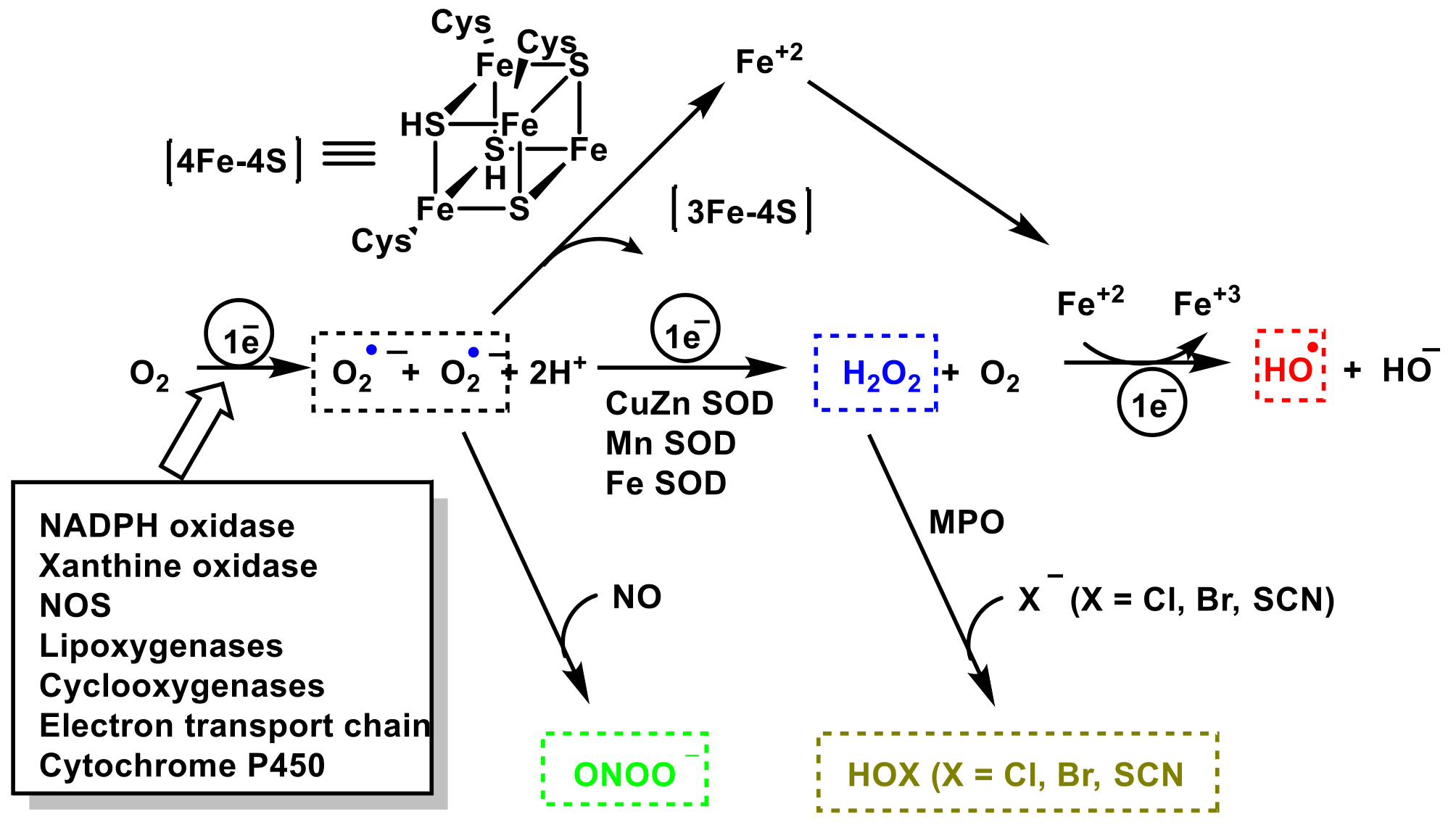
The role of reactive species RS (of oxygen ROS, nitrogen RNS and halogen RHS) on innate immunity is examined. The importance of these species in innate immunity was first recognized in phagocytes that underwent a “respiratory burst” after activation. The anion superoxide O2 and hydrogen peroxide H2O2 are detrimental to the microbial population. NADPH oxidase NOx, as an O2 producer is essential for microbial destruction, and patients lacking this functional oxidase are more susceptible to microbial infections. Reactive nitrogen species RNS (the most important are nitric oxide radical -NO, peroxynitrite ONOO and its derivatives), are also harmful to microorganisms, including bacteria, viruses, and parasites. Hypochlorous acid HOCl and hypothiocyanous acid HOSCN synthesized through the enzyme myeloperoxidase MPO, which catalyzes the reaction between H2O2 and Cl or SCN, are important inorganic bactericidal molecules, effective against a wide range of microbes. 

reactive species ROS RNS and RHS innate immunity antimicrobial

1. Introduction

The immune system can be divided into innate and acquired immunity, which are closely related [1]. Innate immunity is possessed by all types of multicellular organisms and is a primitive prophylactic system in which macrophages, neutrophils, and dendritic cells are primarily responsible for its functioning [2]. In addition, the following are also involved: antimicrobial peptides, natural antibodies, the complement system, NK cells, and gamma delta T lymphocytes [3].
Cells involved in innate immunity recognize foreign substances such as bacteria with toll-like receptors (TLR) and regulate the activation of other cells by the production of various cytokines [4]. There are cells, such as phagocytes, that can invade bodies in a process in which the cell uses its plasma membrane to engulf the large particle, giving rise to an internal compartment called a phagosome, and then activating the acquired immunity system by presenting a portion of the phagocytosed and digested foreign substances from its membrane surface. In the recognition and response process of phagocytosis, reactive oxygen species ROS and reactive nitrogen species RNS are produced [5].
Physiological levels of reactive species (ROS and RNS) are important in cellular signaling, but higher concentrations and prolonged exposure can fight infections by damaging important microbial biomolecules [6]. The chemical changes mediated by reactive species RS are detrimental to cell function because they cause oxidation and nitration, altering the structures of cellular proteins, DNA, and lipids, and impairing their normal function. NO2 and OH can modify proteins by reacting with tyrosine, tryptophan, cysteine, and methionine residues, promoting hydroxylation and nitration in peptides and proteins, impairing their normal function [7].
In the event of bacterial and fungal infection, rapid generation of ROS is essential for host defense. Therefore, ROS generation is important for effective antimicrobial defense, which can prevent inflammation and excessive tissue injury. The human body generates approximately 5 g ROS/day mainly from the leakage of the electron transport chain during oxidative phosphorylation, in the inner membrane of the mitochondrial matrix [8]. O2 and hydrogen peroxide H2O2 are the two primary products of this leakage. However, the generation of any O2 derivative species is dynamically balanced. These radical species have a dual role, at the physiological level they are cellular signalers, and when there is an imbalance between their production and the antioxidant system they are involved in several harmful biological processes, such as protein denaturation and lipid peroxidation.
ROS are generated on the membranes of the endosome of the phagocytosing cells, with the involvement of NOx [9]. Superoxide anion O2 is also produced in the mitochondrial matrix at complexes I to IV during the mitochondrial respiration process [10]. In addition to mitochondria, ROS are produced by a variety of enzymes such as NOx, xanthine oxidase, nitric oxide synthase NOS, and in other cell organelles such as the endoplasmic reticulum, peroxisomes, and cytosol [11]. O2 is unstable and cannot pass through membranes but is rapidly converted to hydrogen peroxide H2O2, a membrane-permeable specie [12]. H2O2, in biochemical reactions, produces the hydroxyl radical OH + OH, highly reactive in the mitochondrial matrix [13].
ROS can also trigger the pathogen defense of phagocytes by non-oxidative means, such as autophagy, receptor signaling, extracellular trapping, and originating lymphocyte action. For example, H2O2 can also modulate genes expression by epigenetic modification and activate transcription factors such as AP-1, NRF2, CREB, HSF1, HIF-1, TP53, NF-κβ, NOTCH, SP1, SCREB-1 and FOXO family [14][15][16][17][18][19][20][21][22].
Nitric oxide NO is a ubiquitous cellular signaling molecule, found in a variety of cell types, including vascular endothelium, platelets, macrophages, and neuronal cells [23]. In the cardiovascular system, NO determines the basal vascular tone and myocardial contractility, inhibits platelet aggregation, limits endothelial adhesion of leukocytes, and regulates myocardial contractility, playing a role in the etiology of cardiovascular disorders: atherosclerosis, hypertension, reperfusion injury and myocardial depression associated with sepsis and septic shock [24]. Vascular endothelial cells continuously produce NO and this basal release regulates the vascular tone. The oxidation of the terminal guanidino-nitrogen atoms of L-arginine produces NO [25].
Peroxynitrite ONOO is a potent oxidizing and nitrating agent, with a short half-life of about 10−2 s [26]. Its derivatives induce lipid peroxidation, inactivation of enzymes and proteins, and mitochondrial dysfunction, among others. ONOO plays an important role in the destruction of foreign pathogens by cells such as macrophages [27]. If its production is deregulated, it contributes to cardiovascular, neurological diseases, and cancer [7]. Secondary reactions of peroxynitrite decomposition produce NO2, OH, and CO3.
The enzyme myeloperoxidase MPO is a hydrogen peroxide oxidoreductase, present in macrophages, in different biological fluids (saliva, synovial fluid, and semen, among others), and in different tissues (heart, kidney, skin, liver, and placenta) [28]. The most common sources are neutrophils, where the enzyme is located at the lysosomal level [29]. The reaction catalyzed by MPO is the oxidation of the Cl anion by H2O2 to give hypochlorous acid HOCl, a very reactive and oxidizing agent, which can also act as a chlorinating agent and is the main strong oxidant generated by neutrophils in appreciable quantities [30].
Another known oxidizing agent involved in the innate immune system is the hypothiocyanite anion OSCN and the hypothiocyanic acid conjugate base HOSCN (a weak acid with a dissociation constant of pKa = 5.3). It is an organic compound that contains the functional group SCN. OSCN is formed by peroxidase enzyme catalysis of hydrogen peroxide and thiocyanate: H2O2 + SCN → OSCN + H2O. Hypothiocyanite occurs naturally as an antimicrobial agent in the human respiratory tract. OSCN is harmless to cells in the human body but is cytotoxic to bacteria, so it has been widely investigated for its capabilities as an alternative antibiotic agent. The chemical and enzymatic scheme for the generation of ROS, RNS, and RHS is represented in Figure 1.
Figure 1. Chemical and enzymatic reactions generating ROS, RNS, and RHS.

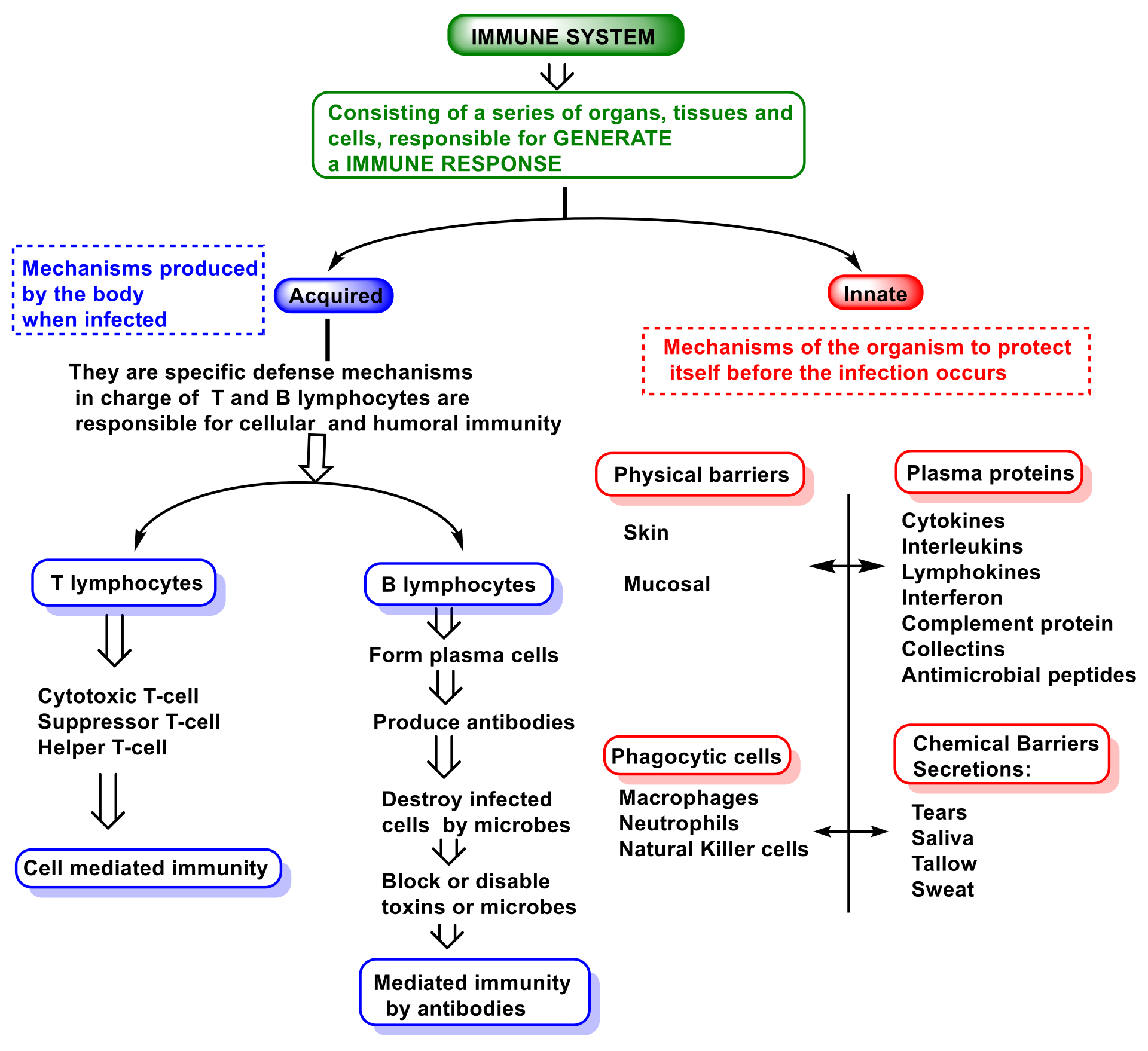
2. Function and Features of Immunity and Innate Immune System

The immune system involves cells, organs, proteins, and tissues throughout the body, and it comprises components such as leukocytes, spleen, bone marrow, lymphatic system, thymus, tonsils, adenoids, and appendix [31]. There are three types of immunity in humans: innate, adaptive, and passive. Figure 2 is a representation of the immune system.
Figure 2. Innate and adaptive immune responses.
Innate immunity is the immunity that people are born with and provides a certain level of immunity that attacks invaders from day one. This innate immunity is the first line of defense against pathogens and includes the skin and mucous membranes of the throat and gut. Innate is non-specific immunity and is an ancient evolutionary defense strategy found in plants, fungi, animals, and primitive multicellular organisms [32].
Adaptive immunity involves specific immune cells and antibodies, and it can prevent disease in the future by remembering what those substances look like and mounting a new immune response, and is carried out by lymphocytes.
Vertebrates, exclusively, have adaptive immunity, which can recognize and destroy specific substances [33]. The adaptive immune response provides vertebrates with the ability to recognize and remember specific pathogens, generating immunity and delivering increasingly potent responses to the re-encountered pathogen. Adaptative immunity includes two parts: one is called humoral and involves a variety of substances found in the body’s humors or fluids, which interfere with the growth of pathogens or clump them together so that they can be eliminated from the body.
The antibody and cell-mediated immune responses are carried out by different classes of lymphocytes, called B cells and T cells, respectively. B cells are activated to secrete antibodies, a type of protein called immunoglobulins, which circulate through the bloodstream and penetrate other body fluids, eventually binding specifically to the foreign antigen that stimulated their production. Antibody binding inactivates viruses and microbial toxins by blocking their ability to bind to receptors on host cells. Antibody binding also marks invading pathogens for destruction by making it easier for the phagocytes of the innate immune system to ingest them [34]. The cell-mediated response is carried out by phagocytes, which ingest and degrade pathogens, as well as by natural killer cells that destroy certain cancer cells [35].
In contrast, adaptive immunity is also responsible for allergic reactions and the rejection of transplanted tissues, which it recognizes as a foreign invader [36].
Passive immunity is a type of temporary immunity that is derived from another person. For example, a newborn receives antibodies from the mother through the placenta before birth and in breast milk after birth. This passive immunity protects the baby from some infections during the first years of life.
Innate and adaptive systems work together to provide vertebrates with increased resistance to micro-organisms, parasites, and potential intruders that may cause harm.
In innate immunity, invaders are identified by pattern recognition receptors that distinguish molecules expressed on microbial surfaces, called pathogen-associated molecular patterns (PAMPs). A second trigger is molecules released from broken or damaged cells, called damage-associated molecular patterns (DAMPs) [37].
Polymorphonuclear leukocytes PMNs recognize secreted molecules produced by bacteria, including peptidoglycan, lipoproteins, lipoteichoic acid, lipopolysaccharide (LPS), CpG-containing DNA, and flagellin. Peptidoglycan recognition protein (PGRP) plays a role in the neutrophil killing of Gram-positive bacteria [38], inhibiting their growth.
A class of pathogen recognition receptors is toll-like receptors (TLRS), a family of at least 10 different receptors found on the surface or in the cytoplasm of cells such as macrophages, intestinal epithelial cells, and mast cells, and which are located on the surface or the membrane of endosomes [39]. Toll receptors bind to PAMPs on extracellular bacteria, such as lipopolysaccharides, flagellin, and lipoproteins [40]. Cytoplasmic TLRs bind to the nucleic acids of intracellular viruses [41]. Once bound to these ligands, TLRs trigger the production of inflammatory cytokines such as interleukin IL-β1 or the tumor necrosis factor TNF-α, triggering what is termed acute inflammation [42].
The presence of inflammatory chemokines controls the recruitment of effector leukocytes in infections, inflammation, tissue injury, and tumors, and has a broad cellular selectivity, acting on cells of both the innate and adaptive immune systems [43]. In this process participates integrins and transmembrane cell adhesion molecules, which regulate cellular growth, proliferation, migration, cellular signaling, cytokine activation, and its release. Therefore, they play important roles in cell proliferation and migration, apoptosis, and tissue repair, as well as in all processes critical to inflammation, infection, and angiogenesis [43][44].
Acute inflammation is the central feature of innate immunity and it is the subsequent step in the early detection of invading organisms or damaged tissues [45]. The inflammatory response is characterized by several features: reddening of the skin (due to increased blood circulation), warmth or increased temperature (sensation of heat around a local infection or systemic fever), swelling of affected tissues (in the throat during the common cold or in joints affected by rheumatoid arthritis), mucus production (runny nose or cough), pain (in sore joints or in the throat) and even possible dysfunction of affected organs and tissues [1]. Inflammation guarantees that leukocytes converge in large numbers towards the site of microbial invasion, attracting these cells from the bloodstream and inducing them to migrate through the tissues to the invasion site [46].
The key to an effective innate response is the rapid recognition of the invasion, for which there are several types of sentinel cells. The most important are macrophages, dendritic cells, and mast and innate lymphoid cells [47]. The first three possess pattern recognition receptors and can detect the presence of PAMPs and DAMPs, so they send a signal through the nuclear factor NF-κβ, to produce cytokines such as IL-1, interferon IFN-α and TNF-α [48]. Molecules such as histamine, leukotrienes, prostaglandin, and specialized peptides are released to initiate the inflammatory process. Three main populations of leukocytes can eliminate invaders: (i) neutrophils, which are particularly effective at killing invading bacteria by engulfing them, activating the respiratory burst, and generating lethal oxidative molecules such as hydrogen peroxide and hypochlorite ions, which kill most invading bacteria; (ii) eosinophils, specialized killers of invading parasites, which for example contain enzymes optimized to kill helminth larvae; and (iii) M1-like macrophages, capable of migrating to areas of microbial invasion more slowly than granulocytes, but able to maintain sustained and effective phagocytosis [49]. They contain the lethal antimicrobial NO and can kill neutrophil-resistant organisms. If inflammation activates macrophages, they secrete a cytokine called IL-23, which acts on the Th17 cell subset, secreting IL-17, which attracts neutrophil granulocytes to sites of inflammation, infection, and tissue damage [50].
Mammals possess at least four populations of innate lymphoid cells ILCs that participate in innate immunity: natural killer (NK) cells, ILC1s, ILC2s, ILC3s, and lymphoid tissue inducer cells [51]. NK natural killer cells are innate lymphoid cells optimized to kill virus-infected cells, and can even kill abnormal cells, which do not express MHC class I major histocompatibility complex molecules [52]. Group I of innate lymphoid cells are found in large numbers in the intestinal wall and secrete macrophage-activating cytokines, playing a key role in antiviral immunity [53]. Group II of innate lymphoid cells are distributed throughout the body and secrete cytokines important for anti-parasitic immunity [54]. Group III of innate lymphoid cells act as Th17 cells and promote inflammation by releasing IL-17 [55]. Lymphoid tissue-inducing cells are hematopoietic and have critical roles in the immune system, both in the embryonic and adult stages [56]. These cells fulfill the following four functions: defense against pathogens, surveillance of tumorigenesis, regulation of homeostasis, and tissue remodeling [57].
When neutrophils arrive at the site of invasion, they bind to invading bacteria and ingest them by phagocytosis, a process mediated by a metabolic pathway called a respiratory burst, which generates oxidative species such as H2O2 and hypochlorous acid HOCl [58]. In contrast, the energy reserves of neutrophils are minimal, and they can only perform a few phagocytic events before they decay. Once the invading microbes are successfully eliminated, the body must repair the damage and eliminate cellular debris and dying cells through the work of macrophages, which originate from monocytes in the blood [59]. Macrophages are attracted to sites of microbial invasion and tissue damage by chemokines, DAMPs, and PAMPs, help kill invaders, remove toxic waste produced in tissues and destroy remaining neutrophils. M1-like macrophages complete the destructive process and are optimized for microbial destruction, while M2-like macrophages are optimized for the removal and repair of damaged tissues [60].
Besides the role played by neutrophils, there is also a parallel mechanism called NETosis, related to the formation of neutrophil extracellular traps (NETs). Various pathogens, antibodies and immune complexes, cytokines, and other physiological stimuli can trigger NETosis. Its induction depends on ROS, the main source being NOx [61]. NOx activation depends on increased Ca2+ concentration in the cytoplasm and, in some cases, on the generation of mitochondrial ROS. NETosis results in the release of granule components into the cytosol, histone modification leading to chromatin decondensation, destruction of the nuclear envelope, as well as the formation of pores in the plasma membrane. Two forms of NETosis have now been described: classical or suicidal NETosis (leading to cell death), and vital NETosis, where the cell retains its viability and many of its effector functions [62].
Classical NETosis is a special form of programmed cell death (PCD), characterized by the release of granule components into the cytosol. Several features of apoptosis, necroptosis, pyroptosis, autophagy, and secondary necrosis are inherent to this form of NETosis. Mitochondrial ROS are involved in NOx activation and in the induction of classical NETosis by various stimuli [63][64].
Vital NETosis helps contain local infections by allowing PMNs to rapidly release NETs and continue to phagocytose live bacteria. In addition, live PMNs that release NETs manage to maintain their membrane integrity, thereby imprisoning the captured bacteria [65][66].
Alongside ROS production, macrophages also employ several directly antimicrobial mechanisms, such as the generation of RNS in the phagosome, and the delivery of cathepsins and other hydrolases into maturing phagosomes [67]. Other indirect antimicrobial mechanisms include: (i) activation of inflammasomes and (ii) secretion of cytokines and chemokines [68]. These mechanisms help orchestrate subsequent innate and adaptive immune responses, as well as major histocompatibility complex MHC-dependent presentation of pathogen-derived antigens [69].
In parallel with acute inflammation, the body has other innate defenses as tissues contain a variety of antimicrobial peptides. These include antimicrobial peptides, such as defensins or cathelicidins, enzymes such as lysozyme that kill many Gram-positive bacteria, and iron-binding proteins such as hepcidin or haptoglobin that prevent the growth of bacteria by depriving them of vital iron [70]. The most important of these defenses is the complement system, a group of about 30 proteins that work together to eliminate invading microbes by covalently and irreversibly binding two proteins called C3 and C4 to microbial surfaces. Once bound, they can lyse microbes via the C5–C8 complex formation and the polymerization of C9 protein forming a membrane attack complex MAC, or participates as opsonins, promoting a quickly and efficiently phagocytosis by leukocytes [71].
This system can be activated in three ways:
(i)
The so-called alternative pathway is activated by the presence of bacterial surfaces that can bind complement protein C3. C3-coated bacteria are rapidly and efficiently phagocytosed and destroyed. C3 can activate other complement components by inducing a protein called C9 to insert itself into the cell walls of bacteria, causing them to rupture;
(ii)
A second pathway of complement activation is triggered when bacterial surface carbohydrates bind to a mannose-binding lectin (MBL), collectin 11 (CL-K1), and ficolins (Ficolin-1, Ficolin-2, and Ficolin-3). Its activation leads to C4 and C2 activation by their serine-proteases; or
(iii)
The classical complement pathway is initiated by antigen-antibody complexes with the antibody isotypes IgG and IgM. Upon activation, several proteins are recruited to generate C3 convertase, which cleaves the C3 protein. The C3b component of cleaved C3 binds to the C3 convertase to generate the C5 convertase, which cleaves the C5 protein. The cleaved products attract phagocytes to the site of infection and mark target cells for elimination by phagocytosis. C5 convertase initiates the terminal phase of the complement system, resulting in the assembly of the MAC membrane attack complex, creating a pore in the target cell membrane, and inducing its lysis [72].
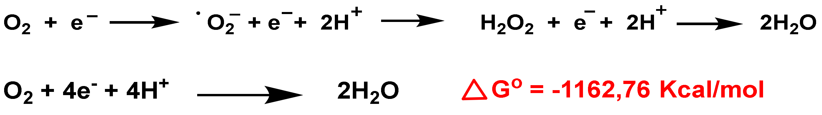
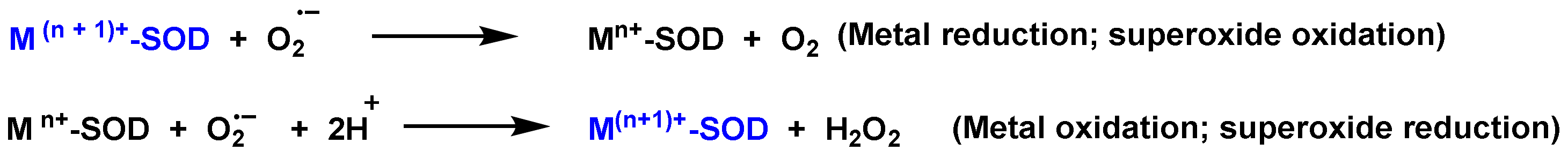
Because of its potential to cause severe tissue damage, the activation of the complement system is carefully controlled through multiple complex regulatory pathways [73].
The complement system plays an important role in mediating tissue injury following the triggering of oxidative stress. Collard et al., 2000, investigated the role of mannose-binding lectin (MBL) and the lectin complement pathway (LCP) in mediating complement activation following endothelial oxidative stress and observed that the LCP lectin complement pathway mediates complement activation following tissue oxidative stress. Thus, they suggest that inhibition of MBL may represent a novel therapeutic strategy for ischaemia/reperfusion injury and other complement-mediated disease states [74].

3. Role of Superoxide Anion O2 and Hydrogen Peroxide H2O2 on Innate Immunity

O2 anion is a by-product of mitochondrial respiration and a crucial element of the innate immune defense system. Biochemically, O2 is generated from two main sources: in the respiratory chain in the mitochondrial matrix and via nicotinamide adenine dinucleotide phosphate. In the electron transport chain, protons introduced by ATP synthase reduce molecular O2 to O2 anion, H2O2, and H2O [75], Figure 3. Consecutive reduction of O2 with H+ and e have a negative Gibbs energy, so it occurs spontaneously, with a ∆Go ≤ 0. The Gibbs free energy is used to calculate the maximum amount of work that can be done by a thermodynamically closed system, with temperature and pressure being constant, and is a necessary condition in processes such as chemical reactions.
Figure 3. O2 reduction chain to O2, H2O2 and H2O.
In the cytosolic SOD-Cu/Zn (it contains Cu and Zn in the catalytic site), SOD transforms the O2 to O2 reducing Cu(II) to Cu(I). Another O2 molecule causes the oxidation of Cu(I) to Cu(II), producing an H2O2 molecule. Zn is monovalent and it only stabilizes the enzyme. The catalytic cycle of Mn SOD is similar, with Mn in the oxidation-reduction reactions, transiting between Mn(III)) and (Mn(II).
Operative roles of H2O2 during inflammation have been observed, modulating protein function by reversible chemical modification of protein thiols [76]. H2O2 induces activation of factor nuclear NF-κβ (the factor that controls DNA transcription), including tyrosine phosphorylation of IkB and activation of IKK [77]. H2O2 can trigger the release of high mobility group 1 protein from macrophages, follow-on increase of proinflammatory stimuli [78].
Polymorphonuclear neutrophils PMN are a critical constituent of the innate immune system. In case of infection, neutrophils are rapidly recruited from the circulation and bone marrow stores by the host- and pathogen-derived components, priming these cells for enhanced antimicrobial activity [79]. One of the most potent biochemical attractants is the interleukin IL-8, produced by cells during the inflammatory process associated with infection [80]. Cells that produce interleukin IL-8 include monocytes, macrophages, mast cells, epithelial cells, keratinocytes, fibroblasts, endothelial cells, and even neutrophils themselves [81]. Bacteria also produce molecules that can directly attract neutrophils, e.g., N-formyl peptides [82]. Neutrophil “priming” is the ability to increase superoxide anion. In fact, this capacity is not limited to O2 production, but also to improved adhesion, phagocytosis, cytokine secretion, leukotriene synthesis, degranulation, and, ultimately, bactericidal activity. In this “priming” effect, neutrophils respond to the release of cytokines, chemokines, growth factors, and lipid-derived signaling molecules. In summary, neutrophils react increasing the release of O2 and inducing the expression of, among others, TNF-α, IFN-γ and -α, several interleukins, C2-ceramide, peroxynitrite, or diamide (thiol oxidizer) [83].
The combination of ROS from neutrophils and granule components is usually effective in killing most bacteria and fungi. PMNs are the most abundant leukocyte in humans and contain a battery of non-specific cytotoxic compounds, so their homeostasis is highly structured. Once neutrophil apoptosis occurs, these cells are eliminated by macrophages, and their apoptosis is accelerated following phagocytosis of bacteria, completing the termination of the infection and associated inflammation [84].
Ultimately, neutrophils use both O2-dependent and O2-independent mechanisms to kill micro-organisms [85]. Phagocytosis triggers the generation of O2 and other ROS and reactive species, such as hydrogen peroxide H2O2, hypochlorous acid HOCl, hydroxyl radical OH and chloramines, as potent microbicidal agents [86]. In parallel, cytoplasmic granules fuse with phagosomes containing bacteria in a process known as degranulation, thereby enriching the vacuole lumen with antimicrobial peptides and proteases [87].
NOx catalyzes the reduction reaction of O2 to O2 and/or H2O2 using NADPH as an electron donor and it is located extracellularly [88]. NOx is involved in pathogen clearance and the regulation of associated inflammation plays an important role in physiological and pathological conditions, such as acute lung injury and bacterial or fungal infections. NOx is electrogenic and allows electron transport across the plasma membrane (altering ionic currents) [89], induces apoptosis (mediating in physiological and pathological processes) [90], regulates cytokine production and T cell death [91], influences gene expression and promotes the formation of extracellular traps [92][93].
The significance of NOx and ROS production is exemplified by a rare inherited disorder known as chronic granulomatous disease CGD. Individuals with CGD have persistent bacterial and fungal infections due to defects in NOx [94].
Following NOx activation, there is a rapid expenditure of O2 in neutrophils, and this mechanism is called the “respiratory burst”. NOx activation by neutrophils occurs in response to stimuli such as formylated peptides, opsonized particles, integrin-dependent adhesion, and the binding of specific pathogen recognition receptors (e.g., dectin-1). SYK tyrosine kinase is a critical component of integrin signaling in neutrophils, mediating NOx activation [95]. SYK tyrosine kinase is a non-receptor kinase that was long considered to exclusively mediate receptor signaling in the adaptive immune response. However, recent studies indicate that it is also involved in innate immunity and non-immune functions. SYK mediates integrin signaling in neutrophils, macrophages, and platelets, signaling by P-selectin glycoprotein ligand 1 (PSGL1), as well as the development of osteoclasts [96]. SYK participates in the innate recognition of fungal and other microbial pathogens, as well as of tissue damage, by C-type lectins. SYK activation by C-type lectins activates the caspase-recruitment domain 9–B cell lymphoma and it is also required for NLR family, pyrin domain-containing 3 (NLRP3) inflammasome activation following fungal infection [97].
It is known that H2O2 forms naturally in living organisms and its attributed physiological role is the capability to induce bacterial killing. It has been estimated that, in lymphocytes, the half-life of the H2O2 is 1 ms while that of the anion O2 is 1 µs [98]. It is not a free radical, but it is a very important reactive form, generating the OH radical in the presence of metals such as iron (Fenton reaction). The hydroxyl radical OH, the most powerful ROS oxidant, is formed during the Haber–Weiss reaction, by the Fenton reaction or by decomposition of peroxynitrite, and has a very short half-life (10−9 s) and high reactivity.
The central source of H2O2 is enzymatically catalyzed by superoxide dismutation through the enzyme superoxide dismutase SOD. SOD is the only enzyme that can clear O2 and it is present at the mitochondrial level as well, in the cytoplasm and extracellular space [99]. It is composed of three isoforms, SOD1 (Cu/Zn-SOD is the predominant O2 scavenger and is localized in the cytoplasm), SOD2 (Mn-SOD, in the mitochondrial intermembrane space, nucleus, and lysosomes) and SOD3 (Cu/Zn-SOD, is localized in the mitochondrion and extracellular matrix) [100].
SOD-catalyzed dismutation of the superoxide radical can be characterized as the next half-reactions, Figure 4.
Figure 4. SOD-catalyzed dismutation of the superoxide radical. M = [Cu (n = 1); Mn and Fe (n = 2)]. The oxidation state of the metal cation varies between n and n + 1.
Oxidative burst is the rapid release of ROS from different cell types, macrophages and neutrophils are especially implicated, and it requires a 10-to-20-fold increase in oxygen consumption through NOx activity. The oxidative burst in phagocytes is commonly associated with bacterial killing, but in the case of alveolar macrophages, they typically produce lower levels of ROS than neutrophils and may require their activation to exhibit their bactericidal properties. Instead, their transient oxidative burst regulates the inflammatory response by inducing cytokine synthesis for redox signaling, resulting in an influx of activated neutrophils and macrophages [101].
In adaptive immunity, ROS-mediated T-cell activation has been suggested to have an immunosuppressive role. T cell activation also requires the help of accessory cells, induction of regulatory T cells Treg by macrophage-derived ROS suppresses other T cells also via ROS. Additionally, localized ROS production drives Treg lineage commitment, while their removal decreases the balance of Treg/T effector cells [102].

References

  1. Marshall, J.S.; Warrington, R.; Watson, W.; Kim, H.L. An introduction to immunology and immunopathology. Allergy Asthma Clin. Immunol. 2018, 14, 1–10.
  2. Malech, H.L.; DeLeo, F.R.; Quinn, M.T. The Role of Neutrophils in the Immune System: An Overview. Methods Mol. Biol 2020, 2087, 3–10.
  3. Hackett, C.J. Innate immune activation as a broad-spectrum biodefense strategy: Prospects and research challenges. J. Allergy Clin. Immunol. 2003, 112, 686–694.
  4. Vijay, K. Toll-like receptors in immunity and inflammatory diseases: Past, present, and future. Int. Immunopharmacol. 2018, 59, 391–412.
  5. Li, H.; Zhou, X.; Huang, Y.; Liao, B.; Cheng, L.; Ren, B. Reactive oxygen species in pathogen clearance: The killing mechanisms, the adaption response, and the side effects. Front. Microbiol. 2021, 11, 622534.
  6. Sharifi-Rad, M.; Anil Kumar, N.V.; Zucca, P.; Varoni, E.M.; Dini, L.; Panzarini, E.; Rajkovic, J.; Tsouh Fokou, P.V.; Azzini, E.; Peluso, I. Lifestyle, oxidative stress, and antioxidants: Back and forth in the pathophysiology of chronic diseases. Front. Physiol. 2020, 11, 694.
  7. Pérez de la Lastra, J.M.; Juan, C.A.; Plou, F.J.; Pérez-Lebeña, E. The Nitration of Proteins, Lipids and DNA by Peroxynitrite Derivatives-Chemistry Involved and Biological Relevance. Stresses 2022, 2, 53–64.
  8. Guo, C.; Sun, L.; Chen, X.; Zhang, D. Oxidative stress, mitochondrial damage and neurodegenerative diseases. Neural Regenerat. Res. 2013, 8, 2003.
  9. Fisher, A.B. Redox signaling across cell membranes. Antioxid Redox Signal. 2009, 11, 1349–1356.
  10. Turrens, J.F. Mitochondrial formation of reactive oxygen species. J. Physiol. 2003, 552, 335–344.
  11. Snezhkina, A.V.; Kudryavtseva, A.V.; Kardymon, O.L.; Savvateeva, M.V.; Melnikova, N.V.; Krasnov, G.S.; Dmitriev, A.A. ROS generation and antioxidant defense systems in normal and malignant cells. Oxidative Med. Cell. Longev. 2019, 2019.
  12. Andrés, C.M.C.; Pérez de la Lastra, J.M.; Juan, C.A.; Plou, F.J.; Pérez-Lebeña, E. Chemistry of Hydrogen Peroxide Formation and Elimination in Mammalian Cells, and Its Role in Various Pathologies. Stresses 2022, 2, 256–274.
  13. Juan, C.A.; Pérez de la Lastra, J.M.; Plou, F.J.; Pérez-Lebeña, E. The chemistry of reactive oxygen species (ROS) revisited: Outlining their role in biological macromolecules (DNA, lipids and proteins) and induced pathologies. Int. J. Mol. Sci. 2021, 22, 4642.
  14. Royer-Pokora, B.; Kunkel, L.M.; Monaco, A.P.; Goff, S.C.; Newburger, P.E.; Baehner, R.L.; Cole, F.S.; Curnutte, J.T.; Orkin, S.H. Cloning the gene for an inherited human disorder—chronic granulomatous disease—On the basis of its chromosomal location. Nature 1986, 322, 32–38.
  15. Dinauer, M.C.; Orkin, S.H.; Brown, R.; Jesaitis, A.J.; Parkos, C.A. The glycoprotein encoded by the X-linked chronic granulomatous disease locus is a component of the neutrophil cytochrome b complex. Nature 1987, 327, 717–720.
  16. Parkos, C.A.; Allen, R.A.; Cochrane, C.G.; Jesaitis, A.J. Purified cytochrome b from human granulocyte plasma membrane is comprised of two polypeptides with relative molecular weights of 91,000 and 22,000. J. Clin. Investig. 1987, 80, 732–742.
  17. Segal, A.W. Absence of both cytochrome b−245 subunits from neutrophils in X-linked chronic granulomatous disease. Nature 1987, 326, 88–91.
  18. Segal, A.W.; Heyworth, P.G.; Cockcroft, S.; Barrowman, M.M. Stimulated neutrophils from patients with autosomal recessive chronic granulomatous disease fail to phosphorylate a Mr-44,000 protein. Nature 1985, 316, 547–549.
  19. Volpp, B.D.; Nauseef, W.M.; Clark, R.A. Two cytosolic neutrophil oxidase components absent in autosomal chronic granulomatous disease. Science 1988, 242, 1295–1297.
  20. Wientjes, F.B.; Hsuan, J.J.; Totty, N.F.; Segal, A.W. p40phox, a third cytosolic component of the activation complex of the NADPH oxidase to contain src homology 3 domains. Biochem J. 1993, 296 (Pt. 3), 557–561.
  21. Abo, A.; Pick, E. Purification and characterization of a third cytosolic component of the superoxide-generating NADPH oxidase of macrophages. J. Biol. Chem. 1991, 266, 23577–23585.
  22. Roberts, A.W.; Kim, C.; Zhen, L.; Lowe, J.B.; Kapur, R.; Petryniak, B.; Spaetti, A.; Pollock, J.D.; Borneo, J.B.; Bradford, G.B.; et al. Deficiency of the Hematopoietic Cell-Specific Rho Family GTPase Rac2 Is Characterized by Abnormalities in Neutrophil Function and Host Defense. Immunity 1999, 10, 183–196.
  23. Pacher, P.; Beckman, J.S.; Liaudet, L. Nitric oxide and peroxynitrite in health and disease. Physiol. Rev. 2007, 87, 315–424.
  24. Förstermann, U.; Xia, N.; Li, H. Roles of Vascular Oxidative Stress and Nitric Oxide in the Pathogenesis of Atherosclerosis. Circ. Res. 2017, 120, 713–735.
  25. Babu, B.R.; Frey, C.; Griffith, O.W. l-arginine binding to nitric-oxide synthase: The role of H-bonds to the nonreactive guanidinium nitrogens. J. Biol. Chem. 1999, 274, 25218–25226.
  26. Radi, R.; Peluffo, G.; Alvarez, M.a.N.; Naviliat, M.; Cayota, A. Unraveling peroxynitrite formation in biological systems. Free Radic. Biol. Med. 2001, 30, 463–488.
  27. Prolo, C.; Álvarez, M.N.; Radi, R. Peroxynitrite, a potent macrophagd-derived oxidizing cytotoxin to combat invading pathogens. Biofactors 2014, 40, 215–225.
  28. Tobler, A.; Koeffler, H.P. Myeloperoxidase: Localization, structure, and function. In Blood Cell Biochemistry Volume 3; Springer: Berlin/Heidelberg, Germany, 1991; pp. 255–288.
  29. Hurst, J.K. What really happens in the neutrophil phagosome? Free Radic. Biol. Med. 2012, 53, 508–520.
  30. Davies, M.J. Myeloperoxidase-derived oxidation: Mechanisms of biological damage and its prevention. J. Clin. Biochem. Nutr. 2010, 48, 8–19.
  31. Janeway, C.A., Jr.; Travers, P.; Walport, M.; Shlomchik, M.J. Principles of innate and adaptive immunity. In Immunobiology: The Immune System in Health and Disease, 5th ed.; Garland Science: New York, NY, USA, 2001.
  32. Kiboneka, A. Principals of innate and adaptive immunity. Immunity to microbes & fundamental concepts in immunology. World J. Adv. Res. Rev. 2021, 10, 188–197.
  33. Zimmerman, L.; Vogel, L.; Bowden, R. Understanding the vertebrate immune system: Insights from the reptilian perspective. J. Exp. Biol. 2010, 213, 661–671.
  34. Alberts, B. Molecular Biology of the Cell; WW Norton & Company: New York, NY, USA, 2017.
  35. Riera Romo, M.; Pérez-Martínez, D.; Castillo Ferrer, C. Innate immunity in vertebrates: An overview. Immunology 2016, 148, 125–139.
  36. LaRosa, D.F.; Rahman, A.H.; Turka, L.A. The innate immune system in allograft rejection and tolerance. J. Immunol. 2007, 178, 7503–7509.
  37. Tanaka, K.; Heil, M. Damage-associated molecular patterns (DAMPs) in plant innate immunity: Applying the danger model and evolutionary perspectives. Annu. Rev. Phytopathol. 2021, 59, 53–75.
  38. DeLeo, F.R.; Diep, B.A.; Otto, M. Host defense and pathogenesis in Staphylococcus aureus infections. Infect. Dis. Clin. N. Am. 2009, 23, 17–34.
  39. Khovidhunkit, W.; Kim, M.-S.; Memon, R.A.; Shigenaga, J.K.; Moser, A.H.; Feingold, K.R.; Grunfeld, C. Effects of infection and inflammation on lipid and lipoprotein metabolism: Mechanisms and consequences to the host. J. Lipid Res. 2004, 45, 1169–1196.
  40. Kieser, K.J.; Kagan, J.C. Multi-receptor detection of individual bacterial products by the innate immune system. Nat. Rev. Immunol. 2017, 17, 376–390.
  41. Finberg, R.W.; Wang, J.P.; Kurt-Jones, E.A. Toll like receptors and viruses. Rev. Med. Virol. 2007, 17, 35–43.
  42. Wu, J.; Niu, P.; Zhao, Y.; Cheng, Y.; Chen, W.; Lin, L.; Lu, J.; Cheng, X.; Xu, Z. Impact of miR-223-3p and miR-2909 on inflammatory factors IL-6, IL-1ß, and TNF-α, and the TLR4/TLR2/NF-κB/STAT3 signaling pathway induced by lipopolysaccharide in human adipose stem cells. PLoS ONE 2019, 14, e0212063.
  43. Moser, B.; Wolf, M.; Walz, A.; Loetscher, P. Chemokines: Multiple levels of leukocyte migration control. Trends Immunol. 2004, 25, 75–84.
  44. Mezu-Ndubuisi, O.J.; Maheshwari, A. The role of integrins in inflammation and angiogenesis. Pediatr. Res. 2021, 89, 1619–1626.
  45. Mortaz, E.; Alipoor, S.D.; Adcock, I.M.; Mumby, S.; Koenderman, L. Update on neutrophil function in severe inflammation. Front. Immunol. 2018, 9, 2171.
  46. Muller, W. Getting leukocytes to the site of inflammation. Vet. Pathol. 2013, 50, 7–22.
  47. Ebbo, M.; Crinier, A.; Vély, F.; Vivier, E. Innate lymphoid cells: Major players in inflammatory diseases. Nat. Rev. Immunol. 2017, 17, 665–678.
  48. Shaikh, P.Z. Cytokines & their physiologic and pharmacologic functions in inflammation: A review. Int. J. Pharm. Life Sci. 2011, 2, 212599524.
  49. Slauch, J.M. How does the oxidative burst of macrophages kill bacteria? Still an open question. Mol. Microbiol. 2011, 80, 580–583.
  50. Tesmer, L.A.; Lundy, S.K.; Sarkar, S.; Fox, D.A. Th17 cells in human disease. Immunol. Rev. 2008, 223, 87–113.
  51. Favaro, R.R.; Phillips, K.; Delaunay-Danguy, R.; Ujčič, K.; Markert, U.R. Emerging Concepts in Innate Lymphoid Cells, Memory, and Reproduction. Front. Immunol. 2022, 13, 824263.
  52. Laskowski, T.J.; Biederstädt, A.; Rezvani, K. Natural killer cells in antitumour adoptive cell immunotherapy. Nat. Rev. Cancer 2022, 22, 1–19.
  53. Ochel, A.; Tiegs, G.; Neumann, K. Type 2 innate lymphoid cells in liver and gut: From current knowledge to future perspectives. Int. J. Mol. Sci. 2019, 20, 1896.
  54. Maazi, H.; Akbari, O. Type two innate lymphoid cells: The Janus cells in health and disease. Immunol. Rev. 2017, 278, 192–206.
  55. Withers, D.R.; Hepworth, M.R. Group 3 innate lymphoid cells: Communications hubs of the intestinal immune system. Front. Immunol. 2017, 8, 1298.
  56. Withers, D.R. Lymphoid tissue inducer cells. Curr. Biol. 2011, 21, R381–R382.
  57. Klose, C.S.; Artis, D. Innate lymphoid cells as regulators of immunity, inflammation and tissue homeostasis. Nat. Immunol. 2016, 17, 765–774.
  58. Ulfig, A.; Leichert, L.I. The effects of neutrophil-generated hypochlorous acid and other hypohalous acids on host and pathogens. Cell. Mol. Life Sci. 2021, 78, 385–414.
  59. Shapouri-Moghaddam, A.; Mohammadian, S.; Vazini, H.; Taghadosi, M.; Esmaeili, S.A.; Mardani, F.; Seifi, B.; Mohammadi, A.; Afshari, J.T.; Sahebkar, A. Macrophage plasticity, polarization, and function in health and disease. J. Cell. Physiol. 2018, 233, 6425–6440.
  60. Atri, C.; Guerfali, F.Z.; Laouini, D. Role of human macrophage polarization in inflammation during infectious diseases. Int. J. Mol. Sci. 2018, 19, 1801.
  61. Azzouz, D.; Khan, M.A.; Palaniyar, N. ROS induces NETosis by oxidizing DNA and initiating DNA repair. Cell Death Discov. 2021, 7, 1–10.
  62. Huang, S.U.-S.; O’Sullivan, K.M. The expanding role of extracellular traps in inflammation and autoimmunity: The new players in casting dark webs. Int. J. Mol. Sci. 2022, 23, 3793.
  63. Vorobjeva, N.; Prikhodko, A.; Galkin, I.; Pletjushkina, O.; Zinovkin, R.; Sud’ina, G.; Chernyak, B.; Pinegin, B. Mitochondrial reactive oxygen species are involved in chemoattractant-induced oxidative burst and degranulation of human neutrophils in vitro. Eur. J. Cell Biol. 2017, 96, 254–265.
  64. Vorobjeva, N.; Galkin, I.; Pletjushkina, O.; Golyshev, S.; Zinovkin, R.; Prikhodko, A.; Pinegin, V.; Kondratenko, I.; Pinegin, B.; Chernyak, B. Mitochondrial permeability transition pore is involved in oxidative burst and NETosis of human neutrophils. Biochim. Biophys. Acta Mol. Basis Dis. 2020, 1866, 165664.
  65. Yipp, B.G.; Kubes, P. NETosis: How vital is it? Blood J. Am. Soc. Hematol. 2013, 122, 2784–2794.
  66. Vorobjeva, N.V.; Chernyak, B.V. NETosis: Molecular Mechanisms, Role in Physiology and Pathology. Biochemistry (Mosc) 2020, 85, 1178–1190.
  67. Schramm, M.; Wiegmann, K.; Schramm, S.; Gluschko, A.; Herb, M.; Utermöhlen, O.; Krönke, M. Riboflavin (vitamin B2) deficiency impairs NADPH oxidase 2 (Nox2) priming and defense against Listeria monocytogenes. Eur. J. Immunol. 2014, 44, 728–741.
  68. Arango Duque, G.; Descoteaux, A. Macrophage cytokines: Involvement in immunity and infectious diseases. Front. Immunol. 2014, 5, 491.
  69. Allan, E.R.; Tailor, P.; Balce, D.R.; Pirzadeh, P.; McKenna, N.T.; Renaux, B.; Warren, A.L.; Jirik, F.R.; Yates, R.M. NADPH oxidase modifies patterns of MHC class II–restricted epitopic repertoires through redox control of antigen processing. J. Immunol. 2014, 192, 4989–5001.
  70. Metz-Boutigue, M.-H.; Shooshtarizadeh, P.; Prevost, G.; Haikel, Y.; Chich, J.-F. Antimicrobial peptides present in mammalian skin and gut are multifunctional defence molecules. Curr. Pharm. Design 2010, 16, 1024–1039.
  71. Sarma, J.V.; Ward, P.A. The complement system. Cell Tissue Res. 2011, 343, 227–235.
  72. Dunkelberger, J.R.; Song, W.-C. Complement and its role in innate and adaptive immune responses. Cell Res. 2010, 20, 34–50.
  73. Zipfel, P.F.; Skerka, C. Complement regulators and inhibitory proteins. Nat. Rev. Immunol. 2009, 9, 729–740.
  74. Collard, C.D.; Väkevä, A.; Morrissey, M.A.; Agah, A.; Rollins, S.A.; Reenstra, W.R.; Buras, J.A.; Meri, S.; Stahl, G.L. Complement activation after oxidative stress: Role of the lectin complement pathway. Am. J. Pathol. 2000, 156, 1549–1556.
  75. Tabassum, N.; Kheya, I.S.; Asaduzzaman, S.; Maniha, S.; Fayz, A.H.; Zakaria, A.; Noor, R. A review on the possible leakage of electrons through the electron transport chain within mitochondria. Life Sci. 2020, 6, 105–113.
  76. Wittmann, C.; Chockley, P.; Singh, S.K.; Pase, L.; Lieschke, G.J.; Grabher, C. Hydrogen peroxide in inflammation: Messenger, guide, and assassin. Adv. Hematol. 2012, 2012, 541471.
  77. Schoonbroodt, S.; Ferreira, V.; Best-Belpomme, M.; Boelaert, J.R.; Legrand-Poels, S.; Korner, M.; Piette, J. Crucial role of the amino-terminal tyrosine residue 42 and the carboxyl-terminal PEST domain of IκBα in NF-κB activation by an oxidative stress. J. Immunol. 2000, 164, 4292–4300.
  78. Tang, D.; Shi, Y.; Kang, R.; Li, T.; Xiao, W.; Wang, H.; Xiao, X. Hydrogen peroxide stimulates macrophages and monocytes to actively release HMGB1. J. Leukoc. Biol. 2007, 81, 741–747.
  79. Mayadas, T.N.; Cullere, X.; Lowell, C.A. The multifaceted functions of neutrophils. Annu. Rev. Pathol. 2014, 9, 181.
  80. Singer, M.; Sansonetti, P.J. IL-8 is a key chemokine regulating neutrophil recruitment in a new mouse model of Shigella-induced colitis. J. Immunol. 2004, 173, 4197–4206.
  81. Filimon, A.; Preda, I.A.; Boloca, A.F.; Negroiu, G. Interleukin-8 in Melanoma Pathogenesis, Prognosis and Therapy—An Integrated View into Other Neoplasms and Chemokine Networks. Cells 2021, 11, 120.
  82. He, H.-Q.; Ye, R.D. The formyl peptide receptors: Diversity of ligands and mechanism for recognition. Molecules 2017, 22, 455.
  83. Kobayashi, S.D.; DeLeo, F.R. Role of neutrophils in innate immunity: A systems biology-level approach. Wiley Interdiscp. Rev. Syst. Biol. Med. 2009, 1, 309–333.
  84. Nguyen, G.; Green, E.; Mecsas, J. Neutrophils to the ROScue: Mechanisms of NADPH oxidase activation and bacterial resistance. Front. Cell Infect. Microbiol. 2017, 7, 373.
  85. Elsbach, P.; Weiss, J. Oxygen-dependent and oxygen-independent mechanisms of microbicidal activity of neutrophils. Immunol. Lett. 1985, 11, 159–163.
  86. Davies, M.J. Reactivity of Peroxidase-Derived Oxidants with Proteins, Glycoproteins and Proteoglycans. In Mammalian Heme Peroxidases; CRC Press: Boca Raton, FL, USA, 2021; pp. 53–77.
  87. Scieszka, D.; Lin, Y.-H.; Li, W.; Choudhury, S.; Yu, Y.; Freire, M. NETome: The molecular characterization of neutrophil extracellular traps (NETs). Cold Spring Harbor Lab. 2020.
  88. Vermot, A.; Petit-Härtlein, I.; Smith, S.M.; Fieschi, F. NADPH oxidases (NOX): An overview from discovery, molecular mechanisms to physiology and pathology. Antioxidants 2021, 10, 890.
  89. Demaurex, N.; Petheö, G.L. Electron and proton transport by NADPH oxidases. Philos. Trans. Royal Soc. B Biol. Sci. 2005, 360, 2315–2325.
  90. Waghela, B.N.; Vaidya, F.U.; Agrawal, Y.; Santra, M.K.; Mishra, V.; Pathak, C. Molecular insights of NADPH oxidases and its pathological consequences. Cell Biochem. Funct. 2021, 39, 218–234.
  91. Purushothaman, D.; Sarin, A. Cytokine-dependent regulation of NADPH oxidase activity and the consequences for activated T cell homeostasis. J. Exp. Med. 2009, 206, 1515–1523.
  92. Manea, S.-A.; Constantin, A.; Manda, G.; Sasson, S.; Manea, A. Regulation of Nox enzymes expression in vascular pathophysiology: Focusing on transcription factors and epigenetic mechanisms. Redox Biol. 2015, 5, 358–366.
  93. Ravindran, M.; Khan, M.A.; Palaniyar, N. Neutrophil extracellular trap formation: Physiology, pathology, and pharmacology. Biomolecules 2019, 9, 365.
  94. Roos, D. Chronic granulomatous disease. Br. Med. Bull. 2016, 118, 50.
  95. Mócsai, A.; Zhou, M.; Meng, F.; Tybulewicz, V.L.; Lowell, C.A. Syk is required for integrin signaling in neutrophils. Immunity 2002, 16, 547–558.
  96. Mócsai, A.; Ruland, J.; Tybulewicz, V.L.J. The SYK tyrosine kinase: A crucial player in diverse biological functions. Nat. Rev. Immunol. 2010, 10, 387–402.
  97. Kerrigan, A.M.; Brown, G.D. Syk-coupled C-type lectins in immunity. Trends Immunol. 2011, 32, 151–156.
  98. Chen, X.; Song, M.; Zhang, B.; Zhang, Y. Reactive oxygen species regulate T cell immune response in the tumor microenvironment. Oxid. Med. Cell. Longev. 2016, 2016.
  99. Fridovich, I. The biology of oxygen radicals: The superoxide radical is an agent of oxygen toxicity; superoxide dismutases provide an important defense. Science 1978, 201, 875–880.
  100. Trachootham, D.; Lu, W.; Ogasawara, M.A.; Nilsa, R.D.; Huang, P. Redox regulation of cell survival. Antioxid. Redox Signal. 2008, 10, 1343–1374.
  101. Forman, H.J.; Torres, M. Reactive oxygen species and cell signaling: Respiratory burst in macrophage signaling. Am. J. Respir. Crit. Care Med. 2002, 166, S4–S8.
  102. Nathan, C.; Cunningham-Bussel, A. Beyond oxidative stress: An immunologist’s guide to reactive oxygen species. Nat. Rev. Immunol. 2013, 13, 349–361.
More
Information
Subjects: Immunology
Contributors MDPI registered users' name will be linked to their SciProfiles pages. To register with us, please refer to https://encyclopedia.pub/register : , , , ,
View Times: 461
Revisions: 2 times (View History)
Update Date: 26 Oct 2022
1000/1000