Submitted Successfully!
To reward your contribution, here is a gift for you: A free trial for our video production service.
Thank you for your contribution! You can also upload a video entry or images related to this topic.
Version Summary Created by Modification Content Size Created at Operation
1 -- 3028 2022-10-13 11:19:11 |
2 update references and layout Meta information modification 3028 2022-10-13 11:45:23 |

Video Upload Options

Do you have a full video?

Confirm

Are you sure to Delete?
Cite
If you have any further questions, please contact Encyclopedia Editorial Office.
Kamaraj, R.;  Drastik, M.;  Maixnerova, J.;  Pavek, P. Allosteric Antagonism of the Pregnane X Receptor. Encyclopedia. Available online: https://encyclopedia.pub/entry/29117 (accessed on 26 April 2024).
Kamaraj R,  Drastik M,  Maixnerova J,  Pavek P. Allosteric Antagonism of the Pregnane X Receptor. Encyclopedia. Available at: https://encyclopedia.pub/entry/29117. Accessed April 26, 2024.
Kamaraj, Rajamanikkam, Martin Drastik, Jana Maixnerova, Petr Pavek. "Allosteric Antagonism of the Pregnane X Receptor" Encyclopedia, https://encyclopedia.pub/entry/29117 (accessed April 26, 2024).
Kamaraj, R.,  Drastik, M.,  Maixnerova, J., & Pavek, P. (2022, October 13). Allosteric Antagonism of the Pregnane X Receptor. In Encyclopedia. https://encyclopedia.pub/entry/29117
Kamaraj, Rajamanikkam, et al. "Allosteric Antagonism of the Pregnane X Receptor." Encyclopedia. Web. 13 October, 2022.
Allosteric Antagonism of the Pregnane X Receptor
Edit

The pregnane X receptor (PXR, NR1I2) is a xenobiotic-activated transcription factor with high levels of expression in the liver. It not only plays a key role in drug metabolism and elimination, but also promotes tumor growth, drug resistance, and metabolic diseases. It has been proposed as a therapeutic target for type II diabetes, metabolic syndrome, and inflammatory bowel disease, and PXR antagonists have recently been considered as a therapy for colon cancer. The identified allosteric sites of the PXR provide new insights into the development of safe and efficient allosteric modulators of the PXR receptor. Researchers therefore propose that novel PXR allosteric sites might be promising targets for treating chronic metabolic diseases and some cancers. 

PXR pregnane X receptor allosteric site CAR colon cancer BF 3 nuclear receptor

1. Introduction

The pregnane X receptor (PXR, NR1I2) is a nuclear receptor superfamily member. Nuclear receptors are mostly activated by exogenous and endogenous compounds. Interestingly, the classified nuclear receptors conserve the DNA binding domain (DBD), but not its ligand-binding domain (LBD). PXR belongs to the NR1I subfamily together with the vitamin D receptor (VDR, NR1I1) and the constitutive androstane receptor (CAR, NR1I3) [1][2].
The PXR protein consists of an N-terminal conserved DBD and activation function domain 1 (AF-1), and the C-terminal contains an LBD with activation function 2 (AF-2). A flexible hinge region intervenes between the DBD and the LBD (Figure 1A). The artificial intelligence application AlphaFold has been used to predict the full length of the PXR structure; moreover, the error plot shows the expected position error for each residue in the sequence (Figure 1B) [3][4][5]. The PXR ligand-binding pocket (LBP) is formed by ~28 amino acids, which are typically hydrophobic residues. In the absence of an agonist, PXR can be found in the cytosol. A ligand-activated PXR forms a heterodimer with retinoid X receptor alpha (RXRα), and the complex binds coactivators (e.g., steroid receptor coactivator-1, SRC-1) (Figure 1C) [6][7][8].
Figure 1. Structure of the PXR receptor. (A) AlphaFold2 prediction of the full-length chain of apo-PXR model (UniProt-O75469). A ribbon model of PXR coloring ranges from blue (high confidence of the predicted structure) to red (low confidence of the predicted structure). (B) Predicted aligned error plot (PAE) for the PXR. The shade of green indicates expected distance error in angstroms. Dark green represents low error, and light green represents high error indicated in the N-terminal/hinge regions of PXR. (C) Crystal structure of the PXR/RXRα LBD complex. The liganded PXR (green) and RXRα (red) bind to the steroid receptor coactivator peptide 1 (SRC-1/blue). The PXR agonist SR12813 is indicated in orange (PDB-4J5W) [7]. (D) List of some prototypical ligands and target genes for the nuclear receptor PXR.
The PXR is expressed in the liver, small intestine, and colon, and is traceable in the brain [9][10][11][12][13][14][15][16]. It functions as the master regulator of drug detoxification in the liver [17]. In a growing number of reports, ligand-activated PXRs alter the metabolic profile of a drug and increase the probability of drug–drug interactions (DDIs) [18][19][20].
Activation of PXRs causes significant clinically relevant DDIs with compounds whose clearance is critically dependent on hepatic biotransformation by inducible cytochrome P450s, such as CYP3A4 (Figure 1D). Under this scenario, the metabolism of the drug is significantly augmented by a PXR ligand (inducer, perpetrator) resulting in decrease in therapeutic efficacy, which may subsequently require shortening dosage intervals or increasing dosage. The most serious DDI interactions mediated by PXR activation have been reported for rifampicin [21][22][23][24].
Activated PXRs play a role in many metabolic pathways, including bile acid metabolism, lipids, and glucose homeostasis, as well as in inflammation [25]. Recently, their potential role in tumor growth, aggressiveness, relapse, and cancer drug resistance was also noted in mouse tumor xenografts for colon cancer [26][27][28][29][30]. These various physiological functions make PXRs a potentially valuable therapeutic target [25][31]. Numerous synthetic or natural ligands, mostly with agonist activities, have been discovered among currently used therapeutics and dietary or natural compounds [32][33]. Activation of PXRs using their ligands has been connected with many metabolic side effects (e.g., hypercholesterolemia or liver steatosis) as well as with intestinal cancer [10][25][26][34][35][36]. Therefore, PXR antagonists could have a significant therapeutic value, although designing a novel PXR antagonist is challenging due to the promiscuous nature of the PXR LBP [37][38][39][40][41][42].

2. The Orthosteric Ligand-Binding Pocket of the PXR

The PXR shares common structural characteristics with other nuclear receptors [43]. The orthosteric binding site of the PXR receptor is large (>1600 Å3), dynamic, and flexible enough to bind bulky ligands [40]. The PXR LBD is characterized by an alpha-helical sandwich structure with a specialized five-stranded beta sheet [44]. The LBP is deeply embedded in the PXR LBD (Figure 2A) [45]. The orthosteric PXR LBP is formed by the helices α3/5/6/7/10/12 (Figure 2B) [46]. The PXR ligand-binding cavity was detected at the bottom of the LBD, with the ligand entrance located between the alpha 2 and 6 helices (Figure 2B) [40][44]. The PXR was observed as a homodimer in solution, and the crystal structure showed linking with β1′ strands and with support by six intermolecular hydrogen bonds in the monomer [43].
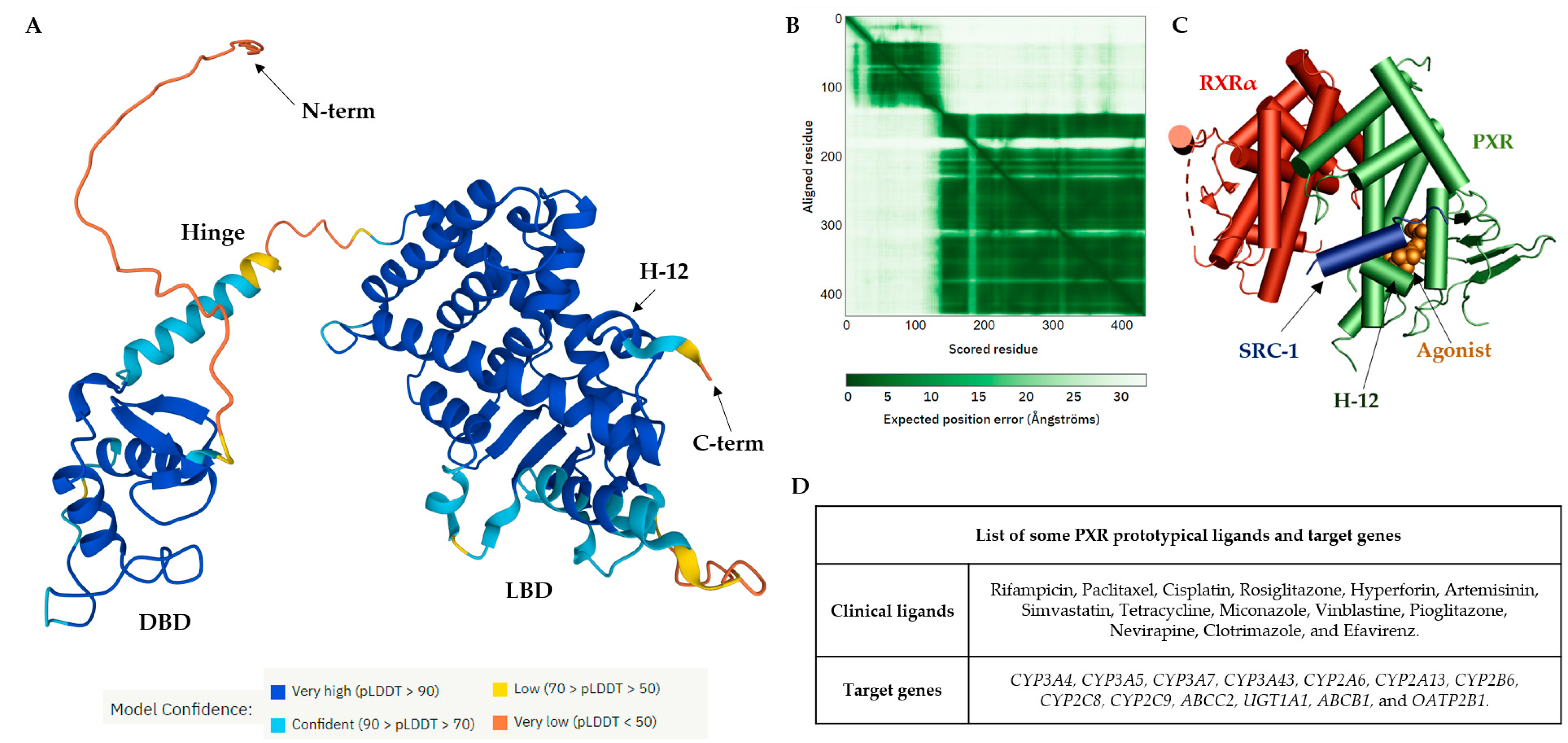
The PXR LBD shares structural similarities with the VDR and CAR, with a sequence homology of 49.4% and 48.6%, respectively. In humans, the main isoform of the PXR (NM_003889.4, variant 1) consists of 434 amino acids, and the main hydrophobic hot spots are the amino acid residues F288, W299, and Y306, which interact with all reported co-crystallized PXR ligands [40]. All available 49 PXR LBD crystal structures represent the PXR receptor in its active state and are available for coactivator interactions through helix 12 (H12) for the following transcriptional activation. Due to the destabilization of the PXR LBD, to date no crystal structure of a PXR antagonist has been reported. Therefore, as an alternative to crystallography, the biophysical technique of hydrogen-deuterium exchange mass spectrometry (HDX-MS) was used to analyze the PXR–antagonist interactions [41].
The structural characteristics of PXR ligands are determined by the nature of the PXR binding pocket: (i) the PXR LBP is large and highly hydrophobic; and (ii) the polar residues Ser247, Gln285, His407, and Arg410 define the ligand binding position in the PXR LBP via strong hydrogen bond (H-bond) interactions. Ser247 and Gln285 residues are involved in the orientation of the ligand binding and the His407 and Arg410 side chains are flexible to accommodate various sizes of ligands in the LBP [47].
Recently, a detailed examination of the PXR antagonist/inverse agonist SPA70 binding has been conducted. The distinction of the PXR antagonist/inverse agonist (SPA70) from agonist (SJB7) binding showed that the agonist should have the hydrogen bond to the polar residue of Ser247 in helix 10 as well as proximity to the helix 12 region. In contrast, the antagonist has interaction with Ser247 residue and/or has weak contacts with helix 12 [39]. Since the PXR agonist SJB7 and the antagonist SPA70 bind to the same ligand-binding pocket in the PXR in the combined agonist and antagonist conformation, it is difficult to discriminate agonist and antagonist binding sites in the PXR LBD [39][41]. Notably, a single PXR LBP mutation (W299A) converts the activity of SPA70 from inhibition to activation [48].
It has recently been reported that the designed PXR molecular glue SJPYT-195, which is composed of SPA70 linked with the CRBN ligand (thalidomide), degraded the GSPT1 translation termination factor instead of the PXR [49], but the loss of GSPT1 decreased the level of PXR protein in human colon cancer cells (SNU-C4) [49]. SJPYT-195 weakly bound the PXR LBD, suggesting that long-length linkers may be more favorable in the design of potent PROTACs for PXR [49][50]. Researchers can also speculate that PXR allosteric sites could be another strategy for the successful targeting of PXR protein degradation (Figure 2) [51].
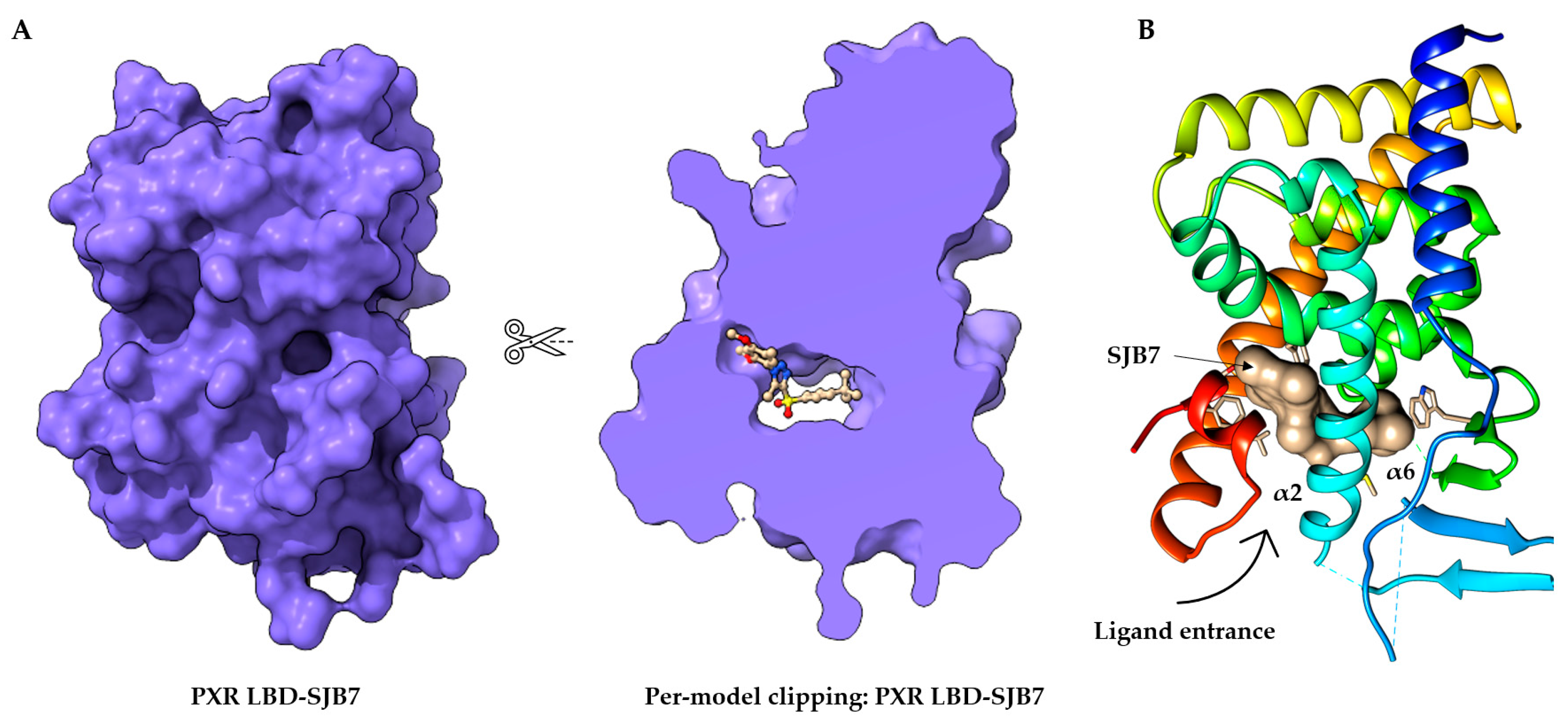
Figure 2. Structure of the orthosteric PXR ligand binding site. (A) Per-model clipping of the PXR ligand binding domain (to visualize invisible portions of a model). The PXR in PDB entry 5X0R has an SJB7 agonist ligand in an interior pocket, visualized using UCSF ChimeraX [41][52]. (B) The crystal structure of the PXR LBD complexed with SJB7. Cartoon representation of PXR- SJB7 binding in the orthosteric site. The PXR agonist SJB7 is indicated in brown, with the arrow indicating the entrance of the ligand. All structures are shown in the same orientation [41][44].

3. Allosteric Modulation of Nuclear Receptors

Allosteric modulators are usually structurally different from orthosteric ligands, and they bind to distinct sites that are spatially distant from orthosteric sites to modulate the activities of orthosteric ligands [53].
The essential features of a receptor allosteric modulation are: (1) orthosteric and allosteric sites not overlapping, i.e., there is no mutual biomolecular interaction in the binding; (2) the allosteric binding of one ligand to its site can affect the binding of the second ligand to the orthosteric site and vice versa; and (3) the effect of allosteric modulation can be positive or negative depending on the existing orthosteric ligand. This phenomenon is known as “probe dependence” [54][55]. Allosteric modulation may be inhibited due to protein–protein interactions (e.g., nuclear receptor–coactivator) or a co-bound receptor, such as in the case of the nuclear receptor homodimerization and heterodimerizations as was reported for G protein-coupled receptors [56].
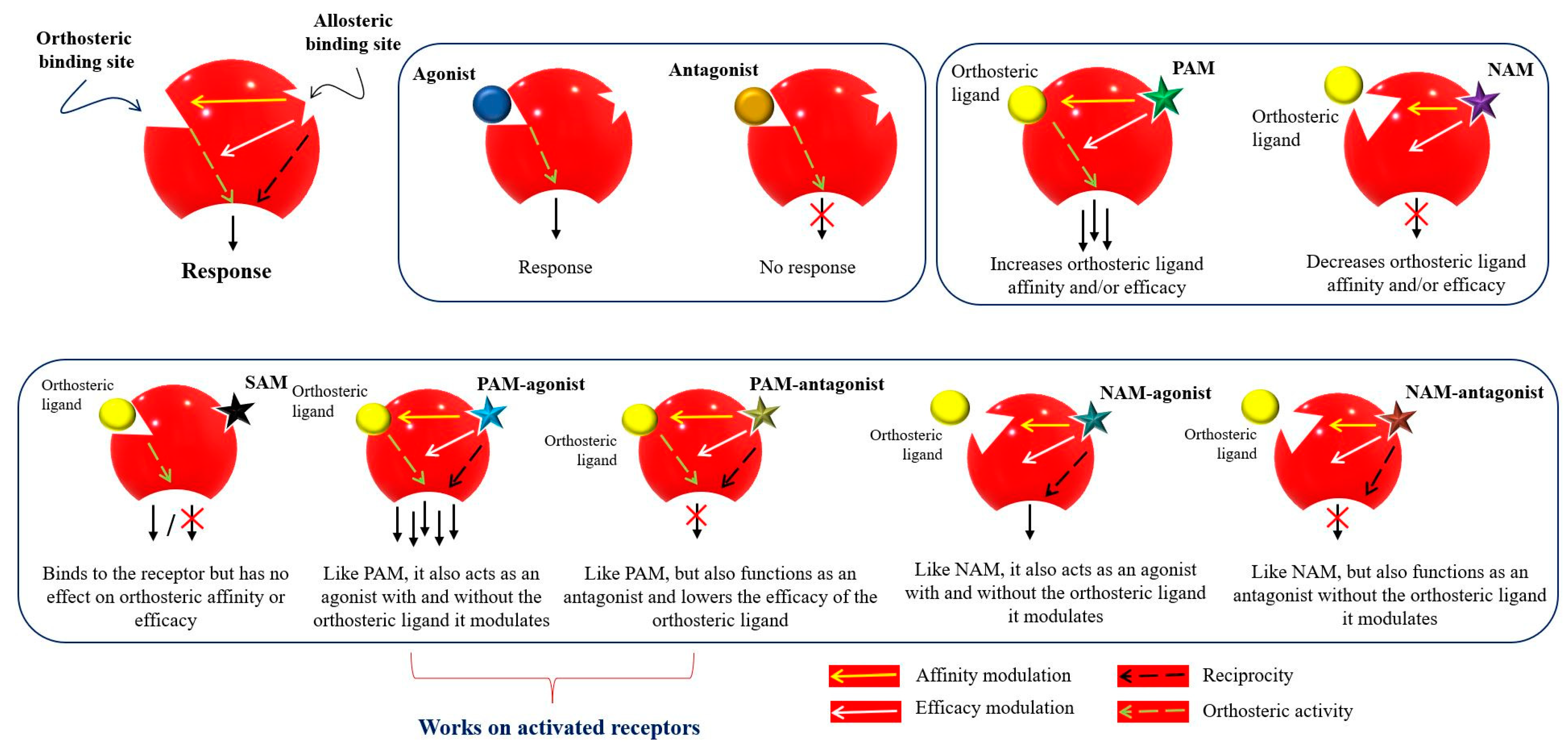
Figure 3 represents the classification of the allosteric modulators and their allosteric properties: affinity modulation, efficacy modulation, reciprocity, and ceiling effect. Affinity modulation indicates the change in the structural conformation of an orthosteric LBP such that the binding affinity of an orthosteric ligand increases or decreases [57]. Efficacy modulation denotes the increase/decrease in intracellular responses (intrinsic efficacy), depending on the orthosteric ligands (agonist or antagonist) [57]. The ceiling effect or the saturability effect means that allosteric modulators are non-competitive and maintain a certain saturation level at a certain concentration [58]. Allosteric modulators may display the possibility of an absolute subtype selectivity for a target protein. For example, a 1700-fold selective allosteric inhibition for phospholipase D1 (PLD1) compared to its subtype PLD2 has been reported [59]. Allosteric modulators may improve the efficacy and potency of an agonist (e.g., from 2 to 100-fold) in a receptor or may have a synergistic effect [60][61]. Furthermore, reciprocity or allosteric activation may occur [54], which means that the receptor is activated directly, without the presence of an orthosteric ligand [62].
Figure 3. Mode of action and classification of allosteric modulators. Graphical representation of orthosteric and allosteric sites of a receptor. Allosteric ligands bind to a topographically distinct site on a receptor to modulate orthosteric ligand affinity (yellow arrow) and/or efficacy (white arrow). Some allosteric ligands can directly activate their signaling (black arrow). Orthosteric ligands (agonist or antagonist) are colored yellow. Star-shaped allosteric modulators are presented with different colors. PAM—positive allosteric modulator; NAM—negative allosteric modulator; SAM—silent allosteric modulator [53][57][63][64].
Currently, 202 allosteric modulators have been reported for nuclear receptors [65], with the following existing allosteric ligand binding surfaces: (i) the AF-2 site; (ii) the binding function 3 (BF-3 site); (iii) the ligand-binding pocket (synergistic); (iv) zinc fingers and response elements; and (v) the AF-1 site [66][67]. Allosteric modulators have been demonstrated for numerous nuclear receptors; for example, Gabler et al. reported imatinib as a first-in-class allosteric farnesoid X receptor (FXR) modulator that enhances agonist-induced FXR activation in a reporter gene expression assay. The imatinib analogues-16 (I-16) possess extraordinary efficacy (EC50 = 1.9 nm) and high selectivity over other nuclear receptors [68][69].

3.1. Allosteric Targeting of the Pregnane X Receptor (PXR)

3.1.1. Duplex—Synergistic Activation of the PXR LBD

The PXR LBD is able to bind two synthetic ligands concomitantly to the orthosteric ligand binding site (Figure 4A) [70], a phenomenon which has been described previously for the PPARγ and ERβ nuclear receptors [71][72]. Two or more compounds accommodate the same binding sites in the receptors and occupy a space within the canonical LBP near the helix 3 region, leading to enhanced coactivator binding, transactivation, and target gene expression [70][71][72].
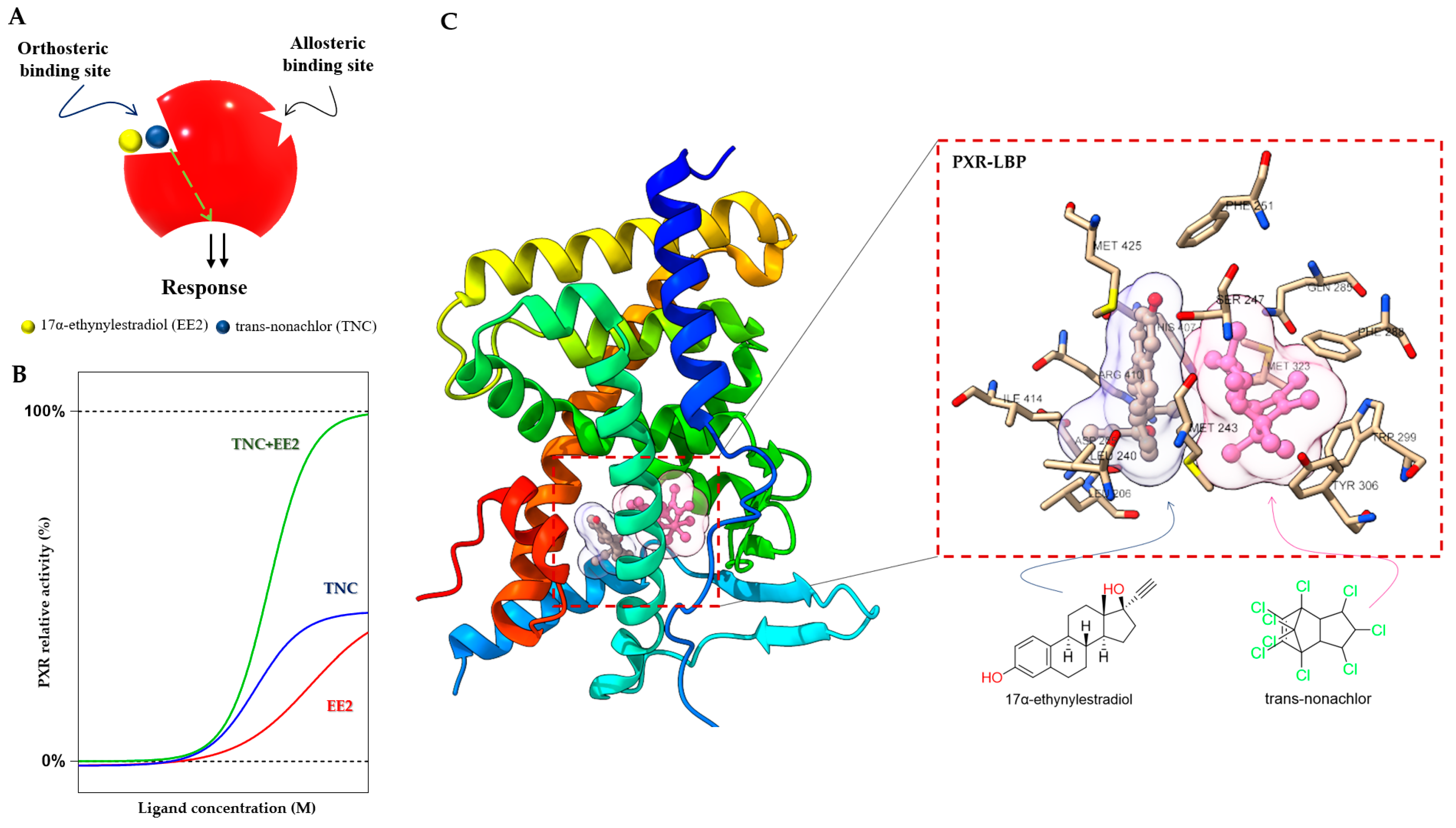
Figure 4. Duplex—synergistic activation of the PXR LBD. (A) Graphical representation of the duplex model; two ligands (EE2 and TNC) bind to the orthosteric ligand binding site on a receptor to increase the efficacy. (B) The dose response simulation shows that the combination of TNC and EE2 produces synergistic effects on PXR activation. (C) Crystal structure of the PXR LBD in complex with EE2 and TNC (PDB-4 × 1 G) and their location highlighted in the ligand binding pocket with interacting amino acid residues [70][73].
In the case of the PXR, it was found that binary cocktails of the pesticide trans-nonachlor (TNC) as well as 17α-ethinylestradiol (EE2), the active component of contraceptive pills, produce synergistic activation of the PXR and increase expression of its target CYP3A4 gene. Interestingly, the single chemicals (EE2 or TNC) were not observed to express the PXR target gene CYP3A4 in the human hepatocyte [70]. Synergistic activation of the PXR by EE2 and TNC reached full agonist activity compared to the potent agonist SR12813, but the compounds act as weak agonists when used separately (Figure 4B).
Small molecules (<500 Da) bind and fill a limited portion of the PXR LBP, leaving empty volume available to accommodate a second compound [73]. This concept has been proposed as a “supramolecular ligand” assembled from two or more compounds that interact with each other within the LBP of a receptor. TNC binds to the LBP, and EE2 binds to the closely adjacent helix 12 in the PXR LBP (Figure 4C). This ectopic site near helix 12 may be considered an allosteric site, with bitopic ligands linking orthosteric and allosteric sites to achieve improved PXR affinity or selectivity [70][74]. The EE2 binding position is a resisted region of the PXR LBP that leaves a significant portion of the pocket unoccupied and available for additional interactions. Supramolecular ligands of the PXR and their properties have been reported to a very limited extent so far, and researchers can expect many more compounds with this activity [70].

3.1.2. The AF-2 Binding Site at the PXR LBD and Its Ligands

It was found that ligands binding to the AF-2 function of the PXR alter the interactions between a coregulator peptide and the PXR. The ligand-dependent groove of the AF-2 is made up of helixes 3, 4, 5, and 12, which are mainly hydrophobic regions [37]. The orthosteric PXR LBP ends with a short helix 12 (H12), which is important for the structural organization of the AF-2 region to recruit transcriptional coregulators. Coregulators play an important functional role in the transduction of PXR signals. Both corepressors and coactivators bind to the AF-2 regions through a short amphipathic helical sequence containing the Leu-Xxx-Xxx-Leu-Leu (LXXLL) motif in coactivators or Ile/Leu-Xxx-Xxx-Ile/Val-Ile motifs in corepressors via an electrostatic interaction [75][76]. When the binding of a ligand is performed, helix H12 undergoes a significant conformational structural change that alters the overall shape of the AF-2 binding site. The PXR ligands alter the AF-2 site after binding into the PXR LBP, and thus modify the recruitment of coactivators or corepressors, resulting in different agonist or antagonist effects of the ligands [77].
All available PXR antagonist molecules act through similar agonist activation mechanisms, except for some AF-2 disruptors (Figure 5). The PXR’s ligand binding site is the same for both agonists and antagonists, but there are small residue differences with helix 12 [39][40][78]. In addition, none of the identified AF-2 site binding antagonists possess allosteric properties (selectivity, efficacy, or affinity) (Table 1) [54]. Targeting of the AF-2 site always involves issues of nuclear receptor selectivity because the AF-2 site is structurally conserved across subtypes of the nuclear receptor family [78][79]. For example, ketoconazole, the first identified PXR AF-2 site allosteric modulator and antagonist, is a common inhibitor of activated PXRs, CARs, LXRα/βs, and FXRs [80][81]. Ketoconazole binding to the surfaces of the AF-2 site suggests that ketoconazole directly blocks a coactivator (e.g., SRC-1) binding, a finding which was confirmed by a double mutant model (T248E/K277Q) in the AF-2 region of the PXR [81].
The phytoestrogen coumestrol is a natural PXR antagonist proposed to bind to a non-LBP. Biochemical binding assays and LBP-filled mutant (obliterating) studies confirm their surface binding is distinct from the orthosteric site of the LBP [82]. Additionally, computational pharmacophore and docking analyses showed that the known PXR antagonists coumestrol and sulforaphane also accommodate the AF-2 ligand-binding site [83].
In another study, the azole compound FLB-12, a derivative of the azole antifungal ketoconazole, antagonized activated PXRs in the hepatocyte cell line and in in vivo models. The triple mutant plasmid of the PXR LBP (S247W/S208W/C284W) was used to confirm its binding sites outside the PXR LBP. FLB-12 disrupts the interaction between the PXR and SRC-1, a finding which was verified by the protein pull-down assay, indicating the location of binding into the AF-2 sites. This antagonist was found to be selective and less toxic as compared to ketoconazole [84].
Ekins et al. reported residues of the PXR AF-2 ligand binding site. The identified AF-2 ligand binding site is predominantly hydrophobic, and it consists of 15 amino acids (Lys252, Ile255, Lys259, Phe264, Ile269, Glu270, Gln272, Ile273, Ser274, Leu276, Lys277, Pro423, Leu424, Glu427, and Leu428). Lys277 probably serves as a “charge clamp” for the interaction between the coactivator SRC-1 (His687) and the PXR, and it may play a significant role in the initial phase of accommodation of azole molecules into the binding groove of the PXR. Two more azole analogs, enilconazole and fluconazole, have also had interactions confirmed with the AF-2 ligand binding site, as well as antagonist activity shown towards the PXR [80][85].
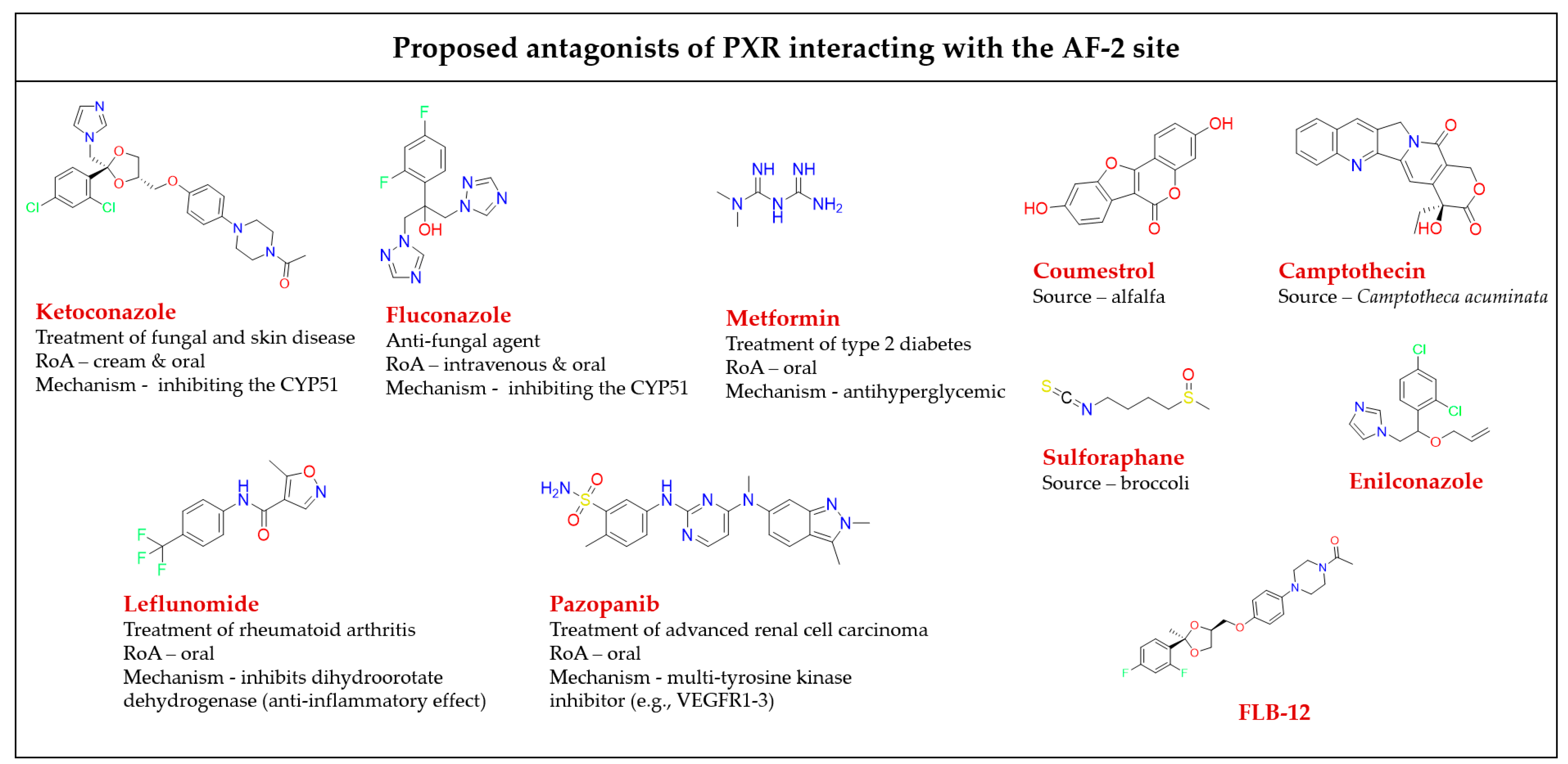
Figure 5. Chemical structures of known AF-2 site-binding PXR antagonists. RoA-routes of administration. The chemical structures were drawn in ChemDraw 20.0 software [80][81][82][83][84][86][87][88][89].
Table 1. A detailed list of antagonists that bind to the PXR AF-2 binding site.
Leflunomide, a drug used clinically for rheumatic arthritis therapy, acts as a PXR antagonist, but as an activator of the CAR [90]. It was shown that it inhibits the PXR/SRC-1 interaction as demonstrated by site-directed mutagenesis of the AF-2 sites [83][91].
The pentacyclic alkaloid camptothecin is known as a topoisomerase I inhibitor, and its analogs are approved for colon cancer therapy. Camptothecin was identified as attenuating CYP3A4 induction by blocking PXR activation via binding outside of the PXR LBP to prevent the recruitment of coactivators (such as SRC-1) [88].

References

  1. Moore, D.D.; Kato, S.; Xie, W.; Mangelsdorf, D.J.; Schmidt, D.R.; Xiao, R.; Kliewer, S.A. International Union of Pharmacology. LXII. The NR1H and NR1I Receptors: Constitutive Androstane Receptor, Pregnene X Receptor, Farnesoid X Receptor α, Farnesoid X Receptor β, Liver X Receptor α, Liver X Receptor β, and Vitamin D Receptor. Pharmacol. Rev. 2006, 58, 742–759.
  2. Smutny, T.; Mani, S.; Pavek, P. Post-translational and Post-transcriptional Modifications of Pregnane X Receptor (PXR) in Regulation of the Cytochrome P450 Superfamily. Curr. Drug Metab. 2013, 14, 1059–1069.
  3. Jumper, J.; Evans, R.; Pritzel, A.; Green, T.; Figurnov, M.; Ronneberger, O.; Tunyasuvunakool, K.; Bates, R.; Žídek, A.; Potapenko, A.; et al. Highly accurate protein structure prediction with AlphaFold. Nature 2021, 596, 583–589.
  4. Perrakis, A.; Sixma, T.K. AI revolutions in biology. EMBO Rep. 2021, 22, e54046.
  5. Monzon, V.; Haft, D.H.; Bateman, A. Folding the unfoldable: Using AlphaFold to explore spurious proteins. Bioinform. Adv. 2022, 2, vbab043.
  6. Hall, A.; Chanteux, H.; Menochet, K.; Ledecq, M.; Schulze, M.E.D. Designing Out PXR Activity on Drug Discovery Projects: A Review of Structure-Based Methods, Empirical and Computational Approaches. J. Med. Chem. 2021, 64, 6413–6522.
  7. Wallace, B.D.; Betts, L.; Talmage, G.; Pollet, R.M.; Holman, N.S.; Redinbo, M.R. Structural and Functional Analysis of the Human Nuclear Xenobiotic Receptor PXR in Complex with RXRα. J. Mol. Biol. 2013, 425, 2561–2577.
  8. Rigalli, J.P.; Theile, D.; Nilles, J.; Weiss, J. Regulation of PXR Function by Coactivator and Corepressor Proteins: Ligand Binding Is Just the Beginning. Cells 2021, 10, 3137.
  9. Bauer, B.; Hartz, A.M.S.; Fricker, G.; Miller, D.S. Pregnane X Receptor Up-Regulation of P-Glycoprotein Expression and Transport Function at the Blood-Brain Barrier. Mol. Pharmacol. 2004, 66, 413–419.
  10. Xing, Y.; Yan, J.; Niu, Y. PXR: A center of transcriptional regulation in cancer. Acta Pharm. Sin. B 2020, 10, 197–206.
  11. Lehmann, J.M.; McKee, D.D.; Watson, M.A.; Willson, T.M.; Moore, J.T.; Kliewer, S.A. The human orphan nuclear receptor PXR is activated by compounds that regulate CYP3A4 gene expression and cause drug interactions. J. Clin. Investig. 1998, 102, 1016–1023.
  12. Kliewer, S.A.; Moore, J.T.; Wade, L.; Staudinger, J.L.; Watson, M.A.; Jones, S.A.; McKee, D.D.; Oliver, B.B.; Willson, T.M.; Zetterström, R.H.; et al. An Orphan Nuclear Receptor Activated by Pregnanes Defines a Novel Steroid Signaling Pathway. Cell 1998, 92, 73–82.
  13. Fukuen, S.; Fukuda, T.; Matsuda, H.; Sumida, A.; Yamamoto, I.; Inaba, T.; Azuma, J. Identification of the novel splicing variants for the hPXR in human livers. Biochem. Biophys. Res. Commun. 2002, 298, 433–438.
  14. Chan, G.N.Y.; Hoque, M.T.; Cummins, C.L.; Bendayan, R. Regulation of P-glycoprotein by orphan nuclear receptors in human brain microvessel endothelial cells. J. Neurochem. 2011, 118, 163–175.
  15. Atlas, P. Nuclear Receptor Subfamily 1 Group I Member 2. Available online: https://www.proteinatlas.org/ENSG00000144852-NR1I2/tissue (accessed on 22 August 2022).
  16. Nishimura, M.; Naito, S.; Yokoi, T. Tissue-specific mRNA Expression Profiles of Human Nuclear Receptor Subfamilies. Drug Metab. Pharmacokinet. 2004, 19, 135–149.
  17. Oladimeji, P.O.; Chen, T. PXR: More Than Just a Master Xenobiotic Receptor. Mol. Pharmacol. 2018, 93, 119–127.
  18. Creusot, N.; Gassiot, M.; Alaterre, E.; Chiavarina, B.; Grimaldi, M.; Boulahtouf, A.; Toporova, L.; Gerbal-Chaloin, S.; Daujat-Chavanieu, M.; Matheux, A.; et al. The Anti-Cancer Drug Dabrafenib Is a Potent Activator of the Human Pregnane X Receptor. Cells 2020, 9, 1641.
  19. Feng, F.; Jiang, Q.; Cao, S.; Cao, Y.; Li, R.; Shen, L.; Zhu, H.; Wang, T.; Sun, L.; Liang, E.; et al. Pregnane X receptor mediates sorafenib resistance in advanced hepatocellular carcinoma. Biochim. Biophys. Acta (BBA)-Gen. Subj. 2018, 1862, 1017–1030.
  20. Gwag, T.; Meng, Z.; Sui, Y.; Helsley, R.N.; Park, S.H.; Wang, S.; Greenberg, R.N.; Zhou, C. Non-nucleoside reverse transcriptase inhibitor efavirenz activates PXR to induce hypercholesterolemia and hepatic steatosis. J. Hepatol. 2019, 70, 930–940.
  21. Sinz, M.W. Evaluation of pregnane X receptor (PXR)-mediated CYP3A4 drug-drug interactions in drug development. Drug Metab. Rev. 2013, 45, 3–14.
  22. Shukla, S.J.; Sakamuru, S.; Huang, R.; Moeller, T.A.; Shinn, P.; VanLeer, D.; Auld, D.S.; Austin, C.P.; Xia, M. Identification of Clinically Used Drugs That Activate Pregnane X Receptors. Drug Metab. Dispos. 2011, 39, 151–159.
  23. Wang, Y.-M.; Ong, S.S.; Chai, S.C.; Chen, T. Role of CAR and PXR in xenobiotic sensing and metabolism. Expert Opin. Drug Metab. Toxicol. 2012, 8, 803–817.
  24. Ma, X.; Shah, Y.M.; Guo, G.L.; Wang, T.; Krausz, K.W.; Idle, J.R.; Gonzalez, F.J. Rifaximin Is a Gut-Specific Human Pregnane X Receptor Activator. J. Pharmacol. Exp. Ther. 2007, 322, 391–398.
  25. Daujat-Chavanieu, M.; Gerbal-Chaloin, S. Regulation of CAR and PXR Expression in Health and Disease. Cells 2020, 9, 2395.
  26. Pondugula, S.R.; Pavek, P.; Mani, S. Pregnane X Receptor and Cancer: Context-Specificity is Key. Nucl. Recep. Res. 2016, 3, 101198.
  27. Bansard, L.; Bouvet, O.; Moutin, E.; Le Gall, G.; Giammona, A.; Pothin, E.; Bacou, M.; Hassen-Khodja, C.; Bordignon, B.; Bourgaux, J.F.; et al. Niclosamide induces miR-148a to inhibit PXR and sensitize colon cancer stem cells to chemotherapy. Stem Cell Rep. 2022, 17, 835–848.
  28. Wang, H.; Venkatesh, M.; Li, H.; Goetz, R.; Mukherjee, S.; Biswas, A.; Zhu, L.; Kaubisch, A.; Wang, L.; Pullman, J.; et al. Pregnane X receptor activation induces FGF19-dependent tumor aggressiveness in humans and mice. J. Clin. Investig. 2011, 121, 3220–3232.
  29. Raynal, C.; Pascussi, J.-M.; Leguelinel, G.; Breuker, C.; Kantar, J.; Lallemant, B.; Poujol, S.; Bonnans, C.; Joubert, D.; Hollande, F.; et al. Pregnane × Receptor (PXR) expression in colorectal cancer cells restricts irinotecan chemosensitivity through enhanced SN-38 glucuronidation. Mol. Cancer 2010, 9, 46.
  30. Planque, C.; Rajabi, F.; Grillet, F.; Finetti, P.; Bertucci, F.; Gironella, M.; Lozano, J.J.; Beucher, B.; Giraud, J.; Garambois, V.; et al. Pregnane X-receptor promotes stem cell-mediated colon cancer relapse. Oncotarget 2016, 7, 56558–56573.
  31. Niu, X.; Wu, T.; Li, G.; Gu, X.; Tian, Y.; Cui, H. Insights into the critical role of the PXR in preventing carcinogenesis and chemotherapeutic drug resistance. Int. J. Biol. Sci. 2022, 18, 742–759.
  32. Lynch, C.; Sakamuru, S.; Huang, R.; Niebler, J.; Ferguson, S.S.; Xia, M. Characterization of human pregnane X receptor activators identified from a screening of the Tox21 compound library. Biochem. Pharmacol. 2021, 184, 114368.
  33. Zhang, J.; Pavek, P.; Kamaraj, R.; Ren, L.; Zhang, T. Dietary phytochemicals as modulators of human pregnane X receptor. Crit. Rev. Food Sci. Nutr. 2021, 1–23.
  34. Karpale, M.; Hukkanen, J.; Hakkola, J. Nuclear Receptor PXR in Drug-Induced Hypercholesterolemia. Cells 2022, 11, 313.
  35. Sayaf, K.; Zanotto, I.; Russo, F.P.; Gabbia, D.; De Martin, S. The Nuclear Receptor PXR in Chronic Liver Disease. Cells 2021, 11, 61.
  36. Pondugula, S.R.; Mani, S. Pregnane xenobiotic receptor in cancer pathogenesis and therapeutic response. Cancer Lett. 2013, 328, 1–9.
  37. Chai, S.C.; Wright, W.C.; Chen, T. Strategies for developing pregnane X receptor antagonists: Implications from metabolism to cancer. Med. Res. Rev. 2020, 40, 1061–1083.
  38. Biswas, A.; Mani, S.; Redinbo, M.R.; Krasowski, M.D.; Li, H.; Ekins, S. Elucidating the ‘Jekyll and Hyde’ Nature of PXR: The Case for Discovering Antagonists or Allosteric Antagonists. Pharm. Res. 2009, 26, 1807–1815.
  39. Li, Y.; Lin, W.; Wright, W.C.; Chai, S.C.; Wu, J.; Chen, T. Building a Chemical Toolbox for Human Pregnane X Receptor Research: Discovery of Agonists, Inverse Agonists, and Antagonists Among Analogs Based on the Unique Chemical Scaffold of SPA70. J. Med. Chem. 2021, 64, 1733–1761.
  40. Watkins, R.E.; Wisely, G.B.; Moore, L.B.; Collins, J.L.; Lambert, M.H.; Williams, S.P.; Willson, T.M.; Kliewer, S.A.; Redinbo, M.R. The human nuclear xenobiotic receptor PXR: Structural determinants of directed promiscuity. Science 2001, 292, 2329–2333.
  41. Lin, W.; Wang, Y.M.; Chai, S.C.; Lv, L.; Zheng, J.; Wu, J.; Zhang, Q.; Wang, Y.D.; Griffin, P.R.; Chen, T. SPA70 is a potent antagonist of human pregnane X receptor. Nat. Commun. 2017, 8, 741.
  42. Mustonen, E.-K.; Pantsar, T.; Rashidian, A.; Reiner, J.; Schwab, M.; Laufer, S.; Burk, O. Target Hopping from Protein Kinases to PXR: Identification of Small-Molecule Protein Kinase Inhibitors as Selective Modulators of Pregnane X Receptor from TüKIC Library. Cells 2022, 11, 1299.
  43. Ong, S.S. Pregnane X Receptor in Drug Development; IntechOpen: London, UK, 2011.
  44. Motta, S.; Callea, L.; Giani Tagliabue, S.; Bonati, L. Exploring the PXR ligand binding mechanism with advanced Molecular Dynamics methods. Sci. Rep. 2018, 8, 16207.
  45. Huang, P.; Chandra, V.; Rastinejad, F. Structural Overview of the Nuclear Receptor Superfamily: Insights into Physiology and Therapeutics. Annu. Rev. Physiol. 2010, 72, 247–272.
  46. Wu, B.; Li, S.; Dong, D. 3D structures and ligand specificities of nuclear xenobiotic receptors CAR, PXR and VDR. Drug Discov. Today 2013, 18, 574–581.
  47. Liu, T.; Beck, J.P.; Hao, J. A concise review on hPXR ligand-recognizing residues and structure-based strategies to alleviate hPXR transactivation risk. RSC Med. Chem. 2022, 13, 129–137.
  48. Huber, A.D.; Wright, W.C.; Lin, W.; Majumder, K.; Low, J.A.; Wu, J.; Buchman, C.D.; Pintel, D.J.; Chen, T. Mutation of a single amino acid of pregnane X receptor switches an antagonist to agonist by altering AF-2 helix positioning. Cell Mol. Life Sci. 2021, 78, 317–335.
  49. Huber, A.D.; Li, Y.; Lin, W.; Galbraith, A.N.; Mishra, A.; Porter, S.N.; Wu, J.; Florke Gee, R.R.; Zhuang, W.; Pruett-Miller, S.M.; et al. SJPYT-195: A Designed Nuclear Receptor Degrader That Functions as a Molecular Glue Degrader of GSPT1. ACS Med. Chem. Lett. 2022, 13, 1311–1320.
  50. Zorba, A.; Nguyen, C.; Xu, Y.; Starr, J.; Borzilleri, K.; Smith, J.; Zhu, H.; Farley, K.A.; Ding, W.; Schiemer, J.; et al. Delineating the role of cooperativity in the design of potent PROTACs for BTK. Proc. Natl. Acad. Sci. USA 2018, 115, E7285–E7292.
  51. Shimokawa, K.; Shibata, N.; Sameshima, T.; Miyamoto, N.; Ujikawa, O.; Nara, H.; Ohoka, N.; Hattori, T.; Cho, N.; Naito, M. Targeting the Allosteric Site of Oncoprotein BCR-ABL as an Alternative Strategy for Effective Target Protein Degradation. ACS Med. Chem. Lett. 2017, 8, 1042–1047.
  52. Pettersen, E.F.; Goddard, T.D.; Huang, C.C.; Meng, E.C.; Couch, G.S.; Croll, T.I.; Morris, J.H.; Ferrin, T.E. UCSF ChimeraX: Structure visualization for researchers, educators, and developers. Protein Sci. 2021, 30, 70–82.
  53. De Smet, F.; Christopoulos, A.; Carmeliet, P. Allosteric targeting of receptor tyrosine kinases. Nat. Biotechnol. 2014, 32, 1113–1120.
  54. Gregory, K.J.; Sexton, P.M.; Christopoulos, A. Overview of receptor allosterism. Curr. Protoc. Pharmacol. 2000, 11, 1–21.
  55. Christopoulos, A.; Kenakin, T. G protein-coupled receptor allosterism and complexing. Pharmacol. Rev. 2002, 54, 323–374.
  56. Smith, N.J.; Milligan, G. Allostery at G Protein-Coupled Receptor Homo- and Heteromers: Uncharted Pharmacological Landscapes. Pharmacol. Rev. 2010, 62, 701–725.
  57. Conn, P.J.; Christopoulos, A.; Lindsley, C.W. Allosteric modulators of GPCRs: A novel approach for the treatment of CNS disorders. Nat. Rev. Drug Discov. 2009, 8, 41–54.
  58. Christopoulos, A. Advances in G Protein-Coupled Receptor Allostery: From Function to Structure. Mol. Pharmacol. 2014, 86, 463–478.
  59. Lewis, J.A.; Scott, S.A.; Lavieri, R.; Buck, J.R.; Selvy, P.E.; Stoops, S.L.; Armstrong, M.D.; Brown, H.A.; Lindsley, C.W. Design and synthesis of isoform-selective phospholipase D (PLD) inhibitors. Part I: Impact of alternative halogenated privileged structures for PLD1 specificity. Bioorg. Med. Chem. Lett. 2009, 19, 1916–1920.
  60. Cao, A.M.; Quast, R.B.; Fatemi, F.; Rondard, P.; Pin, J.P.; Margeat, E. Allosteric modulators enhance agonist efficacy by increasing the residence time of a GPCR in the active state. Nat. Commun. 2021, 12, 5426.
  61. Reddy, G.S.; Kamaraj, R.; Hossain, K.A.; Kumar, J.S.; Thirupataiah, B.; Medishetti, R.; Sushma Sri, N.; Misra, P.; Pal, M. Amberlyst-15 catalysed synthesis of novel indole derivatives under ultrasound irradiation: Their evaluation as serotonin 5-HT2C receptor agonists. Bioorg. Chem. 2021, 116, 105380.
  62. Burford, N.T.; Clark, M.J.; Wehrman, T.S.; Gerritz, S.W.; Banks, M.; O’Connell, J.; Traynor, J.R.; Alt, A. Discovery of positive allosteric modulators and silent allosteric modulators of the mu-opioid receptor. Proc. Natl. Acad. Sci. USA 2013, 110, 10830–10835.
  63. Kenakin, T.; Strachan, R.T. PAM-Antagonists: A Better Way to Block Pathological Receptor Signaling? Trends Pharmacol. Sci. 2018, 39, 748–765.
  64. Fasciani, I.; Petragnano, F.; Aloisi, G.; Marampon, F.; Carli, M.; Scarselli, M.; Maggio, R.; Rossi, M. Allosteric Modulators of G Protein-Coupled Dopamine and Serotonin Receptors: A New Class of Atypical Antipsychotics. Pharmaceuticals 2020, 13, 388.
  65. Allosteric Database (ASD). Nuclear Hormone Receptor >> 202 Modulators. Available online: http://mdl.shsmu.edu.cn/ASD/module/modulators/modulators.jsp (accessed on 14 May 2022).
  66. Changeux, J.P.; Christopoulos, A. Allosteric Modulation as a Unifying Mechanism for Receptor Function and Regulation. Cell 2016, 166, 1084–1102.
  67. Moore, T.W.; Mayne, C.G.; Katzenellenbogen, J.A. Minireview: Not Picking Pockets: Nuclear Receptor Alternate-Site Modulators (NRAMs). Mol. Endocrinol. 2010, 24, 683–695.
  68. Chen, Y.; Li, J.; Wu, Z.; Liu, G.; Li, H.; Tang, Y.; Li, W. Computational Insight into the Allosteric Activation Mechanism of Farnesoid X Receptor. J. Chem. Inf. Model. 2020, 60, 1540–1550.
  69. Gabler, M.; Kramer, J.; Schmidt, J.; Pollinger, J.; Weber, J.; Kaiser, A.; Lohr, F.; Proschak, E.; Schubert-Zsilavecz, M.; Merk, D. Allosteric modulation of the farnesoid X receptor by a small molecule. Sci. Rep. 2018, 8, 6846.
  70. Delfosse, V.; Dendele, B.; Huet, T.; Grimaldi, M.; Boulahtouf, A.; Gerbal-Chaloin, S.; Beucher, B.; Roecklin, D.; Muller, C.; Rahmani, R.; et al. Synergistic activation of human pregnane X receptor by binary cocktails of pharmaceutical and environmental compounds. Nat. Commun. 2015, 6, 8089.
  71. Wang, Y.; Chirgadze, N.Y.; Briggs, S.L.; Khan, S.; Jensen, E.V.; Burris, T.P. A second binding site for hydroxytamoxifen within the coactivator-binding groove of estrogen receptor β. Proc. Natl. Acad. Sci. USA 2006, 103, 9908–9911.
  72. Hughes, T.S.; Giri, P.K.; de Vera, I.M.; Marciano, D.P.; Kuruvilla, D.S.; Shin, Y.; Blayo, A.L.; Kamenecka, T.M.; Burris, T.P.; Griffin, P.R.; et al. An alternate binding site for PPARgamma ligands. Nat. Commun. 2014, 5, 3571.
  73. Delfosse, V.; Huet, T.; Harrus, D.; Granell, M.; Bourguet, M.; Gardia-Parège, C.; Chiavarina, B.; Grimaldi, M.; Le Mével, S.; Blanc, P.; et al. Mechanistic insights into the synergistic activation of the RXR–PXR heterodimer by endocrine disruptor mixtures. Proc. Natl. Acad. Sci. USA 2021, 118, e2020551118.
  74. Lane, J.R.; Sexton, P.M.; Christopoulos, A. Bridging the gap: Bitopic ligands of G-protein-coupled receptors. Trends Pharmacol. Sci. 2013, 34, 59–66.
  75. Zhang, C.; Wu, J.; Chen, Q.; Tan, H.; Huang, F.; Guo, J.; Zhang, X.; Yu, H.; Shi, W. Allosteric binding on nuclear receptors: Insights on screening of non-competitive endocrine-disrupting chemicals. Environ. Int. 2022, 159, 107009.
  76. Pavek, P. Pregnane X Receptor (PXR)-Mediated Gene Repression and Cross-Talk of PXR with Other Nuclear Receptors via Coactivator Interactions. Front. Pharmacol. 2016, 7, 456.
  77. La Sala, G.; Gunnarsson, A.; Edman, K.; Tyrchan, C.; Hogner, A.; Frolov, A.I. Unraveling the Allosteric Cross-Talk between the Coactivator Peptide and the Ligand-Binding Site in the Glucocorticoid Receptor. J. Chem. Inf. Model. 2021, 61, 3667–3680.
  78. Fischer, A.; Smieško, M. Allosteric Binding Sites On Nuclear Receptors: Focus On Drug Efficacy and Selectivity. Int. J. Mol. Sci. 2020, 21, 534.
  79. Wärnmark, A.; Treuter, E.; Wright, A.P.H.; Gustafsson, J.-A.k. Activation Functions 1 and 2 of Nuclear Receptors: Molecular Strategies for Transcriptional Activation. Mol. Endocrinol. 2003, 17, 1901–1909.
  80. Wang, H.; Huang, H.; Li, H.; Teotico, D.G.; Sinz, M.; Baker, S.D.; Staudinger, J.; Kalpana, G.; Redinbo, M.R.; Mani, S. Activated pregnenolone X-receptor is a target for ketoconazole and its analogs. Clin. Cancer Res. 2007, 13, 2488–2495.
  81. Huang, H.; Wang, H.; Sinz, M.; Zoeckler, M.; Staudinger, J.; Redinbo, M.R.; Teotico, D.G.; Locker, J.; Kalpana, G.V.; Mani, S. Inhibition of drug metabolism by blocking the activation of nuclear receptors by ketoconazole. Oncogene 2007, 26, 258–268.
  82. Wang, H.; Li, H.; Moore, L.B.; Johnson, M.D.; Maglich, J.M.; Goodwin, B.; Ittoop, O.R.; Wisely, B.; Creech, K.; Parks, D.J.; et al. The phytoestrogen coumestrol is a naturally occurring antagonist of the human pregnane X receptor. Mol. Endocrinol. 2008, 22, 838–857.
  83. Ekins, S.; Kholodovych, V.; Ai, N.; Sinz, M.; Gal, J.; Gera, L.; Welsh, W.J.; Bachmann, K.; Mani, S. Computational discovery of novel low micromolar human pregnane X receptor antagonists. Mol. Pharmacol. 2008, 74, 662–672.
  84. Venkatesh, M.; Wang, H.; Cayer, J.; Leroux, M.; Salvail, D.; Das, B.; Wrobel, J.E.; Mani, S. In vivo and in vitro characterization of a first-in-class novel azole analog that targets pregnane X receptor activation. Mol. Pharmacol. 2011, 80, 124–135.
  85. Ekins, S.; Chang, C.; Mani, S.; Krasowski, M.D.; Reschly, E.J.; Iyer, M.; Kholodovych, V.; Ai, N.; Welsh, W.J.; Sinz, M.; et al. Human pregnane X receptor antagonists and agonists define molecular requirements for different binding sites. Mol. Pharmacol. 2007, 72, 592–603.
  86. Burk, O.; Kuzikov, M.; Kronenberger, T.; Jeske, J.; Keminer, O.; Thasler, W.E.; Schwab, M.; Wrenger, C.; Windshügel, B. Identification of approved drugs as potent inhibitors of pregnane X receptor activation with differential receptor interaction profiles. Arch. Toxicol. 2018, 92, 1435–1451.
  87. Krausova, L.; Stejskalova, L.; Wang, H.; Vrzal, R.; Dvorak, Z.; Mani, S.; Pavek, P. Metformin suppresses pregnane X receptor (PXR)-regulated transactivation of CYP3A4 gene. Biochem. Pharmacol. 2011, 82, 1771–1780.
  88. Chen, Y.; Tang, Y.; Robbins, G.T.; Nie, D. Camptothecin attenuates cytochrome P450 3A4 induction by blocking the activation of human pregnane X receptor. J. Pharmacol. Exp. Ther. 2010, 334, 999–1008.
  89. Zhou, C.; Poulton, E.J.; Grun, F.; Bammler, T.K.; Blumberg, B.; Thummel, K.E.; Eaton, D.L. The dietary isothiocyanate sulforaphane is an antagonist of the human steroid and xenobiotic nuclear receptor. Mol. Pharmacol. 2007, 71, 220–229.
  90. Carazo, A.; Dusek, J.; Holas, O.; Skoda, J.; Hyrsova, L.; Smutny, T.; Soukup, T.; Dosedel, M.; Pávek, P. Teriflunomide Is an Indirect Human Constitutive Androstane Receptor (CAR) Activator Interacting with Epidermal Growth Factor (EGF) Signaling. Front. Pharmacol. 2018, 9, 993.
  91. Kronenberger, T.; Keminer, O.; Wrenger, C.; Windshügel, B. Nuclear Receptor Modulators—Current Approaches and Future Perspectives. In Drug Discovery and Development: From Molecules to Medicine; InTech: London, UK, 2015.
More
Information
Contributors MDPI registered users' name will be linked to their SciProfiles pages. To register with us, please refer to https://encyclopedia.pub/register : , , ,
View Times: 376
Revisions: 2 times (View History)
Update Date: 13 Oct 2022
1000/1000