Your browser does not fully support modern features. Please upgrade for a smoother experience.
Submitted Successfully!
 +1 credit
Thank you for your contribution! You can also upload a video entry or images related to this topic.
For video creation, please contact our Academic Video Service.
| Version | Summary | Created by | Modification | Content Size | Created at | Operation |
|---|---|---|---|---|---|---|
| 1 | Kinza Abbas | -- | 1090 | 2022-10-01 16:24:35 | | | |
| 2 | Catherine Yang | Meta information modification | 1090 | 2022-10-08 03:46:38 | | |
Video Upload Options
We provide professional Academic Video Service to translate complex research into visually appealing presentations. Would you like to try it?
Cite
If you have any further questions, please contact Encyclopedia Editorial Office.
Abbas, K.; Lu, Y.; Bavishi, S.; Mishra, N.; Tomthundyil, S.; Sawant, S.A.; Shahjouei, S.; Abedi, V.; Zand, R. Associations between CKD, Retina, and CVD. Encyclopedia. Available online: https://encyclopedia.pub/entry/28195 (accessed on 23 July 2026).
Abbas K, Lu Y, Bavishi S, Mishra N, Tomthundyil S, Sawant SA, et al. Associations between CKD, Retina, and CVD. Encyclopedia. Available at: https://encyclopedia.pub/entry/28195. Accessed July 23, 2026.
Abbas, Kinza, Yezhong Lu, Shreya Bavishi, Nandini Mishra, Saumya Tomthundyil, Shreeya Atul Sawant, Shima Shahjouei, Vida Abedi, Ramin Zand. "Associations between CKD, Retina, and CVD" Encyclopedia, https://encyclopedia.pub/entry/28195 (accessed July 23, 2026).
Abbas, K., Lu, Y., Bavishi, S., Mishra, N., Tomthundyil, S., Sawant, S.A., Shahjouei, S., Abedi, V., & Zand, R. (2022, October 01). Associations between CKD, Retina, and CVD. In Encyclopedia. https://encyclopedia.pub/entry/28195
Abbas, Kinza, et al. "Associations between CKD, Retina, and CVD." Encyclopedia. Web. 01 October, 2022.
Copy Citation
The kidney, brain, and retina are highly metabolic organs that require specialized vascular networks to carry out their function. Cohort studies suggest that retinopathy, cerebrovascular disease (CVD), and chronic kidney disease (CKD) frequently coincide. The frequently concurrent prevalence of retinopathy, CVD, and CKD suggests a common basis for pathology.
small vessel disease
cerebrovascular disease
stroke
retinopathy
chronic kidney disease
1. Embryology and Genetics
The retina in the optic cup develops from the diencephalon, which is part of the prosencephalon or forebrain. The shared origin and similar development pattern of vasculature by vasculogenesis and angiogenesis may be the cause of linked pathology in the brain and retina [1][2].
The development of the vasculature supplying the brain begins with the formation of six pairs of primitive branchial arch arteries [3][4]. The combination of the third branchial arch arteries and the distal segment of the paired dorsal aortae forms the internal carotid artery, which then branches into anterior and posterior divisions [4]. The anterior division branches into primitive ophthalmic and olfactory arteries, later giving rise to the anterior cerebral, middle cerebral, and anterior choroidal arteries [5]. In contrast, the posterior division gives rise to the posterior cerebral and posterior choroidal arteries [4].
The hyaloid artery arises from the primitive dorsal ophthalmic artery and supplies the developing inner retina during its maturation, while the vasculogenesis in the choroid epithelium provides oxygen to the outer avascular retinal layer through diffusion [3][6][7]. The angiogenesis in the retina occurs at the late gestation stage by retinal vasculature, which develops as hyaloid vessels regress [3][6][7][8][9][10]. The vascular layer expands and increases in density following the spread of pigmentation in the retinal epithelium and the production of inductive signals from differentiated retinal pigmental epithelium [11][12][13][14].
Similar shared anatomical structures such as fenestrations and regulatory processes, such as the Renin Angiotensin Aldosterone System, may be due to parallel development of glomerular endothelium by vasculogenesis [2]. The retina and kidney also share many developmental pathways, abnormalities, and mutations in molecular pathways, which manifest through features in both organs [15].
Furthermore, cerebral autosomal dominant arteriopathy with subcortical infarcts and leukoencephalopathy (CADASIL) is a rare genetic disorder that primarily affects small vessels in the brain with renal impairment, suggesting a common pathogenetic mechanism of renal and brain lesions in this disease [16]. Retinal axonal loss in CADASIL leads to thinning of the retinal nerve fiber length, retinal vein occlusion, macular edema, and retinal venous disease have also been reported among CADASIL patients [17]. Another autosomal dominant genetic disorder that links these organs is retinal vasculopathy with cerebral leukodystrophy and systemic manifestations (RVCL-S), which presents with cerebroretinal vasculopathy (CRV), WMHs, hereditary vascular retinopathy (HVR), and hereditary endotheliopathy with retinopathy, nephropathy, and stroke (HERNS) [18]. In addition, the growing evidence for the genetic basis of SVD, stemming from MRI studies of WMH, estimate heritability to range between 55–75% [18]. Monogenic SVDs, such as COL4A1/A2-related angiopathies and retinal vasculopathy with cerebral leukodystrophy (RVCL), demonstrate cerebral, ocular, and renal abnormalities, implying that patients with monogenic CSVD have concomitant retinopathy and kidney damage [19]. These provide further indication of shared genetic and developmental similarities in the brain, kidney, and retina.
2. Cardiovascular Physiology
The frictional force of blood in the vessels, otherwise known as wall shear stress, is counteracted by tension and stretch in the vessel wall. These strains and stretch forces exert a vaso-protective role by providing hemostatic balance. Ito et al. proposed the “strain vessel hypothesis” that detailed the concept that high tone vessels evolutionarily developed to maintain perfusion of the brain and kidney. Although they are exposed to high blood pressure, vascular damage from prolonged hemodynamic stress and a perturbation to this biochemical balance results in either physiological adaptation or disease of the vessel walls [20][21][22]. Thus, common vascular risk factors such as high blood pressure and diabetes mellitus may cause damage to these strain vessels in parallel in the brain and kidney [22]. Non-traditional CKD-related risk factors, such as chronic inflammation, endothelial dysfunction, and uremic toxins, can promote cerebrovascular injury by triggering vascular injury and endothelial dysfunction [23].
3. Pathology
The numerous anatomical and functional similarities in the brain, retina, and kidney provide a basis for common pathology. The blood–brain barrier is formed by endothelial cells of the capillary wall with tight junctions, astrocytes with projections sheathing the capillary, and pericytes embedded in the capillary basement membrane [24]. Like the brain, the retina possesses barrier circulation; the blood–retina barrier is arranged as inner and outer barriers [25]. The inner retinal barrier is comprised of endothelial cells with tight junctions and is surrounded by glial cells to maintain retinal homeostasis [25][26].
Similar to the glomerular filtration barrier, the outer retinal barrier formed at the retinal pigment epithelial cell layers functions to regulate the movement of solutes and nutrients from the choroid to the sub-retinal space [25][27]. The arrangement of the choriocapillaris, Bruch’s membrane, and retinal pigment epithelial interface are homologous to the endothelium, glomerular basement membrane, and podocytes in the glomerulus (Figure 1) [28]. Physiologically, the glomerulus and choriocapillaris are both fenestrated, while the podocytes and retinal pigment epithelial cells both function as metabolic barriers, actively mediating the exchange of molecules [27][29]. Additionally, Bruch’s membrane and the glomerular basement membrane both contain a network of a3, a4, and a5 type IV collagen chains [27][30].
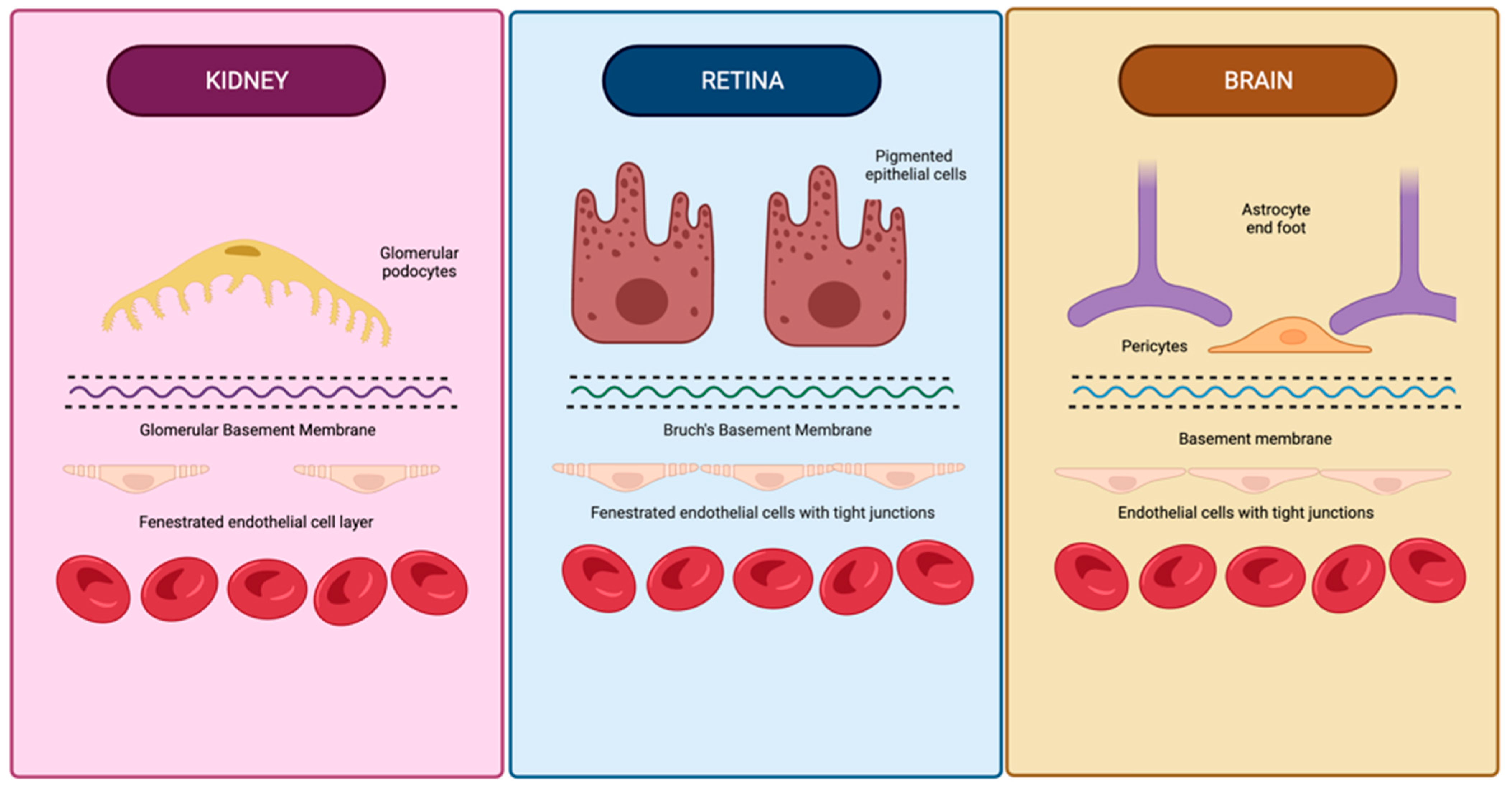
Figure 1. Similarities in anatomical structure of vascular barriers in the brain, retina, and kidneys. The vascular barriers of the brain, retina, and kidneys have many structural similarities, including basement membranes containing type IV collagen, an outer layer with analogous projections (glomerular podocyte foot processes, retinal epithelial cell projections, and astrocyte foot processes), and a fenestrated endothelial cell layer in the glomerulus and retina. Created using BioRender.com on 26 August 2022.
The anatomical and compositional similarities manifest as simultaneous retinopathy and nephropathy in membranoproliferative glomerulonephritis type II and Alport syndrome [31][32][33][34]. The simultaneous pathology and appearance of clinical symptoms in chronic cardiovascular diseases, hypertension, and diabetes mellitus further provide evidence of a link between these organs. Hemodynamic similarities of vascular beds of the brain and kidney are generally noted as the shared mechanism linking cerebral and renal vascular dysfunction biomarkers. The kidney and brain are uniquely low-resistance end-organs exposed to consistent high-volume blood flow [22].
Thus, despite differences in location and responses to injury, concurrent changes are seen in the micro-vasculature of these organs under pathological conditions, which the reserchers hypothesize to be due to their common embryological, functional, histological, and genetic basis (Figure 2).
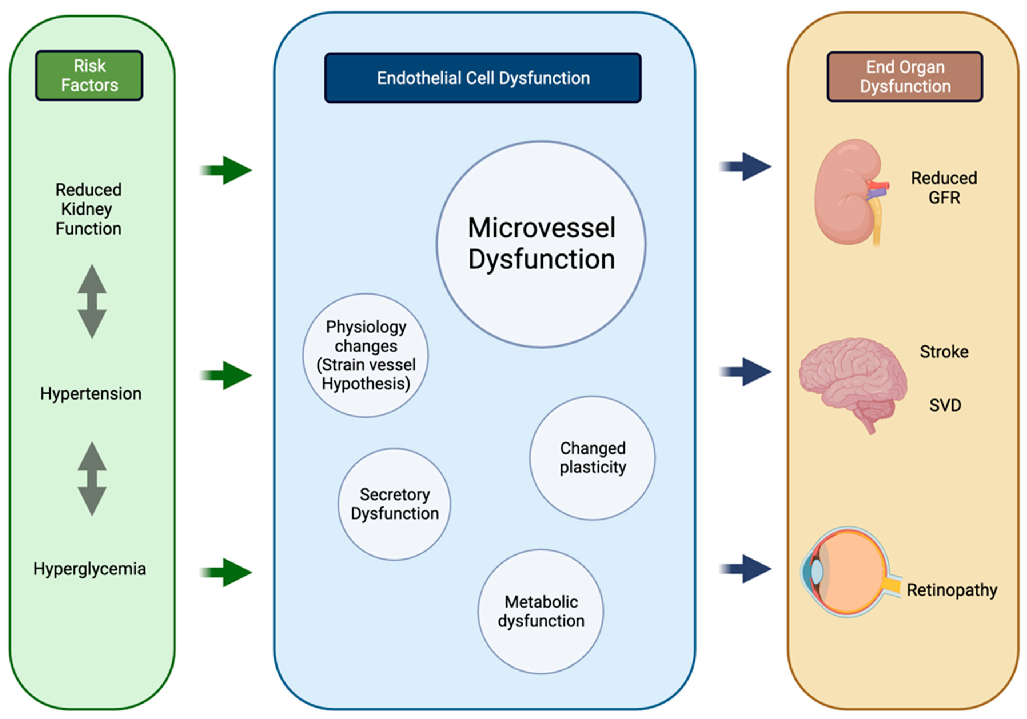
Figure 2. Process of parallel onset end-organ dysfunction. Flowchart demonstrating the interactions between shared systemic risk factors, resulting in endothelial cell changes and end-organ manifestations in the kidneys, brain, and retina. Created using BioRender.com. GFR glomerular filtration rate, SVD small vessel disease. Created using BioRender.com on 25 August 2022.
References
- Patton, N.; Aslam, T.; MacGillivray, T.; Pattie, A.; Deary, I.J.; Dhillon, B. Retinal vascular image analysis as a potential screening tool for cerebrovascular disease: A rationale based on homology between cerebral and retinal microvasculatures. J. Anat. 2005, 206, 319–348.
- Hughes, S.; Yang, H.; Chan-Ling, T. Vascularization of the human fetal retina: Roles of vasculogenesis and angiogenesis. Investig. Ophthalmol. Vis. Sci. 2000, 41, 1217–1228.
- Bertulli, L.; Robert, T. Embryological development of the human cranio-facial arterial system: A pictorial review. Surg. Radiol. Anat. 2021, 43, 961–973.
- Menshawy, K.; Mohr, J.P.; Gutierrez, J.G.A. A Functional Perspective on the Embryology and Anatomy of the Cerebral Blood Supply. J. Stroke 2015, 17, 144–158.
- Yu, J.; Xu, N.; Zhao, Y.; Yu, J. Clinical importance of the anterior choroidal artery: A review of the literature. Int. J. Med Sci. 2018, 15, 368–375.
- Kathuria, S.; Gregg, L.; Chen, J.; Gandhi, D. Normal Cerebral Arterial Development and Variations. Semin. Ultrasound CT MRI 2011, 32, 242–251.
- Saint-Geniez, M.; D’Amore, P.A. Development and pathology of the hyaloid, choroidal and retinal vasculature. Int. J. Dev. Biol. 2004, 48, 1045–1058.
- Lutty, G.A.; McLeod, D.S. Development of the hyaloid, choroidal and retinal vasculatures in the fetal human eye. Prog. Retin. Eye Res. 2017, 62, 58–76.
- Stone, J.; Chan-Ling, T.; Pe’Er, J.; Itin, A.; Gnessin, H.; Keshet, E. Roles of vascular endothelial growth factor and astrocyte degeneration in the genesis of retinopathy of prematurity. Investig. Ophthalmol. Vis. Sci. 1996, 37, 290–299.
- Gregg, L.; Millán, D.S.; Orru’, E.; Tamargo, R.J.; Gailloud, P. Ventral and Dorsal Persistent Primitive Ophthalmic Arteries. Oper. Neurosurg. 2016, 12, 141–152.
- Zhao, S.; Overbeek, P. Regulation of choroid development by the retinal pigment epithelium. Mol. Vis. 2001, 7, 277–282.
- Sakamoto, T.; Sakamoto, H.; Murphy, T.L.; Spee, C.; Soriano, D.; Ishibashi, T.; Hinton, D.R.; Ryan, S.J. Vessel Formation by Choroidal Endothelial Cells In Vitro Is Modulated by Retinal Pigment Epithelial Cells. Arch. Ophthalmol. 1995, 113, 512–520.
- Rousseau, B. Involvement of fibroblast growth factors in choroidal angiogenesis and retinal vascularization. Exp. Eye Res. 2003, 77, 147–156.
- Gogat, K.; Le Gat, L.; Berghe, L.V.D.; Marchant, D.; Kobetz, A.; Gadin, S.; Gasser, B.; Quere, I.; Abitbol, M.; Menasche, M. VEGF and KDR Gene Expression during Human Embryonic and Fetal Eye Development. Investig. Opthalmol. Vis. Sci. 2004, 45, 7–14.
- Mauer, S.M.; Barbosa, J.; Vernier, R.L.; Kjellstrand, C.M.; Buselmeier, T.J.; Simmons, R.L.; Najarian, J.S.; Goetz, F.C. Development of Diabetic Vascular Lesions in Normal Kidneys Transplanted into Patients with Diabetes Mellitus. N. Engl. J. Med. 1976, 295, 916–920.
- Di Donato, I.; Bianchi, S.; De Stefano, N.; Dichgans, M.; Dotti, M.T.; Duering, M.; Jouvent, E.; Korczyn, A.D.; Lesnik-Oberstein, S.A.J.; Malandrini, A.; et al. Cerebral Autosomal Dominant Arteriopathy with Subcortical Infarcts and Leukoencephalopathy (CADASIL) as a model of small vessel disease: Update on clinical, diagnostic, and management aspects. BMC Med. 2017, 15, 41.
- Goldstein, E.D.; Shakoor, A.; Majersik, J.J. Branch Retinal Vein Occlusion and Venous Abnormalities in CADASIL. Neurologist 2020, 25, 178–179.
- Kwa, V.I.; Van Der Sande, J.J.; Stam, J.; Tijmes, N.; Vrooland, J.L. Retinal arterial changes correlate with cerebral small-vessel disease. Neurology 2002, 59, 1536–1540.
- Haffner, C.; Malik, R.; Dichgans, M. Genetic factors in cerebral small vessel disease and their impact on stroke and dementia. J. Cereb. Blood Flow Metab. 2015, 36, 158–171.
- Ito, S.; Nagasawa, T.; Abe, M.; Mori, T. Strain vessel hypothesis: A viewpoint for linkage of albuminuria and cerebro-cardiovascular risk. Hypertens. Res. 2009, 32, 115–121.
- Lu, D.; Kassab, G.S. Role of shear stress and stretch in vascular mechanobiology. J. R. Soc. Interface 2011, 8, 1379–1385.
- Mogi, M.; Horiuchi, M. Clinical Interaction between Brain and Kidney in Small Vessel Disease. Cardiol. Res. Pract. 2011, 2011, 306189.
- Kelly, D.; Rothwell, P.M. Disentangling the multiple links between renal dysfunction and cerebrovascular disease. J. Neurol. Neurosurg. Psychiatry 2019, 91, 88–97.
- Bradbury, M.W.B.; Lightman, S.L. The blood-brain interface. Eye 1990, 4, 249–254.
- Campbell, M.; Humphries, P. The blood-retina barrier tight junctions and barrier modulation. Adv. Exp. Med. Biol. 2013, 763, 70–84.
- Cunha-Vaz, J. The Blood–Retinal Barrier in Retinal Disease. Eur. Ophthalmic Rev. 2009, 3, 105.
- Booij, J.C.; Baas, D.C.; Beisekeeva, J.; Gorgels, T.G.M.F.; Bergen, A.A.B. The dynamic nature of Bruch’s membrane. Prog. Retin. Eye Res. 2010, 29, 1–18.
- Farrah, T.E.; Dhillon, B.; Keane, P.A.; Webb, D.J.; Dhaun, N. The eye, the kidney, and cardiovascular disease: Old concepts, better tools, and new horizons. Kidney Int. 2020, 98, 323–342.
- Pollak, M.R.; Quaggin, S.E.; Hoenig, M.P.; Dworkin, L.D. The Glomerulus: The Sphere of Influence. Clin. J. Am. Soc. Nephrol. 2014, 9, 1461–1469.
- Boutaud, A.; Borza, D.-B.; Bondar, O.; Gunwar, S.; Netzer, K.-O.; Singh, N.; Ninomiya, Y.; Sado, Y.; Noelken, M.E.; Hudson, B.G. Type IV collagen of the glomerular basement membrane. Evidence that the chain specificity of network assembly is encoded by the non-collagenous NC1 domains. J. Biol. Chem. 2000, 275, 30716–30724.
- Savige, J.; Sheth, S.; Leys, A.; Nicholson, A.; Mack, H.G.; Colville, D. Ocular Features in Alport Syndrome: Pathogenesis and Clinical Significance. Clin. J. Am. Soc. Nephrol. 2015, 10, 703–709.
- Colville, D.; Savige, J.; Branley, P.; Wilson, D. Ocular abnormalities in thin basement membrane disease. Br. J. Ophthalmol. 1997, 81, 373–377.
- Dalvin, L.A.; Fervenza, F.C.; Sethi, S.; Pulido, J.S. Manifestations of Complement-Mediated and Immune Complex-Mediated Membranoproliferative Glomerulonephritis. Ophthalmology 2016, 123, 1588–1594.
- Whitmore, S.S.; Sohn, E.H.; Chirco, K.R.; Drack, A.V.; Stone, E.M.; Tucker, B.A.; Mullins, R.F. Complement activation and choriocapillaris loss in early AMD: Implications for pathophysiology and therapy. Prog. Retin. Eye Res. 2014, 45, 1–29.
More
Information
Subjects:
Cardiac & Cardiovascular Systems; Ophthalmology
Contributors
MDPI registered users' name will be linked to their SciProfiles pages. To register with us, please refer to https://encyclopedia.pub/register
:
View Times:
969
Entry Collection:
Hypertension and Cardiovascular Diseases
Revisions:
2 times
(View History)
Update Date:
08 Oct 2022
Table of Contents


Notice
You are not a member of the advisory board for this topic. If you want to update advisory board member profile, please contact office@encyclopedia.pub.
OK
Confirm
Only members of the Encyclopedia advisory board for this topic are allowed to note entries. Would you like to become an advisory board member of the Encyclopedia?
Yes
No
${ textCharacter }/${ maxCharacter }
Submit
Cancel
Back
Comments
${ item }
|
${ item.createdUser.fullName }
${ item.createdAt }
${ item.vote }
${ item.reply }
Delete
${ reply.createdUser.fullName }
${ reply.createdAt }
${ reply.vote }
Delete
There is no reply to this comment~
${ item.replyTextCharacter }/${ item.replyMaxCharacter }
Submit
Cancel
More
No more~
There is no comment~
${ textCharacter }/${ maxCharacter }
Submit
Cancel
${ selectedItem.replyTextCharacter }/${ selectedItem.replyMaxCharacter }
Submit
Cancel
Confirm
Are you sure to Delete?
Yes
No