Your browser does not fully support modern features. Please upgrade for a smoother experience.
Submitted Successfully!
 +1 credit
Thank you for your contribution! You can also upload a video entry or images related to this topic.
For video creation, please contact our Academic Video Service.
| Version | Summary | Created by | Modification | Content Size | Created at | Operation |
|---|---|---|---|---|---|---|
| 1 | Jialing Liu | -- | 2498 | 2022-09-28 18:49:44 | | | |
| 2 | Sirius Huang | Meta information modification | 2498 | 2022-09-29 03:54:28 | | |
Video Upload Options
We provide professional Academic Video Service to translate complex research into visually appealing presentations. Would you like to try it?
Cite
If you have any further questions, please contact Encyclopedia Editorial Office.
Sato, Y.; Falcone-Juengert, J.; Tominaga, T.; Su, H.; Liu, J. Structural Components of the Neurovascular Unit. Encyclopedia. Available online: https://encyclopedia.pub/entry/27915 (accessed on 24 July 2026).
Sato Y, Falcone-Juengert J, Tominaga T, Su H, Liu J. Structural Components of the Neurovascular Unit. Encyclopedia. Available at: https://encyclopedia.pub/entry/27915. Accessed July 24, 2026.
Sato, Yoshimichi, Jaime Falcone-Juengert, Teiji Tominaga, Hua Su, Jialing Liu. "Structural Components of the Neurovascular Unit" Encyclopedia, https://encyclopedia.pub/entry/27915 (accessed July 24, 2026).
Sato, Y., Falcone-Juengert, J., Tominaga, T., Su, H., & Liu, J. (2022, September 28). Structural Components of the Neurovascular Unit. In Encyclopedia. https://encyclopedia.pub/entry/27915
Sato, Yoshimichi, et al. "Structural Components of the Neurovascular Unit." Encyclopedia. Web. 28 September, 2022.
Copy Citation
Formulated as a group effort of the stroke community, the transforming concept of the neurovascular unit (NVU) depicts the structural and functional relationship between brain cells and the vascular structure. Composed of both neural and vascular elements, the NVU forms the blood–brain barrier that regulates cerebral blood flow to meet the oxygen demand of the brain in normal physiology and maintain brain homeostasis. Conversely, the dysregulation and dysfunction of the NVU is an essential pathological feature that underlies neurological disorders spanning from chronic neurodegeneration to acute cerebrovascular events such as ischemic stroke and cerebral hemorrhage.
NVU
blood–brain barrier
pericyte
1. Introduction
The Neurovascular Unit (NVU) is a novel and transforming concept formalized in 2001 by the Stroke Progress Review Group of the National Institute of Neurological Disorders and Stroke [1]. As implied by its name, the NVU is constituted by elements of the nervous and vascular systems. The neural elements consist of astrocytes, pericytes, microglia, peripheral immune cells, and neurons, while the vascular components include Endothelial Cells (ECs), Vascular Smooth Muscle Cells (VSMCs), and pericytes. Together, these cellular networks are responsible for the cellular interplay from neuron-to-vessel communication, Neurovascular Coupling (NVC), vessel-to-neuron signaling, vasculo-neuronal coupling to maintain brain homeostasis, and responding to inflammation and disease. The unique structure of the NVU along with transmembrane proteins forms a barrier that regulates the movement of molecules between the blood and brain, called the blood–brain barrier (BBB) (Figure 1). Many neurological diseases are associated with the breakdown of this interplay, resulting in increased permeability of the BBB and neuronal dysfunction [2][3]. In the event of a stroke, the NVU plays a pivotal role in the progression of stroke injury and remains the main target of neuroprotective therapy.
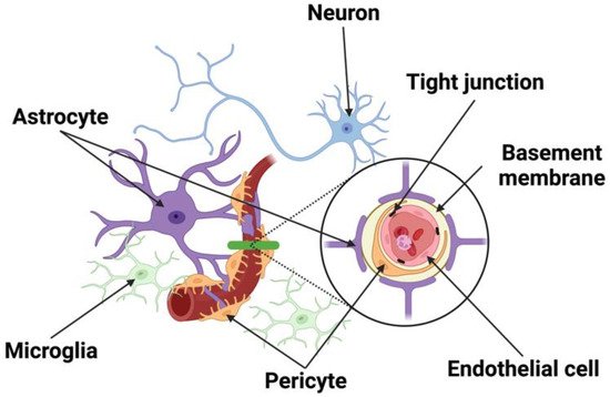
Figure 1. Schematic of the NVU. The NVU is comprised of neurons, vascular cells (endothelial cells, pericytes), basement membrane, and glia (astrocytes, microglia). Neurons make distinct connections with blood vessel and other cells of the NVU. Endothelial cells forming the blood vessels are encased by a basal lamina/basement membrane and are bound by tight junction proteins. Located in the brain parenchyma, astrocytes make contact with both pericytes and endothelial cells at the capillary wall, while pericytes are situated between the end feet of astrocytes and endothelial cells.
2. The Structural Components of the Neurovascular Unit (NVU)
2.1. Neurons
Different types of neurons including noradrenergic [4][5], serotonergic [6], cholinergic [7][8], and GABAergic [9] neurons have been shown to make distinct connections with other cells of the NVU, allowing regional brain activity to be metabolically coupled to blood flow [10]. Evidence shows that BBB opening may be a selective compensatory event rather than a simple anatomical disruption, implicating that interaction between neurons and the brain microvasculature may regulate both blood flow and BBB permeability.
2.2. Endothelial Cells
ECs line blood vessels in the brain and are joined with one another by specialized Tight Junctions (TJs) and Gap Junction (GJ) proteins consisting of various molecular components [11][12][13][14]. These TJs form the BBB, a physical barrier between blood and brain parenchyma [15], and regulate the permeability of the EC layer that keeps unwanted molecules such as toxins from entering the brain. This organization supports the current view that the BBB is not just a physical “barrier”, but a dynamic and metabolic interface [16][17].
Capillaries make up the microcirculation and are divided into three types: continuous, fenestrated, and sinusoidal. Brain capillaries belonging to the continuous type have no perforations on the vessel wall, allowing only small molecules to pass through. In contrast, fenestrated capillaries in the kidneys and GI tract along with sinusoidal capillaries in the lymphoid organs are leakier and have small and large pores, respectively. They allow for the passage of large molecules and even cells compared with the brain capillaries. By directly isolating tissue-specific mRNAs using the RiboTag transgenic mouse model [18], a recent study compared the transcriptome profiles of ECs from the brain, heart, and lung using RNA sequencing. The authors found that each EC type had a distinct genetic signature under normal conditions and an organ-specific response to lipopolysaccharide. Most surprisingly, ECs turned on genes that were expressed by the surrounding tissue upon stimulation, indicating organ-specific endothelial plasticity and adaptation [19]. Specifically in the brain, genes involved in processes akin to neuronal function such as synapse organization, neurotransmitter transport, axon development, and ion transport regulation are also enriched in the brain ECs [19].
Endothelial cells also interact with other components in the brain, such as pericytes, astrocytes, neurons, and the ECM to maintain the normal function of NVU by forming the BBB [16][20]. The interplay among partners in the NVU and the integrity of the BBB is key to brain homeostasis. The interaction between capillaries and neuronal components in neurovascular coupling regulates CBF through the detection of changes in shear stress of the endothelial wall and the supply of oxygen and necessary nutrients in the brain tissue. BBB breakdown leads to an influx of toxic molecules towards the brain, causing neuronal injury and neurodegenerative changes. While the NVU and BBB are critical for the maintenance of Central Nervous System (CNS) homeostasis, they could also interfere with the delivery of therapeutic drugs into the CNS via systemic administration for the treatment of neurological diseases. Further research and clinical trials are needed to successfully deliver necessary drugs to the CNS without causing inadvertent permeability of the BBB.
2.3. Pericytes
Pericytes surround cerebral vessels and are intimately in contact with the ECs embedded in the vascular basement membrane (BM) through gap junctional complexes around pericyte somata [21][22], called peg-socket contacts. Like endothelial cells, pericytes are attached to extracellular matrix proteins of the BM by different integrins that control junctional complex protein expression [23], affecting the functionality of the BBB by controlling both the structure of TJs and the rate of vesicular trafficking. They are also involved in various vascular functions such as BBB formation andmaintenance, angiogenesis, vessel maturation, regulation of blood flow, and immune cell trafficking [24][25][26][27]. Pericytes can inhibit the expression of genes that promote vessel permeability. Pericytes control neuroinflammation by reducing leukocyte trafficking in the regions of blood vessels they cover [24]. A study using pericyte-deficient mice showed a correlation between the reduction of tight junction and adherens junction proteins and the resulting increase in paracellular leakage and eventually BBB breakdown [25]. The resulting permeability leads to an influx of neurotoxic macromolecules, water, and larger molecules that are normally incapable via increased endothelial transcytosis [26][27], and a reduction in capillary blood flow due to microvascular degeneration [28].
Capillary vessels dilate to accommodate oxygen demand in the brain in response to physiological stimulation such as hypercapnia or sensory stimulation. Earlier studies implied that pericytes can contract or relax, resulting in changes in the capillary diameter under various physiological and pathological conditions [29][30][31][32]. To further distinguish which cells in the vessel zones respond to stimulation, a study using optogenetics found that it was SMCs rather than pericytes that contracted when mural cells expressing ChR2 were stimulated [30]. However, another study found that pharmacological inhibitor of myosin contraction signaling along with optical ablation of capillary pericytes resulted in consistent dilation of regions lacking pericyte contact, leading to aberrantly increased flux of blood flow in the uncovered capillary vessels [21][33]. A recent scRNAseq study suggests that pericytes do have the molecular machinery to regulate vessel diameter since capillary pericytes express receptors for vasoactive mediators including L-type voltage-gated calcium channels and those involved in smooth muscle cell actomyosin contraction [34], providing pivotal support for the modulatory role of pericytes in controlling capillary diameters and homogenizing blood flow to facilitate oxygen extraction, particularly during functional hyperemia. Apart from their modulatory role, pericytes seem multipotent and are known to differentiate into both neural and vascular lineage cells after brain ischemia [35][36].
The same group of investigators who found pericytes drive capillary vessel dilation reported that pericyte loss and capillary dilation caused by focal ablation of pericytes with laser was exacerbated in the aged brain, resulting in increased flow heterogeneity in capillary networks. Although the remodeling of neighboring pericytes restored endothelial coverage and vascular tone within days, this process was slower in the aged brain and led to persistent capillary dilation [37]. This suggests that pericytes do communicate with one another and work together to maintain neurovascular coupling. In support of this notion, one study showed that neighboring pericytes in the mouse retina can communicate with each other in response to light stimulation by forming interpericyte tunneling nanotubes that become a functional network with an open-ended proximal side and a closed-ended end-foot that connects with distal pericyte processes via gap junctions, serving as a conduit for intercellular Ca2+ waves between pericytes [38].
2.4. Astrocytes
Linking the vascular and neural systems are perivascular astrocytes, whose astrocytic processes almost completely encapsulate the abluminal EC surface of brain vessels. Astrocytes play a role in the development of junctional complexes and physically link neighboring neurons with blood vessels [39][40][41], allowing them to detect changes in the neuronal microenvironment and adjust the microvasculature appropriately [42][43]. Perivascular astrocytes increase the tightness of TJs [44], promote the expression of endothelial transporters [45], and enzymes associated with the endothelial barrier [40]. Through the secretion of bioactive substances, astrocytes provide physical support and strengthen the BBB, leading to TJ modulation. Gap junctions present between astrocytic end feet and vessel walls mediate intercellular communication and solute movement between them, such as water and ion exchange across the brain microvascular endothelium [40][46]. Water channel aquaporin 4 is noticeably expressed on astrocytic end feet and regulates water movement between the blood and brain [47]. Astrocytes are known for their roles in responding to CNS injury by taking up excess glutamate from the extracellular space and converting it to glutamine [48] to aid in the generation of new neurons, remodeling synapses, and recycling neurotransmitters [24]. Critical to neuronal survival and repair, a large part of this function is mediated by gap junction proteins that connect astrocyte networks into a functional syncytium [13][49][50][51]. Through the secretion of proinflammatory (Interleukin (IL) IL-6 and IL-1β), anti-inflammatory cytokines (IL-10), and chemokines (CCL2, CXCL1, CXCL10, and CXCL12), astrocytes can control microglia differentiation and macrophage activation [52][53][54]. These cytokines lead to hyperplasia of astrocytes, which results in the expression of inflammatory factors that can lead to reactive gliosis and scar formation. Astrocytes can directly restrict the entry of peripheral immune cells through the BBB. They are also the source of MMPs, a family of extracellular proteinases that degrade TJs and the ECM after ischemia, leading to the detachment of astrocytic end feet [55].
2.5. Microglia and Macrophages
Microglia are resident CNS macrophages that originate from the mesoderm during embryonic development and are widely distributed within the CNS. However, the basal ganglia and cerebellum have a higher abundance of microglia than the cerebral cortex [56]. They migrate into the brain and are termed “resting microglia” due to their low phagocytotic properties [57]. These cells communicate with endothelium to help regulate the BBB. As the primary immune cells that account for ~5–15% of all cells in the human brain, microglia can undergo morphological changes that allow them to phagocytose and produce pro-inflammatory cytokines IL-1 and IL-6, and enhance the expression of Intercellular Adhesion Molecule-1, P-selectin, and E-selectin [55][58]. As a result, the accumulation, migration, and adherence of leukocytes across endothelium allow them to mediate inflammatory cascades that further exaggerate the level of infarction.
During disease/trauma microglia become activated, and the extent of their activation is correlated to the severity and type of brain injury [59]. Activation of microglia is associated with dysfunction of the BBB via changes in TJ protein expression and increased BBB permeability [2]. High levels of neurotoxic mediators such as nitric oxide, peroxide, inflammatory cytokines (i.e., Tumor Necrosis Factor-α (TNF-α)), and proteases, as well as complement components [59][60], are produced, ultimately leading to cell injury in the CNS and neuronal cell death. Microglia are adept at sensing any small disturbance in the BBB [61][62] and maintaining BBB integrity during inflammation.
2.6. Junctional Complexes
ECs form the inner lining of blood vessels, creating a barrier between vessels and tissues. Lateral spaces between adjacent ECs called TJs, or Zona Occludins (ZO), and their proteins control the low paracellular permeability and high electrical resistance of the BBB [57]. Common transmembrane TJ proteins include Claudins (primarily Claudin-5) and occludin, which are phosphoproteins with four transmembrane domains that span the intracellular cleft, binding to proteins on adjacent ECs. Claudins and occludin are associated with cytoskeletal signaling proteins such as ZO-1 and ZO-2 and link TJs to the primary cytoskeleton proteins like actin for the maintenance of structural and functional integrity of the endothelium [63]. Junctional adhesion molecules are another family of transmembrane proteins that have a single transmembrane domain and are located at the borders of endothelial cells. These molecules are involved in cell-to-cell adhesion and leukocyte transmigration across the BBB [64]. The regulation of polar solutes and macromolecules across the barrier prevents the passage of unwanted and potentially damaging material such as peptides and proteins between the blood and brain. Through mechanoreceptor properties, ECs can respond and adjust vascular resistance via vasodilation and constriction to compensate for alterations of perfusion pressure and maintain a relatively constant CBF and microvascular pressure that contribute to cerebral autoregulation [65]. Akin to TJs, adherens junctions are protein complexes that occur at cell-cell junctions in endothelial cells. Unlike TJs, AJs join and maintain the connection between actin filaments of the cytoskeleton of neighboring cells. Transmembrane proteins called cadherins are a group of proteins that bind with other cadherins on adjacent ECs to help ECs stick together and regulate the intracellular signaling pathways that control gene transcription [66]. Additionally, gap junctions are intercellular channels that allow ions and molecules to pass through resulting in changes in membrane potential from one cell to another. Contrary to the extremely low permeability of tight junctions, gap junctions allow for the passage of certain molecules between cells. Consisting of connexin proteins, these structures allow for rapid propagation of action potentials along with the slow diffusion of nonorganic ions, secondary messengers, and other small water-soluble molecules [58]. They also transmit chemical signals and metabolites between cells, aiding in the function of vascular, neuronal and glial tissue [58]. Degradation of gap junctions contributes to the release of inflammatory mediators and disrupts the homeostasis of the CNS environment as exemplified under condition of ischemic stroke. These intercellular junctions are important for providing efficient and selective barriers against undesirable environmental conditions, providing the structural integrity of the cells that make up this barrier, and the overall homeostasis of the brain.
2.7. Basement Membrane
The basement membrane (BM) or ECM is an amorphous structure located on the abluminal side of endothelial cells or basal side of epithelial cells [67][68], and is comprised of multiple components such as laminins, collagen, nidogen, and heparan sulfate proteoglycans [67][68][69][70], forming a close contact with the endothelium. The BM plays a crucial role in maintaining vascular integrity and providing a rigid support to vessels and surrounding cells [70] by surrounding the capillaries and separating them from neighboring astrocytes and pericytes [71]. The ECM of the basement membrane limits the transmigration of red blood cells during hemorrhage and leukocytes during inflammation. Dysfunction and degradation of the BM are associated with several neurological disorders and increased barrier breakdown and edema [72][73]. Interestingly, endothelial cells, astrocytes, and pericytes are known to synthesize and deposit specific laminin isoforms in the BM that modulate BBB function [74][75].
References
- Iadecola, C. The Neurovascular Unit Coming of Age: A Journey through Neurovascular Coupling in Health and Disease. Neuron 2017, 96, 17–42.
- Zlokovic, B.V. The Blood-Brain Barrier in Health and Chronic Neurodegenerative Disorders. Neuron 2008, 57, 178–201.
- Obermeier, B.; Daneman, R.; Ransohoff, R.M. Development, maintenance and disruption of the blood-brain barrier. Nat. Med. 2013, 19, 1584–1596.
- Ben-Menachem, E.; Johansson, B.B.; Svensson, T.H. Increased vulnerability of the blood-brain barrier to acute hypertension following depletion of brain noradrenaline. J. Neural Transm. 1982, 53, 159–167.
- Cohen, Z.; Molinatti, G.; Hamel, E. Astroglial and Vascular Interactions of Noradrenaline Terminals in the Rat Cerebral Cortex. J. Cereb. Blood Flow Metab. 1997, 17, 894–904.
- Cohen, Z.; Bonvento, G.; Lacombe, P.; Hamel, E. Serotonin in the regulation of brain microcirculation. Prog. Neurobiol. 1996, 50, 335–362.
- Vaucher, E.; Hamel, E. Cholinergic basal forebrain neurons project to cortical microvessels in the rat: Electron microscopic study with anterogradely transported Phaseolus vulgaris leucoagglutinin and choline acetyltransferase immunocytochemistry. J. Neurosci. 1995, 15, 7427–7441.
- Tong, X.K.; Hamel, E. Regional cholinergic denervation of cortical microvessels and nitric oxide synthase-containing neurons in Alzheimer’s disease. Neuroscience 1999, 92, 163–175.
- Vaucher, E.; Tong, X.K.; Cholet, N.; Lantin, S.; Hamel, E. GABA neurons provide a rich input to microvessels but not nitric oxide neurons in the rat cerebral cortex: A means for direct regulation of local cerebral blood flow. J. Comp. Neurol. 2000, 421, 161–171.
- Buxton, R.B.; Frank, L.R. A Model for the Coupling between Cerebral Blood Flow and Oxygen Metabolism during Neural Stimulation. J. Cereb. Blood Flow Metab. 1997, 17, 64–72.
- Chow, B.W.; Gu, C. The Molecular Constituents of the Blood–Brain Barrier. Trends Neurosci. 2015, 38, 598–608.
- Delaney, C.; Campbell, M. The blood brain barrier: Insights from development and ageing. Tissue Barriers 2017, 5, e1373897.
- De Bock, M.; Leybaert, L.; Giaume, C. Connexin Channels at the Glio-Vascular Interface: Gatekeepers of the Brain. Neurochem. Res. 2017, 42, 2519–2536.
- Nagasawa, K.; Chiba, H.; Fujita, H.; Kojima, T.; Saito, T.; Endo, T.; Sawada, N. Possible involvement of gap junctions in the barrier function of tight junctions of brain and lung endothelial cells. J. Cell. Physiol. 2006, 208, 123–132.
- Reese, T.S.; Karnovsky, M.J. Fine structural localization of a blood-brain barrier to exogenous peroxidase. J. Cell Biol. 1967, 34, 207–217.
- Tso, M.K.; Macdonald, R.L. Subarachnoid hemorrhage: A review of experimental studies on the microcirculation and the neurovascular unit. Transl. Stroke Res. 2014, 5, 174–189.
- Zou, J.; Chen, Z.; Wei, X.; Chen, Z.; Fu, Y.; Yang, X.; Chen, D.; Wang, R.; Jenner, P.; Lu, J.; et al. Cystatin C as a potential therapeutic mediator against Parkinson’s disease via VEGF-induced angiogenesis and enhanced neuronal autophagy in neurovascular units. Cell Death Dis. 2017, 8, e2854.
- Sanz, E.; Yang, L.; Su, T.; Morris, D.R.; McKnight, G.S.; Amieux, P.S. Cell-type-specific isolation of ribosome-associated mRNA from complex tissues. Proc. Natl. Acad. Sci. USA 2009, 106, 13939–13944.
- Jambusaria, A.; Hong, Z.; Zhang, L.; Srivastava, S.; Jana, A.; Toth, P.; Dai, Y.; Malik, A.B.; Rehman, J. Endothelial heterogeneity across distinct vascular beds during homeostasis and inflammation. eLife 2020, 9, e51413.
- Erdő, F.; Krajcsi, P. Age-Related Functional and Expressional Changes in Efflux Pathways at the Blood-Brain Barrier. Front. Aging Neurosci. 2019, 11, 196.
- Hartmann, D.A.; Coelho-Santos, V.; Shih, A.Y. Pericyte Control of Blood Flow Across Microvascular Zones in the Central Nervous System. Annu. Rev. Physiol. 2022, 84, 331–354.
- Ornelas, S.; Berthiaume, A.-A.; Bonney, S.K.; Coelho-Santos, V.; Underly, R.G.; Kremer, A.; Guérin, C.J.; Lippens, S.; Shih, A.Y. Three-dimensional ultrastructure of the brain pericyte-endothelial interface. J. Cereb. Blood Flow Metab. 2021, 41, 2185–2200.
- Winkler, E.A.; Bell, R.D.; Zlokovic, B.V. Central nervous system pericytes in health and disease. Nat. Neurosci. 2011, 14, 1398–1405.
- Potjewyd, G.; Kellett, K.A.; Hooper, N.M. 3D hydrogel models of the neurovascular unit to investigate blood–brain barrier dysfunction. Neuronal Signal. 2021, 5, NS20210027.
- Bell, R.D.; Winkler, E.A.; Sagare, A.P.; Singh, I.; LaRue, B.; Deane, R.; Zlokovic, B.V. Pericytes Control Key Neurovascular Functions and Neuronal Phenotype in the Adult Brain and during Brain Aging. Neuron 2010, 68, 409–427.
- Armulik, A.; Genové, G.; Mäe, M.; Nisancioglu, M.H.; Wallgard, E.; Niaudet, C.; He, L.; Norlin, J.; Lindblom, P.; Strittmatter, K.; et al. Pericytes regulate the blood–brain barrier. Nature 2010, 468, 557–561.
- Winkler, E.A.; Sagare, A.P.; Zlokovic, B.V. The pericyte: A forgotten cell type with important implications for Alzheimer’s disease? Brain Pathol. 2014, 24, 371–386.
- Wang, L.; Xiong, X.; Zhang, L.; Shen, J. Neurovascular Unit: A critical role in ischemic stroke. CNS Neurosci. Ther. 2021, 27, 7–16.
- Hall, C.N.; Reynell, C.; Gesslein, B.; Hamilton, N.B.; Mishra, A.; Sutherland, B.A.; O’Farrell, F.M.; Buchan, A.M.; Lauritzen, M.; Attwell, D. Capillary pericytes regulate cerebral blood flow in health and disease. Nature 2014, 508, 55–60.
- Hill, R.A.; Tong, L.; Yuan, P.; Murikinati, S.; Gupta, S.; Grutzendler, J. Regional Blood Flow in the Normal and Ischemic Brain Is Controlled by Arteriolar Smooth Muscle Cell Contractility and Not by Capillary Pericytes. Neuron 2015, 87, 95–110.
- Cai, W.; Liu, H.; Zhao, J.; Chen, L.; Chen, J.; Lu, Z.; Hu, X. Pericytes in Brain Injury and Repair after Ischemic Stroke. Transl. Stroke Res. 2017, 8, 107–121.
- Yemisci, M.; Gürsoy-Ozdemir, Y.; Vural, A.; Can, A.; Topalkara, K.; Dalkara, T. Pericyte contraction induced by oxidative-nitrative stress impairs capillary reflow despite successful opening of an occluded cerebral artery. Nat. Med. 2009, 15, 1031–1037.
- Hartmann, D.A.; Berthiaume, A.-A.; Grant, R.I.; Harrill, S.A.; Koski, T.; Tieu, T.; McDowell, K.P.; Faino, A.V.; Kelly, A.L.; Shih, A.Y. Brain capillary pericytes exert a substantial but slow influence on blood flow. Nat. Neurosci. 2021, 24, 633–645.
- Vanlandewijck, M.; He, L.; Mäe, M.A.; Andrae, J.; Ando, K.; Del Gaudio, F.; Nahar, K.; Lebouvier, T.; Laviña, B.; Gouveia, L.; et al. A molecular atlas of cell types and zonation in the brain vasculature. Nature 2018, 554, 475–480.
- Sweeney, M.; Ayyadurai, S.; Zlokovic, B.V. Pericytes of the neurovascular unit: Key functions and signaling pathways. Nat. Neurosci. 2016, 19, 771–783.
- Nakagomi, T.; Kubo, S.; Nakano-Doi, A.; Sakuma, R.; Lu, S.; Narita, A.; Kawahara, M.; Taguchi, A.; Matsuyama, T. Brain Vascular Pericytes Following Ischemia Have Multipotential Stem Cell Activity to Differentiate into Neural and Vascular Lineage Cells. Stem Cells 2015, 33, 1962–1974.
- Berthiaume, A.A.; Schmid, F.; Stamenkovic, S.; Santos, V.; Nielson, C.; Weber, B.; Majesky, M.; Shih, A. Deficiency in pericyte remodeling as a basis for impaired capillary flow and structure during brain aging. bioRxiv 2022.
- Alarcon-Martinez, L.; Villafranca-Baughman, D.; Quintero, H.; Kacerovsky, J.B.; Dotigny, F.; Murai, K.K.; Prat, A.; Drapeau, P.; Di Polo, A. Interpericyte tunnelling nanotubes regulate neurovascular coupling. Nature 2020, 585, 91–95.
- VanGilder, R.L.; Rosen, C.L.; Barr, T.L.; Huber, J.D. Targeting the neurovascular unit for treatment of neurological disorders. Pharmacol. Ther. 2011, 130, 239–247.
- Abbott, N.J.; Rönnbäck, L.; Hansson, E. Astrocyte-endothelial interactions at the blood-brain barrier. Nat. Rev. Neurosci. 2006, 7, 41–53.
- Iadecola, C.; Nedergaard, M. Glial regulation of the cerebral microvasculature. Nat. Neurosci. 2007, 10, 1369–1376.
- Attwell, D.; Buchan, A.M.; Charpak, S.; Lauritzen, M.J.; MacVicar, B.A.; Newman, E.A. Glial and neuronal control of brain blood flow. Nature 2010, 468, 232–243.
- He, L.; Linden, D.J.; Sapirstein, A. Astrocyte Inositol Triphosphate Receptor Type 2 and Cytosolic Phospholipase A2 Alpha Regulate Arteriole Responses in Mouse Neocortical Brain Slices. PLoS ONE 2012, 7, e42194.
- Lee, S.-W.; Kim, W.J.; Choi, Y.K.; Song, H.S.; Son, M.J.; Gelman, I.H.; Kim, Y.-J.; Kim, K.-W. SSeCKS regulates angiogenesis and tight junction formation in blood-brain barrier. Nat. Med. 2003, 9, 900–906.
- McAllister, M.S.; Krizanac-Bengez, L.; Macchia, F.; Naftalin, R.; Pedley, K.C.; Mayberg, M.; Marroni, M.; Leaman, S.; Stanness, K.A.; Janigro, D. Mechanisms of glucose transport at the blood–brain barrier: An in vitro study. Brain Res. 2001, 904, 20–30.
- Mathiisen, T.M.; Lehre, K.P.; Danbolt, N.C.; Ottersen, O.P. The perivascular astroglial sheath provides a complete covering of the brain microvessels: An electron microscopic 3D reconstruction. Glia 2010, 58, 1094–1103.
- Simard, M.; Arcuino, G.; Takano, T.; Liu, Q.S.; Nedergaard, M. Signaling at the Gliovascular Interface. J. Neurosci. 2003, 23, 9254–9262.
- Romanos, J.; Benke, D.; Saab, A.S.; Zeilhofer, H.U.; Santello, M. Differences in glutamate uptake between cortical regions impact neuronal NMDA receptor activation. Commun. Biol. 2019, 2, 127.
- Laird, D.W.; Naus, C.C.; Lampe, P.D. SnapShot: Connexins and Disease. Cell 2017, 170, 1260–1260.e1.
- Freitas-Andrade, M.; Wang, N.; Bechberger, J.F.; De Bock, M.; Lampe, P.D.; Leybaert, L.; Naus, C.C. Targeting MAPK phosphorylation of Connexin43 provides neuroprotection in stroke. J. Exp. Med. 2019, 216, 916–935.
- Freitas-Andrade, M.; Bechberger, J.; Wang, J.; Yeung, K.K.; Whitehead, S.N.; Hansen, R.S.; Naus, C.C. Danegaptide Enhances Astrocyte Gap Junctional Coupling and Reduces Ischemic Reperfusion Brain Injury in Mice. Biomolecules 2020, 10, 353.
- Mantovani, A.; Sica, A.; Sozzani, S.; Allavena, P.; Vecchi, A.; Locati, M. The chemokine system in diverse forms of macrophage activation and polarization. Trends Immunol. 2004, 25, 677–686.
- Allaman, I.; Bélanger, M.; Magistretti, P.J. Astrocyte–neuron metabolic relationships: For better and for worse. Trends Neurosci. 2011, 34, 76–87.
- Lan, X.; Han, X.; Li, Q.; Yang, Q.-W.; Wang, J. Modulators of microglial activation and polarization after intracerebral haemorrhage. Nat. Rev. Neurol. 2017, 13, 420–433.
- Jayaraj, R.L.; Azimullah, S.; Beiram, R.; Jalal, F.Y.; Rosenberg, G.A. Neuroinflammation: Friend and foe for ischemic stroke. J. Neuroinflamm. 2019, 16, 142.
- Dickson, D.W.; Mattiace, L.A.; Kure, K.; Hutchins, K.; Lyman, W.D.; Brosnan, C.F. Microglia in human disease, with an emphasis on acquired immune deficiency syndrome. Lab. Investig. 1991, 64, 135–156.
- Jiang, X.; Andjelkovic, A.V.; Zhu, L.; Yang, T.; Bennett, M.V.L.; Chen, J.; Keep, R.F.; Shi, Y. Blood-brain barrier dysfunction and recovery after ischemic stroke. Prog. Neurobiol. 2018, 163–164, 144–171.
- Liang, Z.; Wang, X.; Hao, Y.; Qiu, L.; Lou, Y.; Zhang, Y.; Ma, D.; Feng, J. The Multifaceted Role of Astrocyte Connexin 43 in Ischemic Stroke Through Forming Hemichannels and Gap Junctions. Front. Neurol. 2020, 11, 703.
- Speth, C.; Dierich, M.P.; Sopper, S. HIV-infection of the central nervous system: The tightrope walk of innate immunity. Mol. Immunol. 2005, 42, 213–228.
- Aloisi, F. Immune function of microglia. Glia 2001, 36, 165–179.
- Hines, D.J.; Hines, R.M.; Mulligan, S.J.; Macvicar, B.A. Microglia processes block the spread of damage in the brain and require functional chloride channels. Glia 2009, 57, 1610–1618.
- Petersen, M.A.; Ryu, J.K.; Akassoglou, K. Fibrinogen in neurological diseases: Mechanisms, imaging and therapeutics. Nat. Rev. Neurosci. 2018, 19, 283–301.
- Cummins, P.M. Occludin: One Protein, Many Forms. Mol. Cell. Biol. 2012, 32, 242–250.
- Steinbacher, T.; Kummer, D.; Ebnet, K. Junctional adhesion molecule-A: Functional diversity through molecular promiscuity. Cell. Mol. Life Sci. 2018, 75, 1393–1409.
- Andresen, J.; Shafi, N.I.; Bryan, R.M. Endothelial influences on cerebrovascular tone. J. Appl. Physiol. 2006, 100, 318–327.
- Hartsock, A.; Nelson, W.J. Adherens and tight junctions: Structure, function and connections to the actin cytoskeleton. Biochim. Biophys. Acta 2008, 1778, 660–669.
- Hallmann, R.; Zhang, X.; Di Russo, J.; Li, L.; Song, J.; Hannocks, M.-J.; Sorokin, L. The regulation of immune cell trafficking by the extracellular matrix. Curr. Opin. Cell Biol. 2015, 36, 54–61.
- Kalluri, R. Basement membranes: Structure, assembly and role in tumour angiogenesis. Nat. Rev. Cancer 2003, 3, 422–433.
- Sorokin, L.M. The impact of the extracellular matrix on inflammation. Nat. Rev. Immunol. 2010, 10, 712–723.
- Yurchenco, P.D.; Amenta, P.S.; Patton, B.L. Basement membrane assembly, stability and activities observed through a developmental lens. Matrix Biol. 2004, 22, 521–538.
- Bell, A.H.; Miller, S.L.; Castillo-Melendez, M.; Malhotra, A. The Neurovascular Unit: Effects of Brain Insults During the Perinatal Period. Front. Neurosci. 2020, 13, 1452.
- Cardoso, F.L.; Brites, D.; Brito, M.A. Looking at the blood–brain barrier: Molecular anatomy and possible investigation approaches. Brain Res. Rev. 2010, 64, 328–363.
- del Zoppo, G.; Milner, R.; Mabuchi, T.; Hung, S.; Wang, X.; Koziol, J. Vascular matrix adhesion and the blood–brain barrier. Biochem. Soc. Trans. 2006, 34 Pt 6, 1261–1266.
- Gautam, J.; Zhang, X.; Yao, Y. The role of pericytic laminin in blood brain barrier integrity maintenance. Sci. Rep. 2016, 6, 36450.
- Gautam, J.; Miner, J.H.; Yao, Y. Loss of Endothelial Laminin alpha5 Exacerbates Hemorrhagic Brain Injury. Transl. Stroke Res. 2019, 10, 705–718.
More
Information
Subjects:
Neurosciences
Contributors
MDPI registered users' name will be linked to their SciProfiles pages. To register with us, please refer to https://encyclopedia.pub/register
:
View Times:
3.5K
Entry Collection:
Hypertension and Cardiovascular Diseases
Revisions:
2 times
(View History)
Update Date:
29 Sep 2022
Table of Contents


Notice
You are not a member of the advisory board for this topic. If you want to update advisory board member profile, please contact office@encyclopedia.pub.
OK
Confirm
Only members of the Encyclopedia advisory board for this topic are allowed to note entries. Would you like to become an advisory board member of the Encyclopedia?
Yes
No
${ textCharacter }/${ maxCharacter }
Submit
Cancel
Back
Comments
${ item }
|
${ item.createdUser.fullName }
${ item.createdAt }
${ item.vote }
${ item.reply }
Delete
${ reply.createdUser.fullName }
${ reply.createdAt }
${ reply.vote }
Delete
There is no reply to this comment~
${ item.replyTextCharacter }/${ item.replyMaxCharacter }
Submit
Cancel
More
No more~
There is no comment~
${ textCharacter }/${ maxCharacter }
Submit
Cancel
${ selectedItem.replyTextCharacter }/${ selectedItem.replyMaxCharacter }
Submit
Cancel
Confirm
Are you sure to Delete?
Yes
No