Submitted Successfully!
To reward your contribution, here is a gift for you: A free trial for our video production service.
Thank you for your contribution! You can also upload a video entry or images related to this topic.
Version Summary Created by Modification Content Size Created at Operation
1 -- 2710 2022-09-22 16:29:45 |
2 Reference format revised. -51 word(s) 2659 2022-09-26 04:58:17 | |
3 Remove fef. number from the Description. Meta information modification 2659 2022-09-26 08:21:31 |

Video Upload Options

Do you have a full video?

Confirm

Are you sure to Delete?
Cite
If you have any further questions, please contact Encyclopedia Editorial Office.
Katsandegwaza, B.;  Horsnell, W.;  Smith, K. Pre-Clinical Murine Models of Colitis. Encyclopedia. Available online: https://encyclopedia.pub/entry/27493 (accessed on 02 August 2024).
Katsandegwaza B,  Horsnell W,  Smith K. Pre-Clinical Murine Models of Colitis. Encyclopedia. Available at: https://encyclopedia.pub/entry/27493. Accessed August 02, 2024.
Katsandegwaza, Brunette, William Horsnell, Katherine Smith. "Pre-Clinical Murine Models of Colitis" Encyclopedia, https://encyclopedia.pub/entry/27493 (accessed August 02, 2024).
Katsandegwaza, B.,  Horsnell, W., & Smith, K. (2022, September 22). Pre-Clinical Murine Models of Colitis. In Encyclopedia. https://encyclopedia.pub/entry/27493
Katsandegwaza, Brunette, et al. "Pre-Clinical Murine Models of Colitis." Encyclopedia. Web. 22 September, 2022.
Pre-Clinical Murine Models of Colitis
Edit

Crohn’s disease (CD) and ulcerative colitis (UC) are both highly inflammatory diseases of the gastrointestinal tract, collectively known as inflammatory bowel disease (IBD). Although the cause of IBD is still unclear, several experimental IBD murine models have enabled researchers to make great inroads into understanding human IBD pathology. Murine experimental models of human IBD exhibit immune pathological signatures resembling Crohn’s disease (CD) or ulcerative colitis (UC). These models include the chemical-induced trinitrobenzene sulfonic acid (TNBS) model, oxazolone and dextran sulfate sodium (DSS) models, the gene-deficient I-kappa-B kinase gamma (Iκκ-γ) and interleukin(IL)-10 models, and the CD4+ T-cell transfer model. Although most pre-clinical murine models do not fully recapitulate the complexity of human IBD, these models have added to our knowledge about the causes of disease and have provided targets for developing new treatments. 

inflammatory bowel disease ulcerative colitis Crohn’s disease murine models

1. Introduction

The use of pre-clinical murine models of human IBD is currently employed by researchers to better understand disease etiology. Murine experimental models of human IBD exhibit immune pathological signatures resembling Crohn’s disease (CD) or ulcerative colitis (UC). These models include the chemical-induced trinitrobenzene sulfonic acid (TNBS) model, oxazolone and dextran sulfate sodium (DSS) models, the gene-deficient I-kappa-B kinase gamma (Iκκ-γ) and interleukin(IL)-10 models, and the CD4+ T-cell transfer model [1][2][3][4][5][6]. Although most pre-clinical murine models do not fully recapitulate the complexity of human IBD, these models have added to the knowledge about the causes of disease and have provided targets for developing new treatments.

2. Chemical Induced Colitis

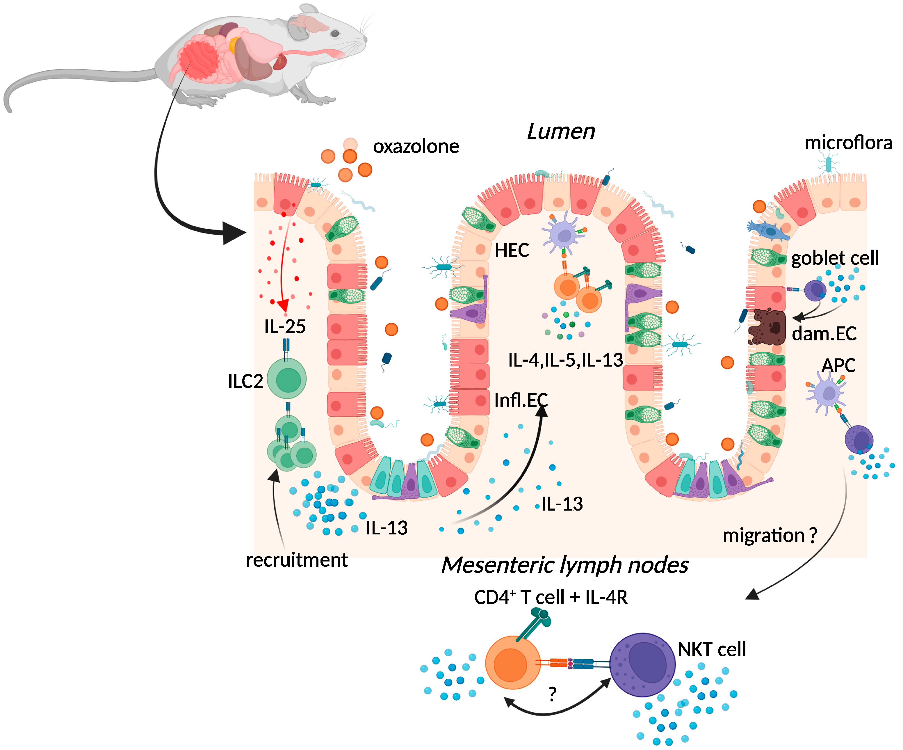
2.1. Oxazolone Colitis

Oxazolone colitis (OC) is induced by the intrarectal administration of a haptenising agent known as oxazolone (4-ethoxymethylene-2-phenyl-2-oxazolin-5-one) in ethanol (Figure 1) [1].
Figure 1. Immune response during oxazolone colitis. Oxazolone administration results in the production of IL-25, activation of ILC2, and production of IL-13, activating CD4+ T cell responses and amplifying type-2 cytokine production. Oxazolone administration also results in the expansion of T cells with surrogate markers of NKT cell function and IL-13 production by populations of CD1-restricted NKT cells. The resulting chronic inflammatory responses result in goblet cell depletion, increased intestinal permeability, and increased adhesion of commensal intestinal microbiota to the epithelium. Key: inflamed epithelial cells (Infl.EC), healthy epithelial cells (HEC), damaged epithelial cell (dam.EC). Created with BioRender.com, accessed on 18 August 2022.
Two methods of OC induction are currently employed to elicit either a self-resolving acute or a chronic response. The former is achieved by the single administration of an oxazolone enema, while the latter is preceded by a dermal pre-sensitisation five days before administration of an oxazolone enema, leading to chronic OC [7][8]. Both acute and chronic OC in mice are characterised by a superficial inflammation of the distal colon mucosa, a high volume of inflammatory infiltrate (neutrophils, macrophages, and lymphocytes), goblet-cell depletion, oedema formation, epithelial cell loss, haemorrhage, and vascular dilation [7][9][10]. These histologic features, as well as the distribution of OC, resemble human UC [1].
Chronic activation of type-2 immune responses has been proposed to be the main driver of human UC [11] (Figure 1). In the murine model, oxazolone activation of the type-2 immune response is characterised by the production of interleukin (IL)-4 in the acute phase, then superseded by the production of IL-13 in the chronic phase [7][12][13][14]. Increased production of IL-13 (and IL-5) was also evident in lamina propria T cells isolated from UC patients [15]. Interestingly, the use of mice deficient in IL-4 receptor-alpha (IL-4Rα), a common receptor for both IL-4 and IL-13, resulted in the exacerbation of OC, which could only be rescued by depletion of IL-13 in these mice [16]. Moreover, OC was prevented in mice given an IL-13 receptor subunit alpha-2 (IL-13Rα2) fusion protein that neutralises IL-13 bioactivity [7], alluding to the possible involvement of IL-13 signalling in the exacerbation of IBD in both pre-clinical models and patients [17]
Although the initial cell source of IL-13 in UC is yet undetermined, both natural killer T cells (NKT) and conventional CD4+ T cells secrete copious amounts of IL-13 during OC [7][16]. CD1d-restricted NKT cells expressing an invariant T-cell receptor can secrete IFN-γ or IL-4, activating CD4+ T cells to become Th1 or Th2 cells (Figure 1) [18].

2.2. TNBS-Induced Colitis

This pre-clinical model also utilises intrarectal administration of a haptenising agent in ethanol: 2,4,6-trinitrobenzene sulfonic acid (TNBS). Administration of 0.5 mg of TNBS in 50% ethanol to mice resulted in chronic transmural colitis, characterised by diarrhoea, weight loss, and rectal prolapse, pathology that mimics some characteristics of CD in humans [4]. Although one administration of TNBS resulted in acute chemical damage to the gut epithelium, inflammation was self-limiting, rather than the chronic inflammation seen in human disease [4]. Differing responses in mice are apparent in this acute model of colitis, varying according to several factors including age, genetic background, and TNBS dose [19]. To achieve chronic colitis, this model was developed by pre-sensitising the skin with 1% TNBS, followed by up to six repeated weekly intrarectal administrations of increasing doses of TNBS [14]. This model resembled the chronic phase of CD and was accompanied by production of IL-23 and IL-17 by lamina propria cells [20].
Isolated lamina propria CD4+ T cells from mice given TNBS secreted high levels of the Th1 cytokine interferon (IFN)-γ, resembling the cytokine profile produced by isolated lamina propria CD4+ T cells from CD patients [21]. This distinguished them from the Th2 profile of the same cells isolated from UC patients, or mice given oxazolone [21]. Although antibodies against IL-12, a pivotal cytokine for Th1 differentiation, abrogated established colitis and the initiation of TNBS-disease in BALB/c mice [4], TNBS-dependent colitis was exacerbated in IFN-γ-deficient mice on a BALB/c background [22].
The clinical importance of the TNBS model is demonstrated by the translation of Neurath’s anti-IL-12 antibody findings from TNBS murine experiments to successful human trials [23]. Importantly, the antibody used in these same studies was later found to react with the promiscuous p40 subunit shared by both IL-12 (a 70 kDa heterodimer of the p40 and a p35 subunit) and IL-23 (heterodimer of the p40 and a p19 subunit) (summarized in [24]). Clinical trials with “brakinumab”, a monoclonal antibody recognising the human p40 subunit, downregulated both IL-12p70 and IL-23 secretion [25] and resulted in a clinical improvement in patients with active CD [26]. Drug development of brakinumab was discontinued due to the existence of another IL-12/IL-23 inhibitor, Stelera (ustekinumab), on the market, which significantly increased the induction and maintenance of clinical remission in patients with UC [27]. Despite the findings of exacerbated TNBS-colitis in mice lacking the p19 subunit of IL-23, novel therapies targeting IL-23 and the IL-23R have been developed and deemed successful when tested in clinical trials of patients with IBD [28].

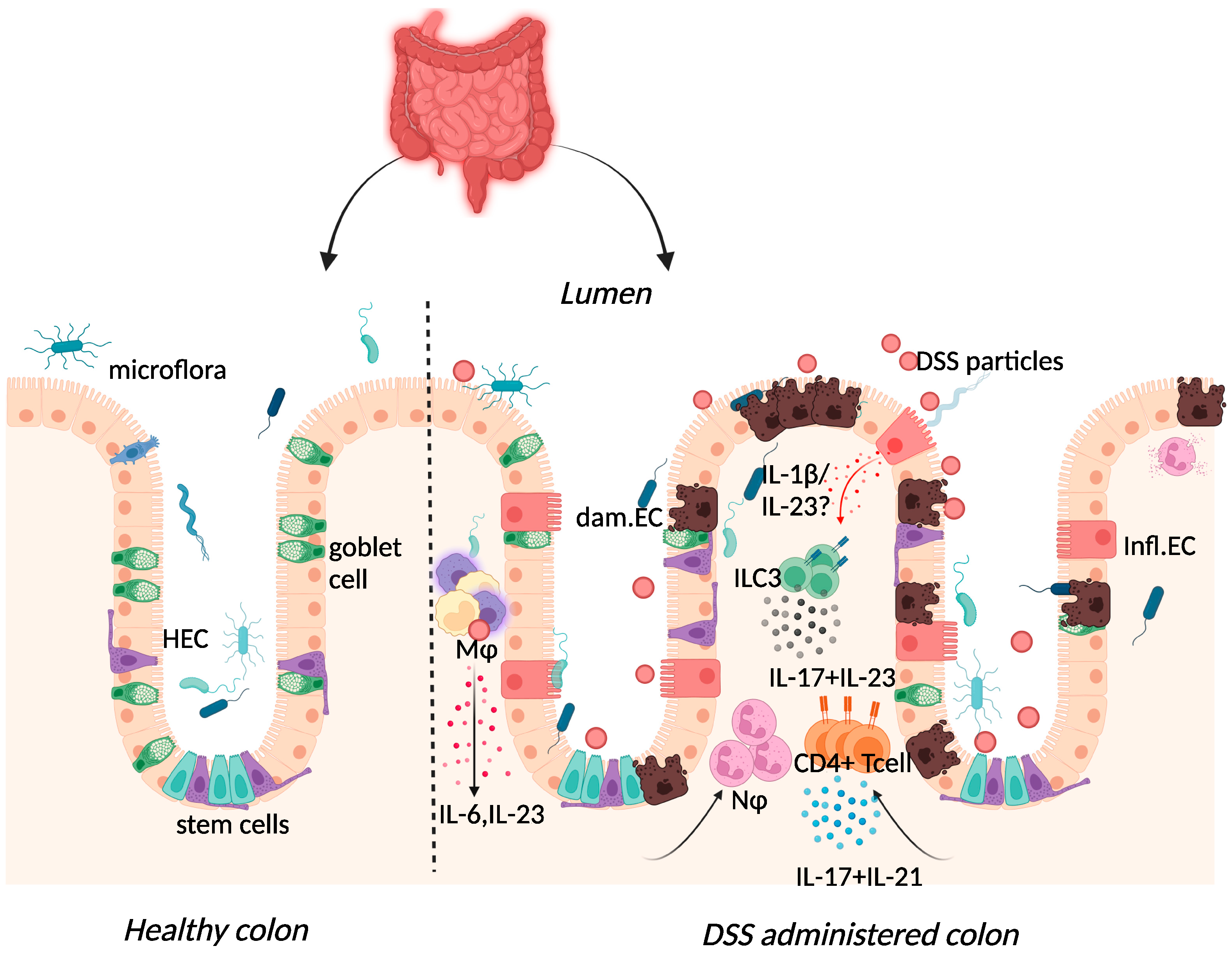
2.3. Dextran-Sulphate-Sodium-Induced Colitis

Dextran sulphate sodium (DSS) colitis is the most widely used experimental murine model of colitis, established by Okayasu in 1990 through the administration of DSS with a molecular weight of 40–50 kDa in drinking water [5]. DSS is thought to form nano-lipid vesicles with medium-chain fatty acids (MCFAs) in the colon, which fuse with colonocyte membranes and increase inflammatory cytokine levels [29]. A high-fat diet rich in MCFAs exacerbated weight loss, inflammatory cytokine expression, and colon shortening in this model, with dodecanoic acid favouring disruption of intestinal barrier function and increased vesicle formation in vitro [29]. One day after administration, DSS particles were present systemically in Kupffer cells of the liver, in macrophages of the mesenteric lymph node, and in the lamina propria of the large intestine [30]. DSS administration was also characterised by erosion of the intestinal epithelium, inflammatory infiltration of the large intestine, and dysbiosis of the intestinal microbiome [5]. While these features are similar to those found in human disease, the transmural inflammation apparent in TNBS-colitis is absent in this model [31]. Although repeated rounds of DSS can be administered to provide the pattern of remitting, relapsing inflammation in human IBD, some of the limitations of this model include inter-batch variability of DSS and the need to optimise DSS dose, given the impact of the intestinal microbiome on disease [31][32].
/media/item_content/202209/6331138e56b68ijms-23-09344-g002.png
Figure 2. Immune response to DSS administration. DSS administration results in epithelial release of IL-1β, activation of ILC3, and release of IL-23. IL-23 release results in the influx of neutrophils and CD4+ T cells, which further respond through enhanced IL-17 signalling. The resulting chronic inflammatory responses result in goblet cell depletion, increased intestinal permeability, and increased adhesion of commensal intestinal microbiota to the epithelium. Key: Inflamed epithelial cells (Infl.EC), healthy epithelial cells (HEC), damaged epithelial cell (dam.EC), macrophages (MΦ), neutrophils (NΦ). Created with BioRender.com, accessed on 18 August 2022.
DSS-colitis can be induced in immunodeficient mice including recombination-activating gene (RAG)-1-deficient and severe combined immune deficient (SCID) mice, suggesting the dispensability of the adaptive immune system in initiating disease [33][34]. Although Kim et al. demonstrated colitis induction in RAG-1-deficient mice, the resultant mild colitis in these mice compared to their wild-type counterparts insinuates that lymphocytes may be necessary for subsequent colitis progression [34]. Histological assessment of biopsy specimens from IBD patients correlated UC and CD with severe mononuclear cell infiltration and basal plasmacytosis (plasma B cells) [35]. In the acute phase of the DSS-colitis model, this infiltrate consisted of innate macrophage, neutrophil, and eosinophil populations recruited following increased cytokine and chemokine expression [36].
Elevated expression of IL-17 and IL-23 was reported in IBD patients and in DSS-colitis, where expression of the two cytokines was intertwined (Figure 2) [25][37][38][39][40][41]. The use of the DSS-colitis model to test the role of these cytokines in disease has revolutionised not only the possible interventions available for patients but has also developed our understanding of mucosal immunology. At steady state, the p19 subunit of IL-23 is highly expressed within Peyer’s patches and the thymus, as well as in polarised Th1 cells, activated macrophages, and dendritic cell populations derived from peripheral blood [42]. IL-23 induced proliferation of memory T cells and elevated secretion of IL-17 in vitro [43]. Subsequently, IL-23 signalling within intestinal epithelial cells was found to play an important role in protection against DSS colitis by regulating regenerating-islet-derived protein 3-beta (Reg3β)-dependent control of flagellated intestinal bacterial abundance and promoting IL-22 production [44]. Indeed DSS-colitis was exacerbated in IL-22-deficient mice [45] and blockade of IL-22 expression delayed recovery from DSS-colitis and exacerbated disease scores [46].

3. Spontaneous Colitis

3.1. Iκκ-γ (NEMO) Deficiency Colitis

Conditional ablation of the NF-kB essential modulator (NEMO), also known as I-kappa-B kinase gamma (Iκκ-γ), within the intestinal epithelium, resulted in spontaneous colitis in mice [3]. Chronic disease in intestinal-epithelial-cell-specific NEMO-deficient mice was associated with TNFR1-dependent colonic epithelial cell death, compromised epithelial integrity, bacterial translocation in the colon, immune cell infiltration, and increased expression of pro-inflammatory cytokines including TNF-α [3]. Absence of disease in double-deficient (NEMOIEC−KO + MYD88−/−) mice, lacking NEMO and the important bacterial sensor myeloid differentiation primary response 88 (MYD88), supported a role for the gut microbiota in driving colitis [3]. Indeed NEMOIEC−KO mice raised under germ-free conditions did not develop spontaneous colitis, whereas co-housing of these mice with specific pathogen-free animals restored disease [47]

3.2. Interleukin-10 (IL-10) Deficiency Colitis

Although IBD is common among adults, childhood IBD constitutes about a quarter of all patients with IBD [48]. Approximately 15% of childhood IBD occurs in children <6 years old and is termed very early onset IBD (VEO-IBD) [49]. While most childhood IBD cases are polygenic in nature, many children with VEO-IBD have an underlying monogenetic disorder that results in severe enterocolitis, including mutations in IL-10 and/or IL-10R (summarized in [50]). Mice deficient in IL-10 develop spontaneous enterocolitis, characterised by progressive cellular infiltration of the cecum, colon, rectum, and small intestine, with transmural lesions and a high incidence of colorectal adenocarcinomas observed in 6-month-old mice [2][51]. Mice lacking IL-10 receptor β also developed spontaneous enterocolitis [52][53]. However, this receptor is shared with other type II cytokine receptors including those specific for IL-22, IL-26, and lambda interferons, in addition to IL-10 [54].
An important role for the gut microbiota in influencing the general enterocolitis seen in IL-10-deficient mice was implicated by a lack of disease in mice housed in specific pathogen-free conditions, or in germ-free conditions [2][55]. Treatment of IL-10-deficient mice with two different antibiotics, both shown to improve scores in patients with Crohn’s disease [56][57], attenuated the development of spontaneous colitis [58]. Similar to IBD patients, colitic IL-10-deficient mice exhibited a markedly reduced species diversity in their faecal microbiome when compared to disease-free controls [55][59][60]. In addition, IL-10/IL-22 double-deficient mice lacking colitis exhibited higher microbial diversity when compared to IL-10-deficient mice [59].

4. Immune Cell Induced Colitis

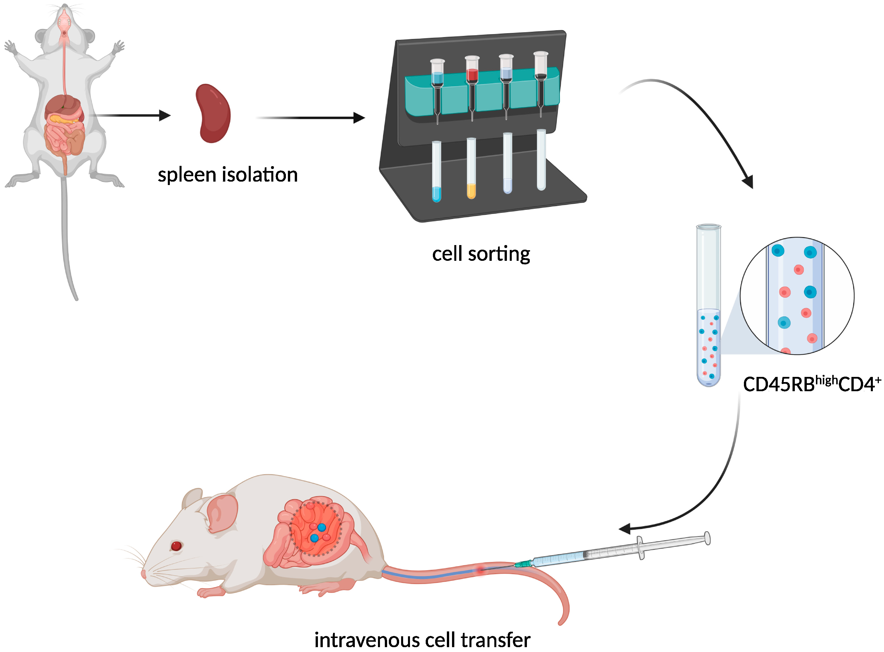
T-Cell Adoptive Transfer Model

The transfer of murine CD45RBhighCD4+ T cells from healthy donors to severe combined immunodeficiency (SCID) (Figure 3) mice resulted in the development of a lethal wasting disease, an influx of inflammatory cells, and increased inflammatory cytokine production in the colon of the recipient [6].
Figure 3. CD45RBhiCD4+ T cell adoptive transfer scheme. Created with BioRender.com, accessed on 18 August 2022.
The resulting chronic colitis that developed within 5–8 weeks was T cell dose-dependent, and despite the presence of transferred T cells in several organs, significant pathology was limited to the large intestine [6]. Interestingly, the transfer of naïve CD45RBhighCD4+ cells into SCID mice with a reduced microbiota significantly reduced colitis when compared to SCID mice housed in SPF conditions [61]. Restoration of T-cell proliferation in germ-free RAG-deficient mice following reconstitution with cecal bacterial lysate-pulsed dendritic cells and induction of colitis in SPF RAG-deficient mice following the transfer of T cells specific for a microbiota flagellin antigen cemented the role of the commensal microbiota in driving disease in this model [62].
Although both CD45RBlow and CD45RBhighCD4+ T cells homed to the intraepithelial and lamina propria compartments of both the small and large intestine [61], CD45RBlow or total unfractionated CD4+ T-cells were incapable of inducing colitis [6]. Following the discovery that the CD45RBlow population comprised a population of CD4+CD25+ regulatory T cells that could cure colitis in SCID or RAG1-deficient recipients of CD4+CD45RBhigh T cells [63], the T-cell transfer model of colitis was further refined by reconstitution of SCID mice with CD25-depleted CD4+ cells [64]. Although colitis in this model was accredited to the production of the Th1 cytokine IFN-γ [6][65], enhanced emergence of IL-17A+IFN-γ population of T cells and suppression of populations of Foxp3+ and IL-10-producing regulatory T cells was attributed to IL-23R signalling in T cells [66]. Similarly, an absence of IL-23 in IL-12p19-deficient RAG-deficient recipients resulted in significantly decreased levels of proinflammatory cytokine production in the intestine [67]. While intestinal inflammation was significantly reduced in the RAG-deficient recipients of IL-23R-deficient CD45RBhighCD4+ T cells, or p19−/−RAG−/− recipients of CD4+CD25CD45RBhigh T cells, these mice still exhibited systemic inflammation and weight loss [66][67], suggesting a tissue-restricted activity of IL-23.
Using IFN-γ-deficient RAG−/− recipients, IFN-γ was subsequently shown to be dispensable for the induction and progression of CD4+CD25CD45RBhigh T cell transfer colitis [68]. Inflammatory responses in these recipients favoured IL-17A production, where neutralisation of IL-17A/F significantly reduced weight loss and histopathological score in these animals [68]. A population of CD4+IL-17F+ T cells was sufficient to induce colitis in RAG1-deficient recipients, where the transition of these precursors to Th1-like cells was an absolute requirement for disease [69]. Interestingly, IL-21 signalling was recently proposed to dampen T-cell transfer colitis by reducing IL-17A production and augmenting IL-22 production in populations of ILC3s [70].
An advantage to this model is the ability to examine early immunological events associated with gut inflammation, including regulatory T cells responses. In addition, the apparent inflammatory response in both the small bowel as well as the colon makes this model ideal for the study of CD [65]. However, the SCID and RAG−/− recipients used in this model spontaneously develop T and B cell populations as they age [71], while NK cells from RAG−/− mice are reported to exhibit altered function and fitness [72]. The lower prevalence of human SCID patients, estimated at 1 in 58,000 new-borns in the United States (0.0017%) [73], compared to a higher incidence of CD (estimated at 10.7 cases per 100,000 individuals in the United States (0.0107%) [48], points to a factorial model of CD induction/development, independent of primary immunodeficiencies in T and B cells. This excludes this model as an exact model of CD, despite the many similarities.

References

  1. Boirivant, M.; Fuss, I.J.; Chu, A.; Strober, W. Oxazolone Colitis: A Murine Model of T Helper Cell Type 2 Colitis Treatable with Antibodies to Interleukin 4. J. Exp. Med. 1998, 188, 1929–1939.
  2. Kühn, R.; Löhler, J.; Rennick, D.; Rajewsky, K.; Müller, W. Interleukin-10-deficient mice develop chronic enterocolitis. Cell 1993, 75, 263–274.
  3. Nenci, A.; Becker, C.; Wullaert, A.; Gareus, R.; Van Loo, G.; Danese, S.; Huth, M.; Nikolaev, A.; Neufert, C.; Madison, B.; et al. Epithelial NEMO links innate immunity to chronic intestinal inflammation. Nature 2007, 446, 557–561.
  4. Neurath, M.F.; Fuss, I.; Kelsall, B.L.; Stüber, E.; Strober, W. Antibodies to interleukin 12 abrogate established experimental colitis in mice. J. Exp. Med. 1995, 182, 1281–1290.
  5. Okayasu, I.; Hatakeyama, S.; Yamada, M.; Ohkusa, T.; Inagaki, Y.; Nakaya, R. A novel method in the induction of reliable experimental acute and chronic ulcerative colitis in mice. Gastroenterology 1990, 98, 694–702.
  6. Powrie, F.; Leach, M.W.; Mauze, S.; Caddie, L.B.; Coffman, R.L. Phenotypically distinct subsets of CD4+ T cells induce or protect from chronic intestinal inflammation in C. B-17 scid mice. Int. Immunol. 1993, 5, 1461–1471.
  7. Heller, F.; Fuss, I.J.; E Nieuwenhuis, E.; Blumberg, R.S.; Strober, W. Oxazolone Colitis, a Th2 Colitis Model Resembling Ulcerative Colitis, Is Mediated by IL-13-Producing NK-T Cells. Immunity 2002, 17, 629–638.
  8. Yang, J.; Zhao, J.; Nakaguchi, T.; Gregersen, H. Biomechanical changes in oxazolone-induced colitis in BALB/C mice. J. Biomech. 2009, 42, 811–817.
  9. Kasaian, M.T.; Page, K.M.; Fish, S.; Brennan, A.; Cook, T.A.; Moreira, K.; Zhang, M.; Jesson, M.; Marquette, K.; Agostinelli, R.; et al. Therapeutic activity of an interleukin-4/interleukin-13 dual antagonist on oxazolone-induced colitis in mice. Immunology 2014, 143, 416–427.
  10. Wang, X.; Ouyang, Q.; Luo, W.J. Oxazolone-induced murine model of ulcerative colitis. Chin. J. Dig. Dis. 2004, 5, 165–168.
  11. Strober, W.; Fuss, I.J. Proinflammatory Cytokines in the Pathogenesis of Inflammatory Bowel Diseases. Gastroenterology 2011, 140, 1756–1767.e1.
  12. Fuss, I.J.; Strober, W. The role of IL-13 and NK T cells in experimental and human ulcerative colitis. Mucosal Immunol. 2008, 1 (Suppl. 1), S31–S33.
  13. Kiesler, P.; Fuss, I.J.; Strober, W. Experimental Models of Inflammatory Bowel Diseases. Cell. Mol. Gastroenterol. Hepatol. 2015, 1, 154–170.
  14. Wirtz, S.; Popp, V.; Kindermann, M.; Gerlach, K.; Weigmann, B.; Fichtner-Feigl, S.; Neurath, M.F. Chemically induced mouse models of acute and chronic intestinal inflammation. Nat. Protoc. 2017, 12, 1295–1309.
  15. Fuss, I.J.; Heller, F.; Boirivant, M.; Leon, F.; Yoshida, M.; Fichtner-Feigl, S.; Yang, Z.; Exley, M.; Kitani, A.; Blumberg, R.S.; et al. Nonclassical CD1d-restricted NK T cells that produce IL-13 characterize an atypical Th2 response in ulcerative colitis. J. Clin. Investig. 2004, 113, 1490–1497.
  16. Hoving, J.C.; Cutler, A.; Leeto, M.; Horsnell, W.G.C.; Dewals, B.G.; Nieuwenhuizen, N.; Brombacher, F. Interleukin 13-mediated colitis in the absence of IL-4Rα signalling. Gut 2017, 66, 2037–2039.
  17. Karmele, E.P.; Pasricha, T.S.; Ramalingam, T.R.; Thompson, R.W.; Iii, R.L.G.; Knilans, K.J.; Hegen, M.; Farmer, M.; Jin, F.; Kleinman, A.; et al. Anti-IL-13Rα2 therapy promotes recovery in a murine model of inflammatory bowel disease. Mucosal Immunol. 2019, 12, 1174–1186.
  18. Kronenberg, M.; Gapin, L. The unconventional lifestyle of NKT cells. Nat. Rev. Immunol. 2002, 2, 557–568.
  19. Velde, A.A.T.; Verstege, M.I.; Hommes, D.W. Critical appraisal of the current practice in murine TNBS-induced colitis. Inflamm. Bowel Dis. 2006, 12, 995–999.
  20. Fichtner-Feigl, S.; Fuss, I.J.; Preiß, J.; Strober, W.; Kitani, A. Treatment of murine Th1- and Th2-mediated inflammatory bowel disease with NF- B decoy oligonucleotides. J. Clin. Investig. 2005, 115, 3057–3071.
  21. Fuss, I.J.; Neurath, M.; Boirivant, M.; Klein, J.S.; De La Motte, C.; A Strong, S.; Fiocchi, C.; Strober, W. Disparate CD4+ lamina propria (LP) lymphokine secretion profiles in inflammatory bowel disease. Crohn’s disease LP cells manifest increased secretion of IFN-gamma, whereas ulcerative colitis LP cells manifest increased secretion of IL-5. J. Immunol. 1996, 157, 1261–1270.
  22. Dohi, T.; Fujihashi, K.; Rennert, P.D.; Iwatani, K.; Kiyono, H.; McGhee, J.R. Hapten-induced Colitis Is Associated with Colonic Patch Hypertrophy and T Helper Cell 2–Type Responses. J. Exp. Med. 1999, 189, 1169–1180.
  23. Croxford, A.L.; Kulig, P.; Becher, B. IL-12-and IL-23 in health and disease. Cytokine Growth Factor Rev. 2014, 25, 415–421.
  24. Langrish, C.L.; McKenzie, B.S.; Wilson, N.J.; Malefyt, R.D.W.; Kastelein, R.A.; Cua, D.J. IL-12 and IL-23: Master regulators of innate and adaptive immunity. Immunol. Rev. 2004, 202, 96–105.
  25. Fuss, I.J.; Becker, C.; Yang, Z.; Groden, C.; Hornung, R.L.; Heller, F.; Neurath, M.F.; Strober, W.; Mannon, P.J. Both IL-12p70 and IL-23 are synthesized during active Crohnʼs disease and are down-regulated by treatment with anti-IL-12 p40 monoclonal antibody. Inflamm. Bowel Dis. 2006, 12, 9–15.
  26. Mannon, P.J.; Fuss, I.J.; Mayer, L.; Elson, C.O.; Sandborn, W.J.; Present, D.; Dolin, B.; Goodman, N.; Groden, C.; Hornung, R.L.; et al. Anti–Interleukin-12 Antibody for Active Crohn’s Disease. N. Engl. J. Med. 2004, 351, 2069–2079.
  27. Sands, B.E.; Sandborn, W.J.; Panaccione, R.; O’Brien, C.D.; Zhang, H.; Johanns, J.; Adedokun, O.J.; Li, K.; Peyrin-Biroulet, L.; Van Assche, G.; et al. Ustekinumab as Induction and Maintenance Therapy for Ulcerative Colitis. N. Engl. J. Med. 2019, 381, 1201–1214.
  28. Sewell, G.W.; Kaser, A. Interleukin-23 in the Pathogenesis of Inflammatory Bowel Disease and Implications for Therapeutic Intervention. J. Crohn’s Colitis 2022, 16, ii3–ii19.
  29. Laroui, H.; Ingersoll, S.A.; Liu, H.C.; Baker, M.T.; Ayyadurai, S.; Charania, M.A.; Laroui, F.; Yan, Y.; Sitaraman, S.V.; Merlin, D. Dextran Sodium Sulfate (DSS) Induces Colitis in Mice by Forming Nano-Lipocomplexes with Medium-Chain-Length Fatty Acids in the Colon. PLoS ONE 2012, 7, e32084.
  30. Kitajima, S.; Takuma, S.; Morimoto, M. Tissue Distribution of Dextran Sulfate Sodium(DSS) in the Acute Phase of Murine DSS-Induced Colitis. J. Vet. Med. Sci. 1999, 61, 67–70.
  31. Chassaing, B.; Aitken, J.D.; Malleshappa, M.; Vijay-Kumar, M. Dextran Sulfate Sodium (DSS)-Induced Colitis in Mice. Curr. Protoc. Immunol. 2014, 104, 15.25.1–15.25.14.
  32. Perše, M.; Cerar, A. Dextran Sodium Sulphate Colitis Mouse Model: Traps and Tricks. J. Biomed. Biotechnol. 2012, 2012, 718617.
  33. Dieleman, L.A.; Ridwan, B.U.; Tennyson, G.S.; Beagley, K.W.; Bucy, R.; Elson, C.O. Dextran sulfate sodium-induced colitis occurs in severe combined immunodeficient mice. Gastroenterology 1994, 107, 1643–1652.
  34. Kim, T.W.; Seo, J.N.; Suh, Y.H.; Park, H.J.; Kim, J.H.; Kim, J.Y.; Oh, K.I. Involvement of lymphocytes in dextran sulfate sodium-induced experimental colitis. World J. Gastroenterol. 2006, 12, 302–305.
  35. Tanaka, M.; Riddell, R.H.; Saito, H.; Soma, Y.; Hidaka, H.; Kudo, H. Morphologic Criteria Applicable to Biopsy Specimens for Effective Distinction of Inflammatory Bowel Disease from Other Forms of Colitis and of Crohn’s Disease from Ulcerative Colitis. Scand. J. Gastroenterol. 1999, 34, 55–67.
  36. Stevceva, L.; Pavli, P.; Husband, A.; Ramsay, A.; Doe, W. Dextran sulphate sodium-induced colitis is ameliorated in interleukin 4 deficient mice. Genes Immun. 2001, 2, 309–316.
  37. Fujino, S.; Andoh, A.; Bamba, S.; Ogawa, A.; Hata, K.; Araki, Y.; Bamba, T.; Fujiyama, Y. Increased expression of interleukin 17 in inflammatory bowel disease. Gut 2003, 52, 65–70.
  38. Gheita, T.A.; El Gazzar, I.I.; El-Fishawy, H.S.; Aboul-Ezz, M.A.; Kenawy, S.A. Involvement of IL-23 in enteropathic arthritis patients with inflammatory bowel disease: Preliminary results. Clin. Rheumatol. 2014, 33, 713–717.
  39. Jiang, W.; Su, J.; Zhang, X.; Cheng, X.; Zhou, J.; Shi, R.; Zhang, H. Elevated levels of Th17 cells and Th17-related cytokines are associated with disease activity in patients with inflammatory bowel disease. Agents Actions 2014, 63, 943–950.
  40. Lucaciu, L.A.; Ilieș, M.; Vesa, C.; Seicean, R.; Din, S.; Iuga, C.A.; Seicean, A. Serum Interleukin (IL)-23 and IL-17 Profile in Inflammatory Bowel Disease (IBD) Patients Could Differentiate between Severe and Non-Severe Disease. J. Pers. Med. 2021, 11, 1130.
  41. Mirsattari, D.; Seyyedmajidi, M.; Zojaji, H.; Haghazali, M.; Orimi, P.G.; Shoushtarizadeh, T.; Almasi, S. The relation between the level of interleukin-23 with duration and severity of ulcerative colitis. Gastroenterol. Hepatol. Bed Bench 2012, 5, 49–53.
  42. Oppmann, B.; Lesley, R.; Blom, B.; Timans, J.C.; Xu, Y.; Hunte, B.; Vega, F.; Yu, N.; Wang, J.; Singh, K.; et al. Novel p19 Protein Engages IL-12p40 to Form a Cytokine, IL-23, with Biological Activities Similar as Well as Distinct from IL-12. Immunity 2000, 13, 715–725.
  43. Aggarwal, S.; Ghilardi, N.; Xie, M.-H.; de Sauvage, F.J.; Gurney, A.L. Interleukin-23 Promotes a Distinct CD4 T Cell Activation State Characterized by the Production of Interleukin-17. J. Biol. Chem. 2003, 278, 1910–1914.
  44. Aden, K.; Rehman, A.; Falk-Paulsen, M.; Secher, T.; Kuiper, J.; Tran, F.; Pfeuffer, S.; Sheibani-Tezerji, R.; Breuer, A.; Luzius, A.; et al. Epithelial IL-23R Signaling Licenses Protective IL-22 Responses in Intestinal Inflammation. Cell Rep. 2016, 16, 2208–2218.
  45. Zenewicz, L.A.; Yancopoulos, G.D.; Valenzuela, D.M.; Murphy, A.J.; Stevens, S.; Flavell, R.A. Innate and Adaptive Interleukin-22 Protects Mice from Inflammatory Bowel Disease. Immunity 2008, 29, 947–957.
  46. Sugimoto, K.; Ogawa, A.; Mizoguchi, E.; Shimomura, Y.; Andoh, A.; Bhan, A.K.; Blumberg, R.S.; Xavier, R.J.; Mizoguchi, A. IL-22 ameliorates intestinal inflammation in a mouse model of ulcerative colitis. J. Clin. Investig. 2008, 118, 534–544.
  47. Vlantis, K.; Polykratis, A.; Welz, P.-S.; van Loo, G.; Pasparakis, M.; Wullaert, A. TLR-independent anti-inflammatory function of intestinal epithelial TRAF6 signalling prevents DSS-induced colitis in mice. Gut 2015, 65, 935–943.
  48. Loftus, E.V., Jr. Clinical epidemiology of inflammatory bowel disease: Incidence, prevalence, and environmental influences. Gastroenterology 2004, 126, 1504–1517.
  49. Heyman, M.B.; Kirschner, B.S.; Gold, B.D.; Ferry, G.; Baldassano, R.; Cohen, S.A.; Winter, H.S.; Fain, P.; King, C.; Smith, T.; et al. Children with early-onset inflammatory bowel disease (IBD): Analysis of a pediatric IBD consortium registry. J. Pediatr. 2005, 146, 35–40.
  50. Zhu, L.; Shi, T.; Zhong, C.; Wang, Y.; Chang, M.; Liu, X. IL-10 and IL-10 Receptor Mutations in Very Early Onset Inflammatory Bowel Disease. Gastroenterol. Res. 2017, 10, 65–69.
  51. Berg, D.; Davidson, N.; Kühn, R.; Muller, W.; Menon, S.; Holland, G.; Thompson-Snipes, L.; Leach, M.W.; Rennick, D. Enterocolitis and colon cancer in interleukin-10-deficient mice are associated with aberrant cytokine production and CD4(+) TH1-like responses. J. Clin. Investig. 1996, 98, 1010–1020.
  52. Shouval, D.S.; Biswas, A.; Goettel, J.A.; McCann, K.; Conaway, E.; Redhu, N.S.; Mascanfroni, I.D.; Al Adham, Z.; Lavoie, S.; Ibourk, M.; et al. Interleukin-10 Receptor Signaling in Innate Immune Cells Regulates Mucosal Immune Tolerance and Anti-Inflammatory Macrophage Function. Immunity 2014, 40, 706–719.
  53. Spencer, S.D.; Di Marco, F.; Hooley, J.; Pitts-Meek, S.; Bauer, M.; Ryan, A.M.; Sordat, B.; Gibbs, V.C.; Aguet, M. The Orphan Receptor CRF2-4 Is an Essential Subunit of the Interleukin 10 Receptor. J. Exp. Med. 1998, 187, 571–578.
  54. Mendoza, J.L.; Schneider, W.M.; Hoffmann, H.-H.; Vercauteren, K.; Jude, K.M.; Xiong, A.; Moraga, I.; Horton, T.M.; Glenn, J.S.; de Jong, Y.P.; et al. The IFN-λ-IFN-λR1-IL-10Rβ Complex Reveals Structural Features Underlying Type III IFN Functional Plasticity. Immunity 2017, 46, 379–392.
  55. Sellon, R.K.; Tonkonogy, S.; Schultz, M.; Dieleman, L.A.; Grenther, W.; Balish, E.; Rennick, D.M.; Sartor, R.B. Resident Enteric Bacteria Are Necessary for Development of Spontaneous Colitis and Immune System Activation in Interleukin-10-Deficient Mice. Infect. Immun. 1998, 66, 5224–5231.
  56. Colombel, J.-F.; Lémann, M.; Cassagnou, M.; Bouhnik, Y.; Duclos, B.; Dupas, J.-L.; Notteghem, B.; Mary, J.-Y. A Controlled Trial Comparing Ciprofloxacin With Mesalazine for The Treatment of Active Crohn’s Disease. Am. J. Gastroenterol. 1999, 94, 674–678.
  57. Sutherland, L.; Singleton, J.; Sessions, J.; Hanauer, S.; Krawitt, E.; Rankin, G.; Summers, R.; Mekhjian, H.; Greenberger, N.; Kelly, M. Double blind, placebo controlled trial of metronidazole in Crohn’s disease. Gut 1991, 32, 1071–1075.
  58. Madsen, K.L.; Doyle, J.S.; Tavernini, M.M.; Jewell, L.D.; Rennie, R.P.; Fedorak, R.N. Antibiotic therapy attenuates colitis in interleukin 10 gene–deficient mice. Gastroenterology 2000, 118, 1094–1105.
  59. Gunasekera, D.C.; Ma, J.; Vacharathit, V.; Shah, P.; Ramakrishnan, A.; Uprety, P.; Shen, Z.; Sheh, A.; Brayton, C.F.; Whary, M.T.; et al. The development of colitis in Il10−/− mice is dependent on IL-22. Mucosal Immunol. 2020, 13, 493–506.
  60. Ott, S.J.; Musfeldt, M.; Wenderoth, D.F.; Hampe, J.; Brant, O.; Fölsch, U.R.; Timmis, K.N.; Schreiber, S. Reduction in diversity of the colonic mucosa associated bacterial microflora in patients with active inflammatory bowel disease. Gut 2004, 53, 685–693.
  61. Aranda, R.; Sydora, B.C.; McAllister, P.L.; Binder, S.W.; Yang, H.Y.; Targan, S.R.; Kronenberg, M. Analysis of intestinal lymphocytes in mouse colitis mediated by transfer of CD4+, CD45RBhigh T cells to SCID recipients. J. Immunol. 1997, 158, 3464–3473.
  62. Feng, T.; Wang, L.; Schoeb, T.R.; Elson, C.O.; Cong, Y. Microbiota innate stimulation is a prerequisite for T cell spontaneous proliferation and induction of experimental colitis. J. Exp. Med. 2010, 207, 1321–1332.
  63. Mottet, C.; Uhlig, H.H.; Powrie, F. Cutting Edge: Cure of Colitis by CD4+CD25+ Regulatory T Cells. J. Immunol. 2003, 170, 3939–3943.
  64. Kjellev, S.; Lundsgaard, D.; Poulsen, S.S.; Markholst, H. Reconstitution of Scid mice with CD4+CD25− T cells leads to rapid colitis: An improved model for pharmacologic testing. Int. Immunopharmacol. 2006, 6, 1341–1354.
  65. Powrie, F.; Leach, M.W.; Mauze, S.; Menon, S.; Caddle, L.B.; Coffman, R.L. Inhibition of Thl responses prevents inflammatory bowel disease in scid mice reconstituted with CD45RBhi CD4+ T cells. Immunity 1994, 1, 553–562.
  66. Ahern, P.P.; Schiering, C.; Buonocore, S.; McGeachy, M.J.; Cua, D.J.; Maloy, K.J.; Powrie, F. Interleukin-23 Drives Intestinal Inflammation through Direct Activity on T Cells. Immunity 2010, 33, 279–288.
  67. Hue, S.; Ahern, P.; Buonocore, S.; Kullberg, M.C.; Cua, D.J.; McKenzie, B.S.; Powrie, F.; Maloy, K.J. Interleukin-23 drives innate and T cell–mediated intestinal inflammation. J. Exp. Med. 2006, 203, 2473–2483.
  68. Brasseit, J.; Chung, C.K.C.K.; Noti, M.; Zysset, D.; Hoheisel-Dickgreber, N.; Genitsch, V.; Corazza, N.; Mueller, C. Divergent Roles of Interferon-γ and Innate Lymphoid Cells in Innate and Adaptive Immune Cell-Mediated Intestinal Inflammation. Front. Immunol. 2018, 9, 23.
  69. Harbour, S.N.; Maynard, C.L.; Zindl, C.L.; Schoeb, T.R.; Weaver, C.T. Th17 cells give rise to Th1 cells that are required for the pathogenesis of colitis. Proc. Natl. Acad. Sci. USA 2015, 112, 7061–7066.
  70. Poholek, C.; Dulson, S.J.; Zajac, A.J.; Harrington, L.E. IL-21 Controls ILC3 Cytokine Production and Promotes a Protective Phenotype in a Mouse Model of Colitis. ImmunoHorizons 2019, 3, 194–202.
  71. Bosma, G.C.; Fried, M.; Custer, R.P.; Carroll, A.; Gibson, D.M.; Bosma, M.J. Evidence of functional lymphocytes in some (leaky) scid mice. J. Exp. Med. 1988, 167, 1016–1033.
  72. Karo, J.M.; Schatz, D.G.; Sun, J.C. The RAG Recombinase Dictates Functional Heterogeneity and Cellular Fitness in Natural Killer Cells. Cell 2014, 159, 94–107.
  73. Kwan, A.; Abraham, R.S.; Currier, R.; Brower, A.; Andruszewski, K.; Abbott, J.; Baker, M.; Ballow, M.; Bartoshesky, L.E.; Bonilla, F.A.; et al. Newborn Screening for Severe Combined Immunodeficiency in 11 Screening Programs in the United States. JAMA 2014, 312, 729–738.
More
Information
Subjects: Immunology
Contributors MDPI registered users' name will be linked to their SciProfiles pages. To register with us, please refer to https://encyclopedia.pub/register : , ,
View Times: 718
Revisions: 3 times (View History)
Update Date: 26 Sep 2022
1000/1000
Video Production Service