Submitted Successfully!
To reward your contribution, here is a gift for you: A free trial for our video production service.
Thank you for your contribution! You can also upload a video entry or images related to this topic.
Version Summary Created by Modification Content Size Created at Operation
1 -- 2264 2022-09-18 08:00:47 |
2 Reference format revised. -13 word(s) 2251 2022-09-19 04:57:15 |

Video Upload Options

Do you have a full video?

Confirm

Are you sure to Delete?
Cite
If you have any further questions, please contact Encyclopedia Editorial Office.
Najib, M.A.;  Selvam, K.;  Khalid, M.F.;  Ozsoz, M.;  Aziah, I. Quantum Dot-Based Lateral Flow Immunoassay. Encyclopedia. Available online: https://encyclopedia.pub/entry/27276 (accessed on 25 April 2024).
Najib MA,  Selvam K,  Khalid MF,  Ozsoz M,  Aziah I. Quantum Dot-Based Lateral Flow Immunoassay. Encyclopedia. Available at: https://encyclopedia.pub/entry/27276. Accessed April 25, 2024.
Najib, Mohamad Ahmad, Kasturi Selvam, Muhammad Fazli Khalid, Mehmet Ozsoz, Ismail Aziah. "Quantum Dot-Based Lateral Flow Immunoassay" Encyclopedia, https://encyclopedia.pub/entry/27276 (accessed April 25, 2024).
Najib, M.A.,  Selvam, K.,  Khalid, M.F.,  Ozsoz, M., & Aziah, I. (2022, September 18). Quantum Dot-Based Lateral Flow Immunoassay. In Encyclopedia. https://encyclopedia.pub/entry/27276
Najib, Mohamad Ahmad, et al. "Quantum Dot-Based Lateral Flow Immunoassay." Encyclopedia. Web. 18 September, 2022.
Quantum Dot-Based Lateral Flow Immunoassay
Edit

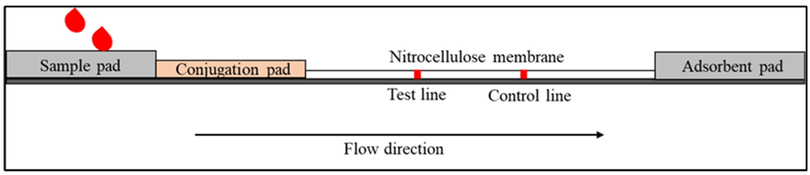
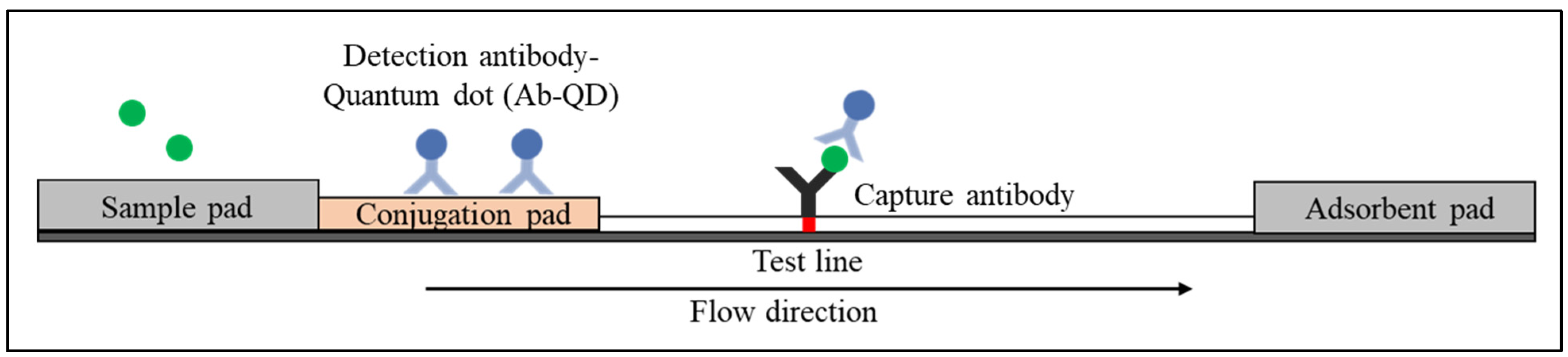
Point-of-care testing (POCT) technology has exhibited an outstanding capability for the detection of several disease biomarkers owing to the fact that such techniques are fast, easy to perform, efficient, and low cost. The lateral flow immunoassay (LFIA) is one such strategy for POCT. LFIA is a well-established platform and a potent assay for fast and inexpensive testing, as this technology is instrumentation independent and allows the visualisation of test results by the naked eye. In order to enhance the sensitivity and specificity of the LFIAs, as well as to allow the quantitation of results, fluorescence immunochromatographic assays have been developed by utilising fluorescent reporters. Fluorescence immunochromatographic assays have advantages over conventional approaches in regard to sensitivity as it produces a higher intensity band on the test and control lines. One such promising fluorescent reporter is quantum dots (QDs). QDs are tiny semiconducting nanocrystals with diameters ranging from 2 to 10 nanometers. QDs have unique electronic characteristics that are intermediate between those of bulk semiconductors and discrete molecules, which is due in part to their high surface-to-volume ratios. The most visible result is fluorescence, in which the nanocrystals emit distinct colours determined by particle size.

quantum dot lateral flow immunoassay infectious diseases

1. Lateral Flow Immunoassay

Lateral flow immunoassay (LFIA) is an immunochromatographic paper-based assay for detecting targets in complex mixtures [1]. LFIAs have several key features. First, the assays are rapid, where the visualisation of the test results can be performed in less than 30 min. Second, the assays can automatically separate the target analytes from the biological samples without sophisticated extra steps such as washing steps in enzyme-linked immunosorbent assays (ELISA). The assay is usually based on antigen–antibody interaction, and sample movement across the membrane occurs via capillary force [2]. Finally, the assays can be operated without the need for expensive equipment and highly trained staff to perform sophisticated analytical procedures, making them suitable for POCT and field-based diagnostic uses. Since the LFIA is an antibody-based approach, other compounds with similar structures may impact its specificity and sensitivity, producing false positive results. In addition, the Kd (dissociation constant) of the antibody–antigen and kinetic rate constant are especially important since LFIA is an in-flow system where the antigen–nanoparticle complex is in contact with the capture antibody for a limited time. The Kd of antibody–antigen conjugate and the colourimetric readout serve as constraints on test sensitivity. Readers and unique biochemical approaches have been developed to enhance product quality and customer comfort in order to overcome these constraints.
The standard LFIA device comprises four components [3]. The sample pad is the first component for sample loading. The sample pad serves to absorb the sample and control the distribution of the sample to the second part. In addition, the sample pad also acts as a filter to separate the whole blood to remove undesired elements such as red and white blood cells from the plasma, which contain antibodies and other proteins that are shed by viruses or bacteria [4]. The second component is the conjugate pad, where conjugate antibodies labelled with biorecognition elements (reporter particles) are immobilised. When the sample reaches the conjugate pad, the conjugated antibodies bind to the target analytes. The antibody–target complex then flows through the nitrocellulose membrane (via capillary force) to where the reaction happens on the test and control lines. The test and control lines comprise immobilised antibodies or proteins (depending on the type of target analytes) that will bind with the antibody–target complex and produce a signal attributed to the reporter particles. The remaining fluid is absorbed by the adsorbent pad (also known as the wicking pad), which is designed to capture the remaining sample and avoid backflow. The Schematic of LFIA is shown in Figure 1.
Figure 1. Schematic diagram shows the basic structure of LFI that consists of a sample pad, conjugation pad, nitrocellulose membrane and adsorbent pad.
Once the sample has reached the test line on the membrane, the signal that appears on the test line can be visualised and interpreted to identify the presence of the analyte. The signal strength on the test line correlates with the number of targets that bind to the detector conjugate. Colloidal gold nanoparticles (AuNPs) have been utilised as reporter particles in LFIAs because of the vibrant colours emitted by their interaction with visible light. The optical properties of AuNPs are tunable by changing the size. For small (~30 nm) monodisperse AuNPs, the surface plasmon resonance phenomenon causes absorption of light in the blue–green portion of the spectrum (~450 nm) while red light (~700 nm) is reflected, producing a rich red colour. As the AuNPs size increases (~100 nm), a longer wavelength of surface plasmon resonance is absorbed, producing solutions with a pale blue or purple colour. The standard size of gold nanoparticles that have been used for LFIA is 40 nm.
The two main configurations of LFIA are primarily divided into the sandwich and competitive formats. The sandwich format is used to detect large targets that have at least two epitopes (binding sites). In contrast, the competitive format is used to detect small targets with a single epitope [5].

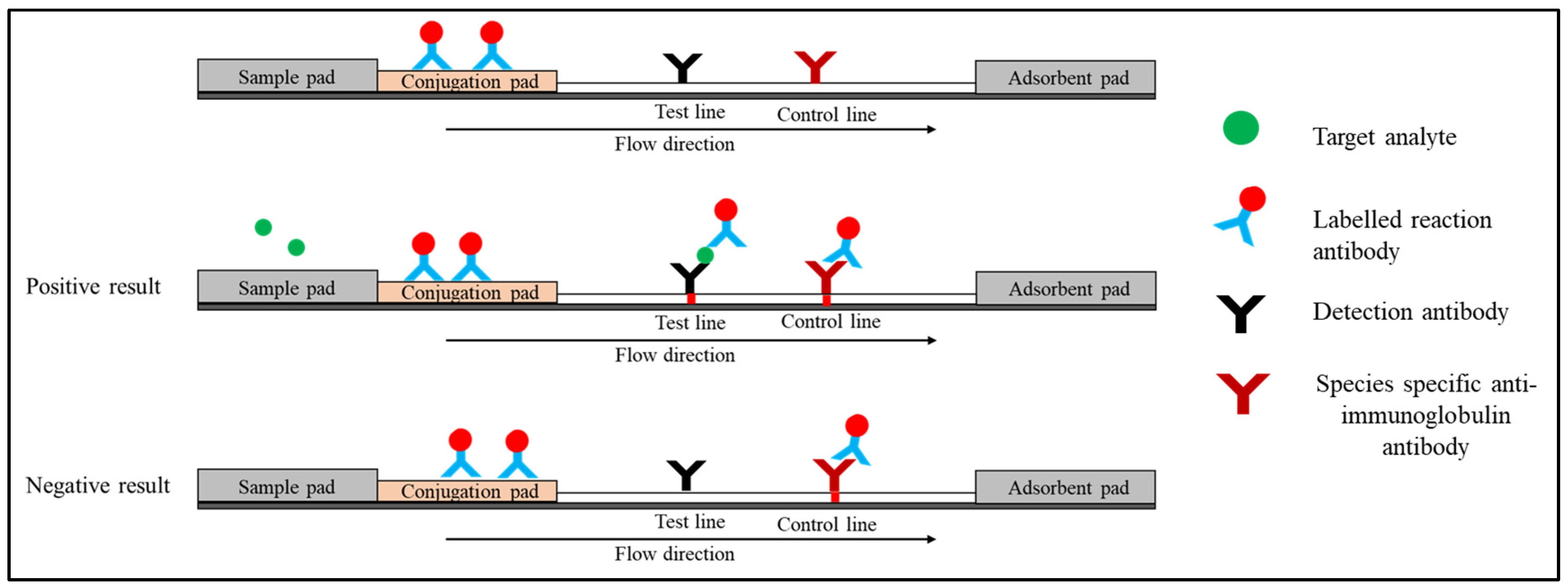
1.1. Sandwich Immunoassay

Sandwich LFIAs have been widely used for the detection of various disease biomarkers as well as small molecules, such as vitamin D [6]. Three different antibodies are usually used in this format [7]. (i) Conjugate antibodies recognise one of the target analyte’s epitopes, and they are immobilised on a conjugation pad linked to the reporter particles. When the sample is added, the conjugate antibody rehydrates and migrates to the test and control lines via capillary force. (ii) Capture antibodies are immobilised on the nitrocellulose membrane at the test line and are specific to another epitope of the target analyte. (iii) Species-specific anti-immunoglobulin antibodies that are immobilised on the control line membrane interact with the reaction antibody.
When the specimen is applied to the sample pad, the target analyte binds with the conjugate antibody on the conjugate pad (analyte–Ab complex) and it is directed to the nitrocellulose membrane. This complex reacts with detection antibodies on the test line, resulting in the sandwich shown in Figure 2. On the control line, excess reaction antibody reacts with species-specific anti-immunoglobulin antibodies. Two red lines will appear at the test and control lines, indicating the presence of the target analyte [8]. In the absence of a target analyte, the reaction antibody only reacts with the species-specific anti-immunoglobulin antibody on the control line. At the control line, only one red line will appear. The presence of a control line indicates that the flow is completed and the test is valid. There are various strategies have been employed at the control line. The most popular approach is by utilising species-specific anti-IgG (Figure 2). Alternatively, researchers have also used an independent antibody–antigen complex such as biotinylated antibodies or oligonucleotides and bovine serum albumin-biotin conjugate [9][10][11].
Figure 2. Schematic diagram shows the sandwich format of LFI.

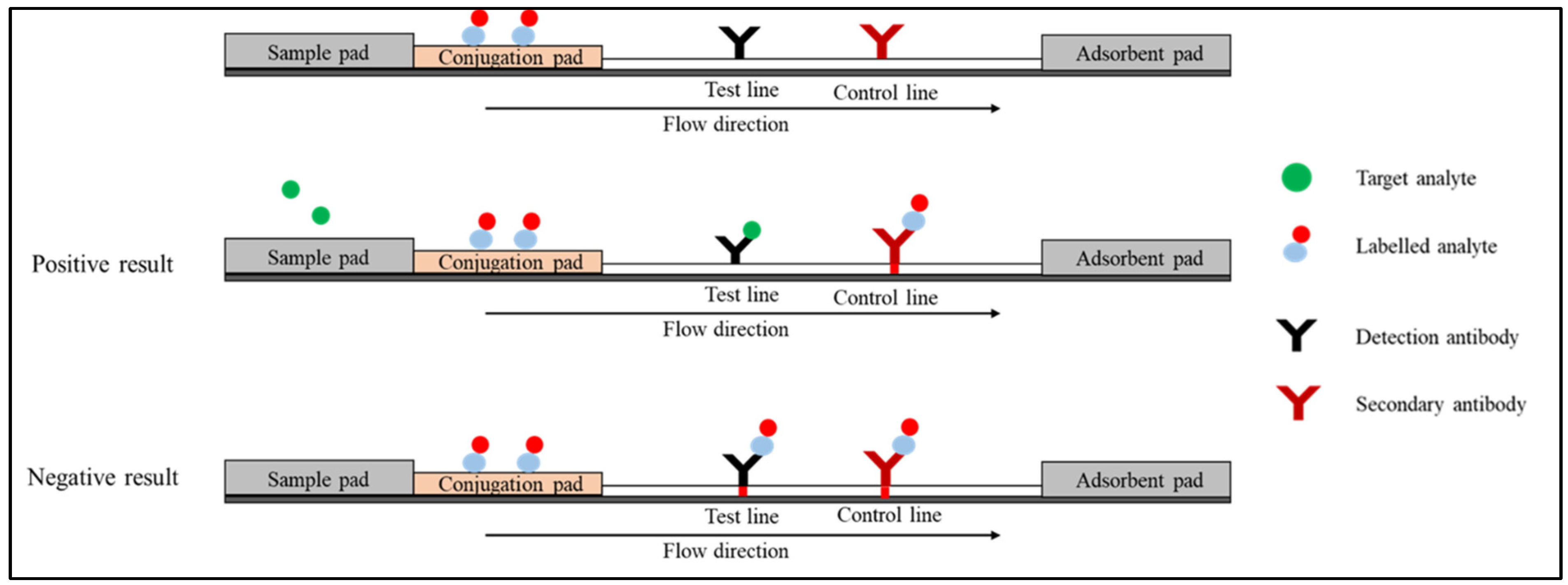
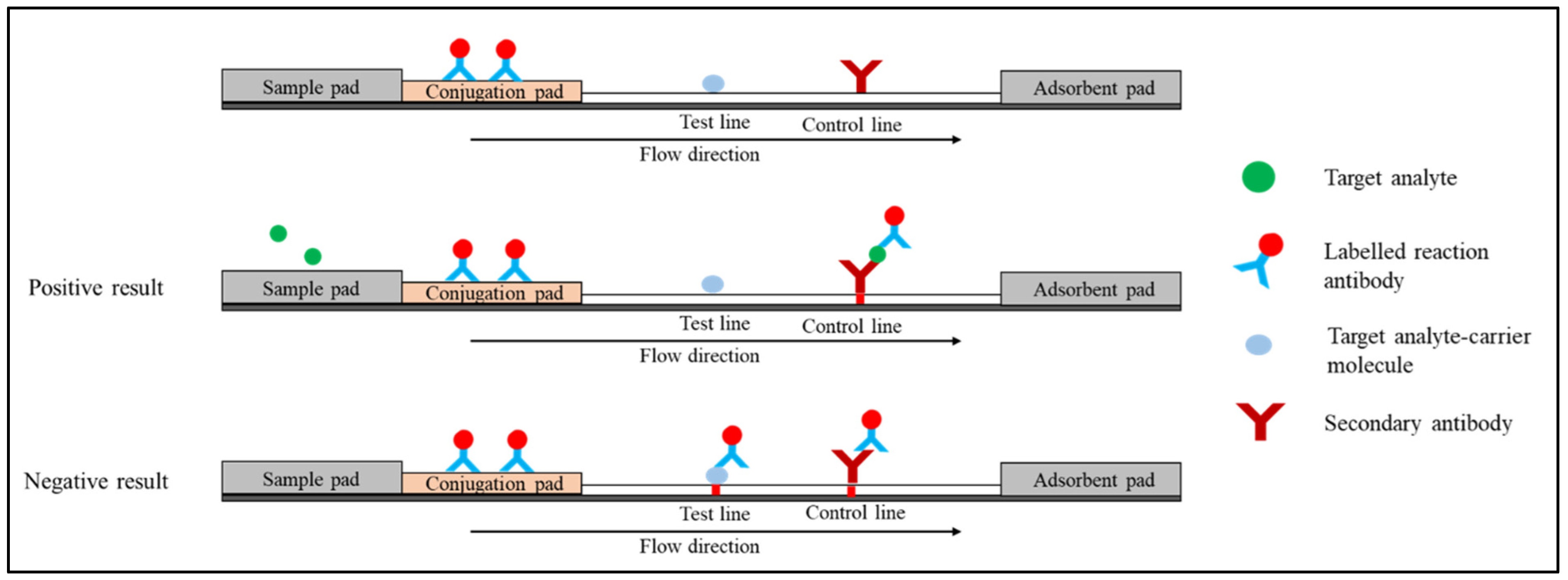
1.2. Competitive Immunoassay (or Inhibition Immunoassay)

The competitive immunoassay format, also known as the inhibition immunoassay, can be conducted in two types of arrangements. In the first arrangement (Figure 3), the labelled analyte is attached to the conjugation pad. In the absence of the target analyte in the specimen, the labelled analyte moves through the strip bind detection antibody on the test line and the secondary antibody on the control line. The red colour will appear on both the test and control lines. In the presence of the target analyte, the unlabelled analyte in the specimen competes with the labelled analyte and binds to the test line. In the control line, the labelled analyte binds to the secondary antibody. Only a red line is formed on the control line [12].
Figure 3. Schematic diagram shows the competitive format (first arrangement).
In another arrangement (Figure 4), the conjugate pad is immobilised with a labelled reaction antibody. Target analyte-carrier molecule conjugate and secondary antibody are used in the test and control lines, respectively. The target analyte in the sample and the target analyte-carrier molecule on the test line compete for binding to the labelled reaction antibody. In the absence of the target analyte, the labelled reaction antibody moves through the strip and binds to the test line’s target analyte-carrier molecule conjugate and the control line’s secondary antibody. The red colour will appear on both test and control lines. In the presence of the target analyte, the labelled reaction antibodies react with the analyte in the sample and move to the control line. One red line is monitored at the control line on the strip [13].
Figure 4. Schematic diagram shows competitive format (second arrangement).

2. Quantum Dot-Based Lateral Flow Immunoassay

Quantum dots (QDs) are the most auspicious fluorescent reporters due to their unique properties, which include high stability, high extinction coefficients, high quantum yields, and a long fluorescence lifespan [14][15]. These characteristics make QDs excellent reporters that can be functionalised with conjugate antibodies for the development of highly sensitive LFIAs. A schematic illustration of the QDs functionalised with conjugate antibodies in sandwich LFIAs is illustrated in Figure 5. The use of QDs as a reporter in sandwich LFIAs requires two antibodies wherein both the conjugate and capture antibodies are specific to the target of interest. Conjugate antibodies labelled with QDs are immobilised on the conjugate pad to enable measurable fluorescence detection. Meanwhile, on a nitrocellulose membrane, capture antibodies are immobilised to capture the target of interest, forming the QD-labelled antibody–target–antibody complex. This complex produces a bright fluorescent band in response to ultraviolet excitation. Similarly, QDs can also be used in competitive LFIAs format by functionalising the nanoparticles with reaction antibodies or analytes on the conjugate pad.
Figure 5. The configuration of the sandwich QD-based LFIA method.

2.1. Types of Quantum Dots

Quantum dots (QDs) can be grouped into different types based on their components and structure. There are three main types of QDs, which include core-type QDs, core-shell QDs, and alloyed QDs.

2.2. Strategies for Conjugating Quantum Dot with Antibodies

The process of conjugating antibodies to the surface of QDs can be performed using several methods. Passive adsorption is the established technique for conjugating antibodies to the surface of QDs and is still widely used. Utilising the interactions (forces) between molecules and surfaces at a certain pH (e.g., van der Waals and ionic forces), antibodies can be directed to spontaneously bind to QDs to form a conjugate. The antibody is normally added in excess to certify that the entire surface of the QDs is covered. After the conjugation is finished, any antibody that is still free in the solution is removed using centrifugation or filtration.
Covalent binding is the most commonly used method for conjugating antibodies to the QDs surface. Covalent binding of QD to conjugate antibodies offers several advantages. Fewer numbers of antibodies are needed to increase the sensitivity, thus lowering the overall cost. Furthermore, covalent conjugates exhibit high stability, allowing their use in difficult sample matrices and high-salt buffering solutions. Additionally, conjugates are easily prepared without the need for extensive salt or pH optimisations, thus, meaning antibody screening experiments can be performed faster.
One common strategy for employing covalent binding is by functionalising the QD surfaces with carboxyl (carboxylic acid). However, obtaining QDs functionalised with carboxyl groups could be tricky. There are several methods to synthesise carboxyl-functionalised QDs have been previously reported. Mansur et al. [16] used acid-functionalised poly (vinyl alcohol) (PVA–COOH) polymer as a capping ligand to synthesise CdSe nanoparticles. The synthesis method was performed using the colloidal chemistry technique via the aqueous route at room temperature. Alternatively, Chen et al. [17] dissolved QDs in deionised water using an ultrasonic bath. Then, HCl solution was added to the solution and mechanically stirred to replace Na+ ions with H+, producing carboxyl-functionalised QD. Once the QDs functionalised with carboxyl groups, the carboxylic groups on QD surfaces can be linked to a primary amine in lysine residues of antibody using 1-Ethyl-3-(3-dimethylaminopropyl) carbodiimide (EDC) and N-hydroxysulfosuccinimide (NHS) reagents to form amide bonds [18][19]. Another approach is via reductive amination, by oxidising the oligosaccharides on the Fc region of the antibody followed by coupling to the amine- or hydrazide-functionalised QDs [20].

2.3. Performance of Quantum Dot-Based Lateral Flow Immunoassay

Syphilis is a sexually transmitted infection (STI) that causes serious health problems worldwide. In 2010, Yang et al. [21] developed a QD-LFIA to be used for the screening of syphilis. The QD-LFIA was designed to detect anti-TP47 polyclonal antibodies by visualising the emission of CdTe under a portable ultraviolet lamp. Thioglycolic acid (TGA) was used to link the QDs with Staphylococcal Protein A (SPA). In the presence of anti-TP47 polyclonal antibodies, the QD-labelled SPA will form a complex with the antibodies. Then, the anti-TP47 antibodies will bind to the TP47 antigen immobilised on the test line, producing a signal that can be visualised under UV light. The assay is suitable for rapid indirect screening of syphilis as the turnaround time is only 10 min. With regard to the limit of detection, the QD-LFIA could detect as low as 2 ng/mL, which was tenfold higher than that of the AuNPs–based method. 

Tuberculosis, an infectious disease caused by the Mycobacterium tuberculosis, is a global public health problem and among the top 10 leading causes of death worldwide, particularly in low- and middle-income countries [22]. In an effort to prevent the spread of the disease, a QD-LFIA device based on a double-antibody sandwich format labelled with core–shell quantum dots (CdSe/ZnS) coupled with streptavidin was developed for the detection of M. tuberculosis fprA proteins [23]. It reported that the device was able to detect fprA proteins in liquid samples at the lowest dilution of 12.5 pg/µL. The sensitivity of LFIA improved as compared to other immunochromatographic tests following the use of QDs labelled antibodies.

References

  1. Koczula, K.M.; Gallotta, A. Lateral Flow Assays. Essays Biochem. 2016, 60, 111–120.
  2. Singh, J.; Sharma, S.; Nara, S. Evaluation of Gold Nanoparticle Based Lateral Flow Assays for Diagnosis of Enterobacteriaceae Members in Food and Water. Food Chem. 2015, 170, 470–483.
  3. Mao, X.; Wang, W.; Du, T.-E. Rapid Quantitative Immunochromatographic Strip for Multiple Proteins Test. Sensors Actuators B Chem. 2013, 186, 315–320.
  4. Biagini, R.E.; Sammons, D.L.; Smith, J.P.; MacKenzie, B.A.; Striley, C.A.F.; Snawder, J.E.; Robertson, S.A.; Quinn, C.P. Rapid, Sensitive, and Specific Lateral-Flow Immunochromatographic Device To Measure Anti-Anthrax Protective Antigen Immunoglobulin G in Serum and Whole Blood. Clin. Vaccine Immunol. 2006, 13, 541–546.
  5. Gupta, Y.; Ghrera, A.S. Recent Advances in Gold Nanoparticle-Based Lateral Flow Immunoassay for the Detection of Bacterial Infection. Arch. Microbiol. 2021, 203, 3767–3784.
  6. Vemulapati, S.; Rey, E.; O’Dell, D.; Mehta, S.; Erickson, D. A Quantitative Point-of-Need Assay for the Assessment of Vitamin D3 Deficiency. Sci. Rep. 2017, 7, 14142.
  7. Bahadır, E.B.; Sezgintürk, M.K. Lateral Flow Assays: Principles, Designs and Labels. TrAC Trends Anal. Chem. 2016, 82, 286–306.
  8. Mosley, G.L.; Pereira, D.Y.; Han, Y.; Lee, S.Y.; Wu, C.M.; Wu, B.M.; Kamei, D.T. Improved Lateral-Flow Immunoassays for Chlamydia and Immunoglobulin M by Sequential Rehydration of Two-Phase System Components within a Paper-Based Diagnostic. Microchim. Acta 2017, 184, 4055–4064.
  9. Lin, L.; Guo, J.; Liu, H.; Jiang, X. Rapid Detection of Hepatitis B Virus in Blood Samples Using a Combination of Polymerase Spiral Reaction With Nanoparticles Lateral-Flow Biosensor. Front. Mol. Biosci. 2021, 7, 578892.
  10. Kim, C.; Yoo, Y.K.; Han, S.I.; Lee, J.; Lee, D.; Lee, K.; Hwang, K.S.; Lee, K.H.; Chung, S.; Lee, J.H. Battery Operated Preconcentration-Assisted Lateral Flow Assay. Lab Chip 2017, 17, 2451–2458.
  11. Gwyn, S.; Mitchell, A.; Dean, D.; Mkocha, H.; Handali, S.; Martin, D.L. Lateral Flow-Based Antibody Testing for Chlamydia Trachomatis. J. Immunol. Methods 2016, 435, 27–31.
  12. Sajid, M.; Kawde, A.-N.; Daud, M. Designs, Formats and Applications of Lateral Flow Assay: A Literature Review. J. Saudi Chem. Soc. 2015, 19, 689–705.
  13. Alhussien, M.N.; Dang, A.K. Sensitive and Rapid Lateral-Flow Assay for Early Detection of Subclinical Mammary Infection in Dairy Cows. Sci. Rep. 2020, 10, 11161.
  14. Hwang, E.; Hwang, H.M.; Shin, Y.; Yoon, Y.; Lee, H.; Yang, J.; Bak, S.; Lee, H. Chemically Modulated Graphene Quantum Dot for Tuning the Photoluminescence as Novel Sensory Probe. Sci. Rep. 2016, 6, 39448.
  15. Schroeder, K.L.; Goreham, R.V.; Nann, T. Graphene Quantum Dots for Theranostics and Bioimaging. Pharm. Res. 2016, 33, 2337–2357.
  16. Mansur, H.S.; Mansur, A.A.P. CdSe Quantum Dots Stabilized by Carboxylic-Functionalized PVA: Synthesis and UV–Vis Spectroscopy Characterization. Mater. Chem. Phys. 2011, 125, 709–717.
  17. Chen, L.; Zhang, C.; Du, Z.; Li, H.; Zhang, L.; Zou, W. Fabrication of Carboxyl Group-Functionalized Carbon Quantum Dots and Its Transparent and Luminescent Epoxy Matrix Nanocomposites for White LED Encapsulation. Macromol. Mater. Eng. 2015, 300, 1232–1237.
  18. Wang, S.; Liu, Y.; Jiao, S.; Zhao, Y.; Guo, Y.; Wang, M.; Zhu, G. Quantum-Dot-Based Lateral Flow Immunoassay for Detection of Neonicotinoid Residues in Tea Leaves. J. Agric. Food Chem. 2017, 65, 10107–10114.
  19. Li, M.; Ma, M.; Hua, X.; Shi, H.; Wang, Q.; Wang, M. Quantum Dots-Based Fluoroimmunoassay for the Simultaneous Detection of Clothianidin and Thiacloprid in Environmental and Agricultural Samples. RSC Adv. 2015, 5, 3039–3044.
  20. Foubert, A.; Beloglazova, N.V.; Rajkovic, A.; Sas, B.; Madder, A.; Goryacheva, I.Y.; De Saeger, S. Bioconjugation of Quantum Dots: Review & Impact on Future Application. TrAC Trends Anal. Chem. 2016, 83, 31–48.
  21. Yang, H.; Li, D.; He, R.; Guo, Q.; Wang, K.; Zhang, X.; Huang, P.; Cui, D. A Novel Quantum Dots–Based Point of Care Test for Syphilis. Nanoscale Res. Lett. 2010, 5, 875–881.
  22. López-López, J.P.; Posada-Martínez, E.L.; Saldarriaga, C.; Wyss, F.; Ponte-Negretti, C.I.; Alexander, B.; Miranda-Arboleda, A.F.; Martínez-Sellés, M.; Baranchuk, A. Tuberculosis and the Heart. J. Am. Heart Assoc. 2021, 10, e019435.
  23. Cimaglia, F.; Aliverti, A.; Chiesa, M.; Poltronieri, P.; De Lorenzis, E.; Santino, A.; Sechi, L.A. Quantum Dots Nanoparticle-Based Lateral Flow Assay for Rapid Detection of Mycobacterium Species Using Anti-FprA Antibodies. Nanotechnol. Dev. 2012, 2, 5.
More
Information
Contributors MDPI registered users' name will be linked to their SciProfiles pages. To register with us, please refer to https://encyclopedia.pub/register : , , , ,
View Times: 1.7K
Revisions: 2 times (View History)
Update Date: 19 Sep 2022
1000/1000