Submitted Successfully!
To reward your contribution, here is a gift for you: A free trial for our video production service.
Thank you for your contribution! You can also upload a video entry or images related to this topic.
Version Summary Created by Modification Content Size Created at Operation
1 -- 2097 2022-08-17 14:52:45 |
2 Reference format revised. Meta information modification 2097 2022-08-18 10:54:48 |

Video Upload Options

Do you have a full video?

Confirm

Are you sure to Delete?
Cite
If you have any further questions, please contact Encyclopedia Editorial Office.
Chevalier, K.;  Mauget-Faÿsse, M.;  Vasseur, V.;  Azar, G.;  Obadia, M.A.;  Poujois, A. Eye Involvement in Wilson’s Disease. Encyclopedia. Available online: https://encyclopedia.pub/entry/26244 (accessed on 20 April 2024).
Chevalier K,  Mauget-Faÿsse M,  Vasseur V,  Azar G,  Obadia MA,  Poujois A. Eye Involvement in Wilson’s Disease. Encyclopedia. Available at: https://encyclopedia.pub/entry/26244. Accessed April 20, 2024.
Chevalier, Kevin, Martine Mauget-Faÿsse, Vivien Vasseur, Georges Azar, Michaël Alexandre Obadia, Aurélia Poujois. "Eye Involvement in Wilson’s Disease" Encyclopedia, https://encyclopedia.pub/entry/26244 (accessed April 20, 2024).
Chevalier, K.,  Mauget-Faÿsse, M.,  Vasseur, V.,  Azar, G.,  Obadia, M.A., & Poujois, A. (2022, August 17). Eye Involvement in Wilson’s Disease. In Encyclopedia. https://encyclopedia.pub/entry/26244
Chevalier, Kevin, et al. "Eye Involvement in Wilson’s Disease." Encyclopedia. Web. 17 August, 2022.
Eye Involvement in Wilson’s Disease
Edit

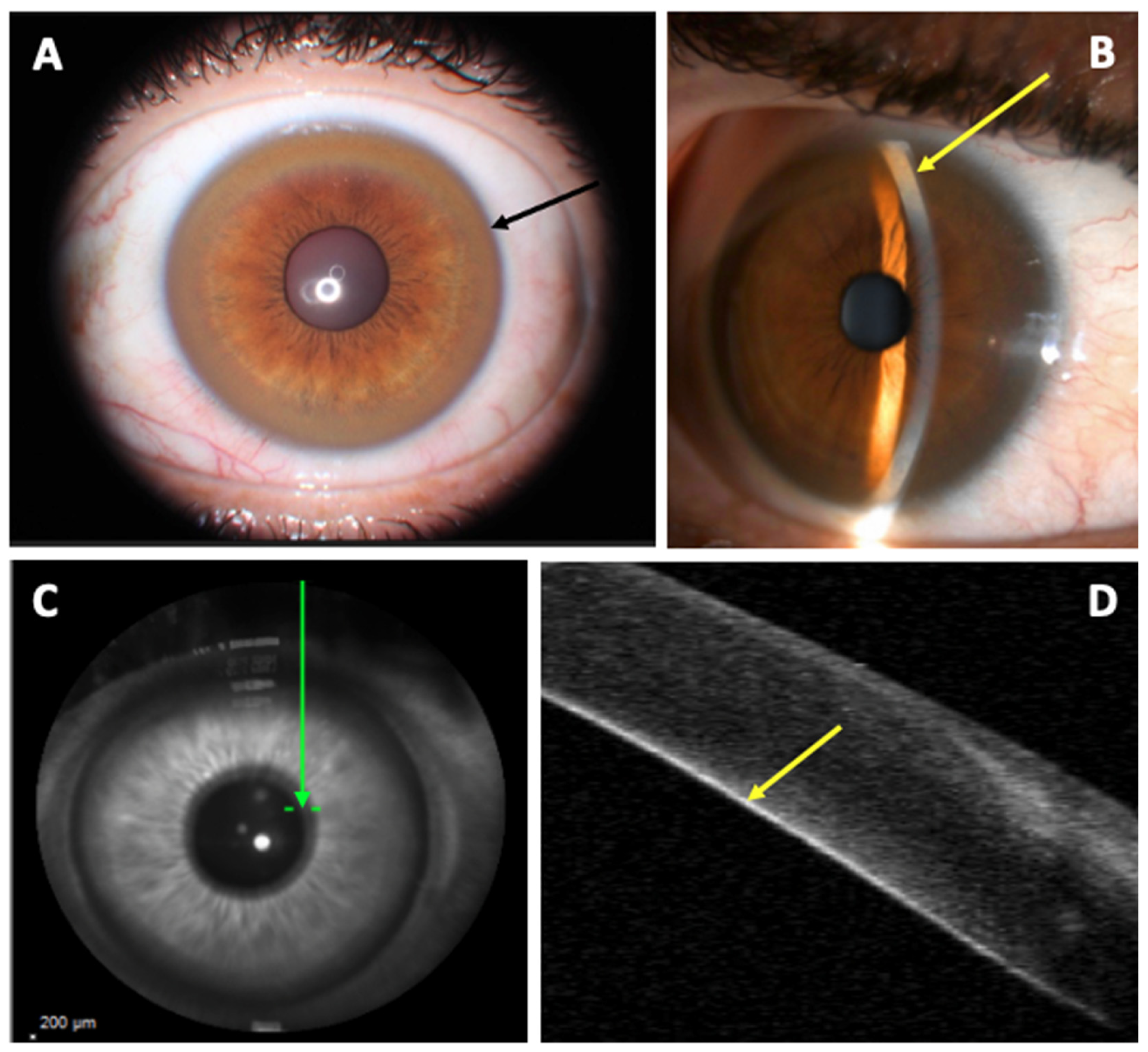
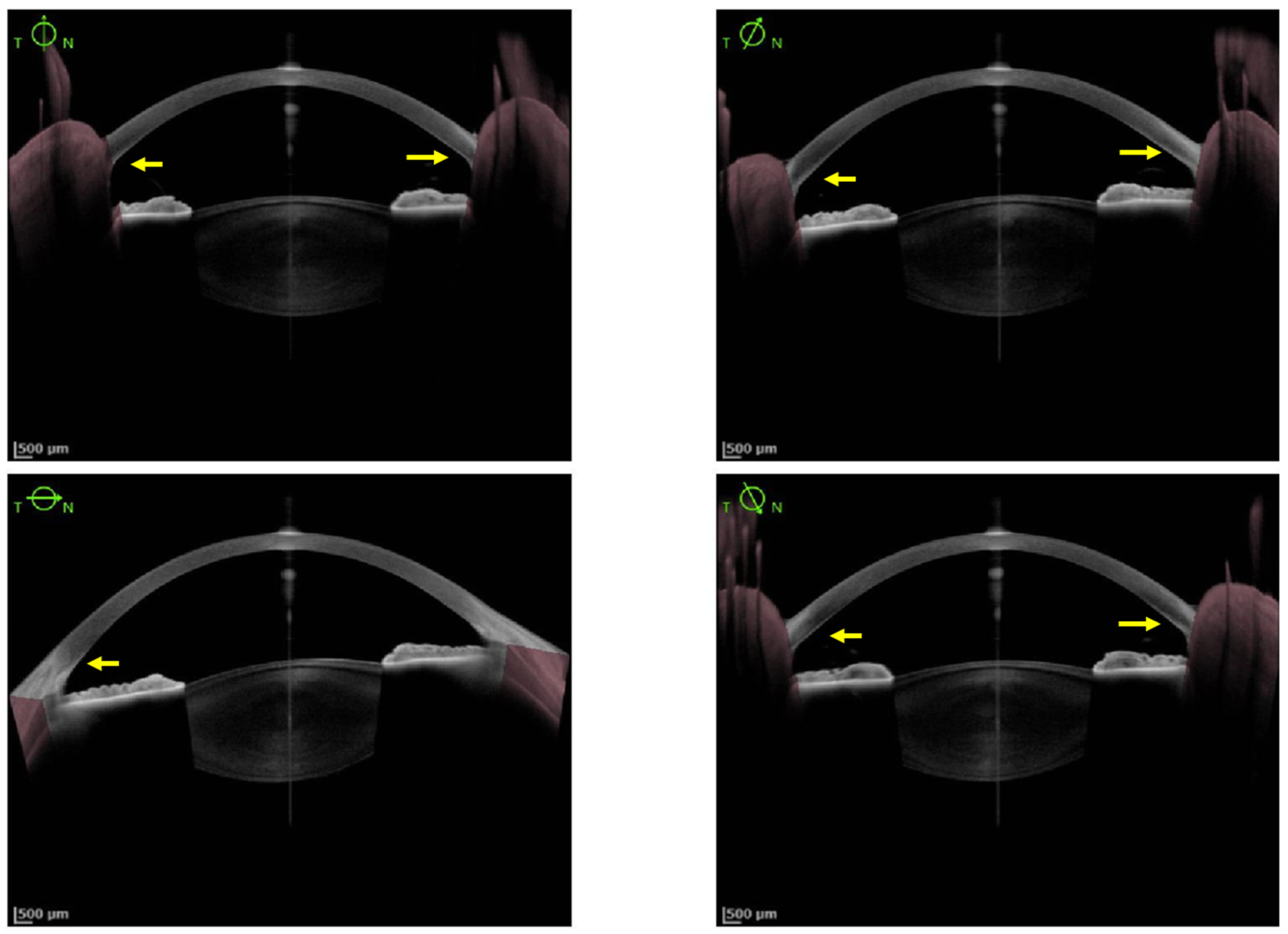
Wilson’s disease (WD) is an autosomal recessive genetic disorder due to a mutation of the ATP7B gene, resulting in impaired hepatic copper excretion and accumulation in various tissues. Ocular findings are one of the hallmarks of the disease. Many ophthalmological manifestations have been described and new techniques are currently available to improve their diagnosis and to follow their evolution. The most common ocular findings seen in WD patients are Kayser–Fleischer ring (KFR) and sunflower cataracts. Other ocular manifestations may involve retinal tissue, visual systems and eye mobility. Diagnosis and follow-up under decoppering treatment of these ocular findings are generally easily performed with slit-lamp examination (SLE). However, new techniques are available for the precocious detection of ocular findings due to WD and may be of great value for non-experimented ophthalmologists and non-ophthalmologists practitioners. 

Wilson’s disease Kayser–Fleischer ring eye involvement sunflower cataract copper

1. Introduction

Wilson’s disease (WD), also known as hepatolenticular degeneration, is a rare genetic condition due to a recessive mutation of the ATP7B gene. The disease consists of a continuous copper accumulation in many tissues and requires life-long treatment. With fewer than 1000 cases in France [1], this condition is mainly characterized by hepatic, ophthalmological and neurological features due to copper accumulation in those organs. Treatments available for this disease rely mainly on copper chelators (D-Penicillamine and trientine salts) and zinc salts.
The ophthalmological manifestations are one of the hallmarks of the disease. Corneal deposition of copper, called Kayser–Fleischer ring (KFR), is typical in WD and constitutes one of the diagnostic criteria for this disease [2]. A sign of extra-hepatic copper accumulation, KFR is a useful biomarker that allows the evolution under chelator treatments. It is a reversible sign and may disappear under treatment. Apart from KFR, many other ophthalmological manifestations have been described. In addition, new techniques have made it possible to improve the diagnosis of these manifestations and to follow their evolution [1].

2. Eye Involvement in Wilson’s Disease

2.1. Cornea Involvement

KFR was first described in 1902 and 1903, respectively, by two German ophthalmologists, Benhard Kayser and Bruno Fleischer. It consists of a ring-shaped copper deposit in the anterior chamber angle within the internal corneal layer of the Descemet’s membrane, at the Schwalbe’s line (Figure 1) [3][4]. In fact, copper is deposited throughout the cornea, and sulfur-copper complexes producing the visible copper deposits are formed only in Descemet’s membrane [5]. On SLE, KFR appears like a golden-brown, golden-green, green-yellow, golden-yellow, bronze or reddish-brown coloring ring in the limbic area of the cornea [6]. It first develops in the superior part of the cornea (at the 12-o’clock position), then inferiorly, and finally in the horizontal meridian forming a closed ring. [3][7][8][9] Thus a closed KFR is evidence of a long-term disease [3]. This pattern of formation and resolution of the KFR could be explained by the vertical flow of aqueous fluid in the anterior chamber of the eye [10]. It is usually bilateral [3] but a unilateral case was reported in 1986 [11].
Figure 1. Kayser–Fleischer Ring. (A): Slip-lamp examination showing a diffuse circumferential Kayser–Fleischer ring in the left eye (black arrow); (B): Slit-lamp examination: visualization of the copper deposit at the posterior part of the cornea in fine slit (yellow arrow); (C): Corneal B-scan localization (Spectralis; Heildelberg Engineering) (green arrow); (D): marked hyperreflectivity of the posterior part of the cornea corresponding to the copper deposit (yellow arrow).
KFR confirms the presence of excess free copper in the bloodstream but is not pathognomonic for WD, as it may occur in any disorder with impaired biliary copper excretion [7][12][13][14][15][16]. KFR is not constantly detected by the classical SLE in WD; its prevalence is estimated between 36–62% in patients with hepatic manifestation, between 77.8–85.2% in patients with neurological manifestation [3][5][17] and between 10–30% in asymptomatic WD patients [3][4][5][9][17][18][19][20][21][22][23]. The incidence of KFR also varies according to the age of the diagnosis. Indeed, the largest pediatric cohort in WD has been recently published by Couchonnal et al. describing 182 children with WD. In this cohort, at diagnosis, 149 (81.8%) children had an ophthalmologic evaluation [24]. Among them, 58 (38.9%) had a detectable KFR: 40/129 (31.0%) were hepatic patients and 18/19 (94.7%) were neurological patients. The youngest patient with a detectable KFR was a seven-year-old, and a total of eight patients (13.7%) with detectable KFR were younger than 10 years, all were hepatic patients. The incidence of KFR in children is much lower than in adults. Nevertheless, it is puzzling that KFR is more frequent in neurological patients (like in adults) but more early in hepatic patients.
In vivo confocal microscopy (IVCM) shows that KFR consists of granular, bright particles that increase in density toward Descemet’s membrane and is associated with a decreased number of keratocytes and peculiar dark, and round areas in all stromal layers. When the ring is not visible in subjects with WD, changes to the corneal microstructure are insignificant [25].
The presence of KFR, identified with SLE by a skilled examiner or rarely with the naked eye, is included in the current diagnosis criteria of WD [2][26][27]. KFR usually disappears progressively with effective treatment of WD, fading initially from lateral and medial aspects of the cornea, then finally from its superior part. As its recurrence suggests a non-adherence to treatment, close monitoring of the ocular status during the follow-up is highly recommended [3][6][9][23][25][26][28][29][30][31][32]. Nevertheless, its disappearance is not correlated with the resolution of the other signs or symptoms of the disease [3][6][8][14].
The sensitivity (Se) of SLE to detect a KFR is known to be low (KFR was missed in more than 50% of hepatic WD patients) [2][33]. That could be explained by the predominance of copper deposits in the anterior chamber angle that cannot be detected with a standard SLE. Therefore, a thorough ophthalmological exam with gonioscopy, which permits a detailed examination of the iridocorneal angle structures, remains crucial [3]. SLE and gonioscopy both required experiment operators, as non-trained ophthalmologists could miss the KFR [3]. These findings lead to the proposal of other methods that could be used to assess the presence and progression of copper deposits in the cornea.
Anterior segment optical coherence tomography (AS-OCT) could be used for the detection of KFR. The KFR appears on a grey scale as a hyper-reflective layer at the level of Descemet’s membrane in the peripheral cornea (Figure 2). On a color scale, it appears as a green/green-yellow/yellow/yellow-orange band. KFR can be easily measured using the gray scale of AS-OCT [3][28][29]. In a study of 29 patients with WD, 15 had normal slit-lamp evaluation but abnormal AS-OCT (p < 0.001) hypothesizing that AS-OCT is a more accurate diagnostic tool that could detect significantly more cases of KFR as compared to the slit-lamp evaluation in participants with hepatic and neurological WD manifestations [3]. This technique could permit more easy recognition of KFR for non-experimented ophthalmologists, as well as non-ophthalmologists practitioners [28][30]. Moreover, it is also useful in children and non-cooperative patients because the imaging process involves fixation for only a few seconds without exposure to bright light [34]. AS-OCT could possibly determine the density of copper deposit in KFR and help the clinician determine the severity of the disease. Further studies are needed to know if AS-OCT can distinguish KFR from pigmented corneal rings in non-Wilsonian liver disease or arcus senilis and if repetitive AS-OCT could help assess the good response of chelator treatments in WD [28].
/media/item_content/202208/62fdfbcc6a93ejcm-11-02528-g003.png
Figure 3. High-resolution Swept Source technology (Anterion®, Heidelberg Engineering, Heidelberg, Germany) provides images of very high quality allowing the detection of a faint Kayser–Fleischer ring (KFR) hardly visible on slit-lamp examination (yellow arrow).
Pentacam HR Scheimpflug imaging is a device that can provide three-dimensional image representations of the anterior segment, which may be useful for screening narrow angles. 
Aside from KFR, corneal nerve fibers are also impacted by copper deposits.

2.2. Lens Involvement

Sunflower cataract is another classical ocular manifestation of WD, the frequency of which varies widely in the literature (between 2 and 20%) [10]. A recent study performed by Langwinska-Wosko et al. on 81 consecutive newly diagnosed WD patients reported that such cataract was detected in only one (1.2%) of all patients, suggesting that sunflower cataract is a very rare ocular sign of WD in de novo and untreated patients. After a year of treatment, the cataract fully disappeared [17]. This manifestation was first described by Siemerling and Oloff in 1922 as “cataracts like rays of the sun” [35]. The scholars already noted the similarities between the cataract seen in their patients with WD to the one produced by an intraocular foreign body containing copper [9]. Sunflower cataract consists of copper deposition in the lens capsule and not within the lens cortex or nucleus itself, and has an aspect of a central disk with radiating petal-like spokes that give its name [7].
Sunflower cataracts usually do not impair vision, cannot be seen with the unaided eye or with an ophthalmoscope, and require slit-lamp evaluation for detection [23]. Like KFR, sunflower cataracts usually regress with copper-chelating treatment [17][36].

2.3. Macula, Retinal Nerve Fiber Layer and Visual Pathways Involvement

Correlation between brain MRI lesions and impairment of visual pathways, macula and retinal nerve fiber layer (RNFL) was found by Langwinska-Wosko et al. [37]. They compared 58 WD patients mean age 38.7 years, with or without brain lesions on MRI (39 MRI+ and 19 MRI−, respectively) and 30 healthy controls (mean age 39.6 years). Total RNFL measured spectral-domain optical coherence tomography (SD-OCT) was thinner in WD patients MRI+ than WD patients MRI− (p = 0.001). Central macular thickness (CMT) was also significantly thinner in WD patients MRI+ than WD patients MRI− (p < 0.001). No significant difference was found in RNFL or CMT between WD patients MRI− and controls. Latency of visual evoked potentials (PEV) and electroretinography (ERG) were prolonged in WD patients MRI+ compared to WD patients MRI− (p < 0.001 and p < 0.001, respectively). Interestingly, some WD patients MRI− had electrophysiological abnormalities. These results confirmed what had been already demonstrated by a German team in 2012 [38]. An Indian team has also shown prolonged latencies in PEV and ERG in WD patients with neurological manifestations compared to controls and the improvement of PEV and ERG latencies after treatment of WD [39].

2.4. Eye Mobility

WD is responsible for eye movement abnormalities such as slow horizontal and vertical saccades [23][40][41], abnormal vertical smooth pursuit [42], increased antisaccadic latency and error rate [43]. Ingster-Moati et al. found that 91% of 34 WD patients (mean age 29 years, 24 neurological forms, 9 hepatic forms and 1 asymptomatic patient) had abnormalities of ocular motility detected by electro-oculography. Here, 29 patients (85%) had an abnormal vertical smooth pursuit, 41% a vertical optokinetic nystagmus and 41% an impaired horizontal smooth pursuit. Among the 27 who underwent MRI, seven patients had normal brainstem and lenticular nuclei images despite the detection of ocular motility abnormalities [42]. Brain MRI of WD patients showed a strong association between prolonged latencies of prosaccades and the brainstem atrophy (r = −0.53 and p = 0,02 for horizontal latencies and r = 0.47 and p = 0.004 for vertical maximum speed in prosaccades, respectively). Impairment in eye movement is probably secondary to the lesions induced by the copper deposit in the brainstem as the nerve centers responsible for vertical and horizontal eye tracking are located in the midbrain and the pons, respectively.

3. Conclusions

Ocular manifestations may be the first presenting symptoms of WD, which must be recognized to prevent fatal outcomes. 
As KFR is an essential criterion for the diagnosis of WD, multiple methods have been studied to improve its diagnosis. So far, SLE is the gold standard for detection of KFR, but its Se is low and it requires experimented ophthalmologists. Since 2016, AS-OCT studies demonstrated a better Se and Sp to diagnose KFR compared to SLE. Its interpretation appears easier and more accessible for non-experimented ophthalmologists and non-ophthalmologist practitioners [28][30]. The density of copper deposit in the cornea at the diagnosis and during the follow-up of WD could determine the severity of the disease and the response under chelator treatment [28]. Pentacam HR Scheimpflug imaging using ImageJ software and calculation of the ratio between anterior and posterior peak signal presents also a good Se (96%) and specificity (Sp, 95%) to detect KFR. Nevertheless, the utilization of those methods to assess therapeutic efficacy needs further evaluation.
It appears that ophthalmological involvement is frequent in WD patients, in particular for the KFR and, to a lesser extent, sunflower cataracts. Other manifestations involving retinal and visual systems, eye mobility or other structures of the eye have been described with various frequencies. AV is nearly often preserved despite corneal or neurological involvement. The evolution of ophthalmologic manifestations seems to be correlated with decoppering treatment, especially for KFR and sunflower cataracts. New methods like AS-OCT and Scheimpflug imaging are alternatives to traditional SLE. These methods allow non-ophthalmologists to look for and quantify KFR more easily and are useful tools to follow the evolution of these abnormalities under chelating treatment. In the near future, the recent development of AI in the analysis of ophthalmic imaging will probably be helpful for the screening of WD anomalies.

References

  1. Poujois, A.; Woimant, F.; Samson, S.; Chaine, P.; Girardot-Tinant, N.; Tuppin, P. Characteristics and prevalence of Wilson’s disease: A 2013 observational population-based study in France. Clin. Res. Hepatol. Gastroenterol. 2018, 42, 57–63.
  2. Roberts, E.A.; Schilsky, M.L.; American Association for Study of Liver Diseases (AASLD). Diagnosis and treatment of Wilson disease: An update. Hepatology 2008, 47, 2089–2111.
  3. Broniek-Kowalik, K.; Dzieżyc, K.; Litwin, T.; Członkowska, A.; Szaflik, J.P. Anterior segment optical coherence tomography (AS-OCT) as a new method of detecting copper deposits forming the Kayser–Fleischer ring in patients with Wilson disease. Acta Ophthalmol. 2019, 97, e757–e760.
  4. Kelly, C.; Pericleous, M. Wilson disease: More than meets the eye. Postgrad. Med. J. 2018, 94, 335–347.
  5. Pfeiffer, R.F. Wilson’s Disease. Skull Base 2007, 27, 123–132.
  6. Fenu, M.; Liggi, M.; Demelia, E.; Sorbello, O.; Civolani, A. Kayser–Fleischer ring in Wilson’s disease: A cohort study. Eur. J. Intern. Med. 2012, 23, e150–e156.
  7. Prasad, D.; Bhriguvanshi, A. Ocular manifestations of liver disease in children: Clinical aspects and implications. Ann. Hepatol. 2020, 19, 608–613.
  8. Vitiello, L.; De Bernardo, M.; Nuzio, S.G.; Mandato, C.; Rosa, N.; Vajro, P. Pediatric liver diseases and ocular changes: What hepatologists and ophthalmologists should know and share with each other. Dig. Liver Dis. 2019, 52, 1–8.
  9. Walshe, J.M. The eye in Wilson disease. QJM 2010, 104, 451–453.
  10. Członkowska, A.; Litwin, T.; Chabik, G. Wilson Disease. In Handbook of Clinical Neurology; Elsevier: Amsterdam, The Netherlands, 2017; Volume 142, pp. 101–119. ISBN 978-0-444-63625-6.
  11. Innes, J.R.; Strachan, I.M.; Triger, D.R. Unilateral Kayser-Fleischer ring. Br. J. Ophthalmol. 1986, 70, 469–470.
  12. Al-Khaier, A.; Nischal, K.K. The eye in metabolic disease. Hosp. Med. 2003, 64, 609–612.
  13. Alakus, M.F.; Caglayan, M.; Ekin, N.; Oncul, H.; Arac, E.; Dag, U.; Diri, H. Investigation of corneal topographic and densitometric properties of Wilson’s disease patients with or without a Kayser-Fleischer ring. Eye Vis. 2021, 8, 1–8.
  14. Gitlin, J.D. Wilson disease. Gastroenterology 2003, 125, 1868–1877.
  15. Walshe, J.M. The Kayser–Fleischer Ring. Br. J. Hosp. Med. 2014, 75, 2.
  16. Sridhar, U.; Tripathy, K. Commentary: Kayser-Fleischer-like rings in patients with hepatic disease. Indian J. Ophthalmol. 2021, 69, 1088.
  17. Langwińska-Wośko, E.; Litwin, T.; Dzieżyc, K.; Członkowska, A. The sunflower cataract in Wilson’s disease: Pathognomonic sign or rare finding? Acta Neurol. Belg. 2015, 116, 325–328.
  18. Youn, J.; Kim, J.S.; Kim, H.-T.; Lee, J.-Y.; Lee, P.H.; Ki, C.-S.; Cho, J.W. Characteristics of neurological Wilson’s disease without Kayser–Fleischer ring. J. Neurol. Sci. 2012, 323, 183–186.
  19. Demirkiran, M.; Jankovic, J.; Lewis, R.A.; Cox, D.W. Neurologic presentation of Wilson disease without Kayser-Fleischer rings. Neurology 1996, 46, 1040–1043.
  20. Medici, V.; Rossaro, L.; Sturniolo, G. Wilson disease—A practical approach to diagnosis, treatment and follow-up. Dig. Liver Dis. 2007, 39, 601–609.
  21. Willeit, J.; Kiechl, S. Wilson’s disease with neurological impairment but no Kayser-Fleischer rings. Lancet 1991, 337, 1426.
  22. Imam, L.; Haboubi, H.N. G-Eye: Ocular manifestations of gastrointestinal disease. Front. Gastroenterol. 2019, 11, 162–167.
  23. Lorincz, M.T. Neurologic Wilson’s disease. Ann. N. Y. Acad. Sci. 2010, 1184, 173–187.
  24. Couchonnal, E.; Lion-François, L.; Guillaud, O.; Habes, D.; Debray, D.; Lamireau, T.; Broué, P.; Fabre, A.; Vanlemmens, C.; Sobesky, R.; et al. Pediatric Wilson’s Disease: Phenotypic, Genetic Characterization and Outcome of 182 Children in France. J. Pediatr. Gastroenterol. Nutr. 2021, 73, e80–e86.
  25. Grupchev, D.I.; Radeva, M.N.; Georgieva, M.; Grupcheva, C.N. In vivo confocal microstructural analysis of corneas presenting Kayser-Fleischer rings in patients with Wilson’s disease. Arq. Bras. Oftalmol. 2018, 81, 137–143.
  26. European Association for Study of Liver. EASL Clinical Practice Guidelines: Wilson’s disease. J. Hepatol. 2012, 56, 671–685.
  27. Socha, P.; Janczyk, W.; Dhawan, A.; Baumann, U.; D’Antiga, L.; Tanner, S.; Iorio, R.; Vajro, P.; Houwen, R.; Fischler, B.; et al. Wilson’s Disease in Children: A Position Paper by the Hepatology Committee of the European Society for Paediatric Gastroenterology, Hepatology and Nutrition. J. Pediatr. Gastroenterol. Nutr. 2018, 66, 334–344.
  28. Sridhar, M.S. Advantages of Anterior Segment Optical Coherence Tomography Evaluation of the Kayser–Fleischer Ring in Wilson Disease. Cornea 2017, 36, 343–346.
  29. Sridhar, M.S.; Rangaraju, A.; Anbarasu, K.; Reddy, S.P.; Daga, S.; Jayalakshmi, S.; Shaik, B. Evaluation of Kayser–Fleischer ring in Wilson disease by anterior segment optical coherence tomography. Indian J. Ophthalmol. 2017, 65, 354–357.
  30. Sridhar, M.S.; Pineda, R. Anterior segment optical coherence tomography to look for Kayser-Fleischer rings. Pract. Neurol. 2017, 17, 222–223.
  31. Telinius, N.; Ott, P.; Hjortdal, J. Detection of Kayser-Fleischer ring using Scheimpflug imaging. Acta Ophthalmol. 2016, 95, e248–e249.
  32. Zhao, T.; Fang, Z.; Tian, J.; Liu, J.; Xiao, Y.; Li, H.; Chen, B. Imaging Kayser-Fleischer Ring in Wilson Disease Using In Vivo Confocal Microscopy. Cornea 2018, 38, 332–337.
  33. Ceresara, G.; Fogagnolo, P.; Zuin, M.; Zatelli, S.; Bovet, J.; Rossetti, L. Study of Corneal Copper Deposits in Wilson’s Disease by in vivo Confocal Microscopy. Ophthalmologica 2014, 231, 147–152.
  34. Rathi, A.; Takkar, B.; Gaur, N.; Maharana, P.K. Optical coherence tomography of the Kayser-Fleischer ring: An ancillary diagnostic tool for Wilson’s disease in children. BMJ Case Rep. 2017, 2017, bcr-2017-220007.
  35. Siemerling, E.; Oloff, H. Pseudosklerose (Westphal-Strümpell). Klin. Wochenschr. 1922, 1, 1087–1089.
  36. Litwin, T.; Langwińska-Wośko, E.; Dzieżyc, K.; Członkowska, A. Sunflower cataract: Do not forget Wilson’s disease. Pract. Neurol. 2015, 15, 385–386.
  37. Langwińska-Wośko, E.; Litwin, T.; Szulborski, K.; Członkowska, A. Optical coherence tomography and electrophysiology of retinal and visual pathways in Wilson’s disease. Metab. Brain Dis. 2015, 31, 405–415.
  38. Albrecht, P.; Müller, A.-K.; Ringelstein, M.; Finis, D.; Geerling, G.; Cohn, E.; Aktas, O.; Hartung, H.-P.; Hefter, H.; Methner, A. Retinal Neurodegeneration in Wilson’s Disease Revealed by Spectral Domain Optical Coherence Tomography. PLoS ONE 2012, 7, e49825.
  39. Satishohandra, P.; Naik, K.R. Visual pathway abnormalities Wilson’s disease: An electrophysiological study using electroretinography and visual evoked potentials. J. Neurol. Sci. 2000, 176, 13–20.
  40. Jung, H.-K.; Choi, S.Y.; Kim, J.-M.; Kim, J.-S. Selective slowing of downward saccades in Wilson’s disease. Park. Relat. Disord. 2013, 19, 134–135.
  41. Kirkham, T.H.; Kamin, D.F. Slow saccadic eye movements in Wilson’s disease. J. Neurol. Neurosurg. Psychiatry 1974, 37, 191–194.
  42. Ingster-Moati, I.; Quoc, E.B.; Pless, M.; Djomby, R.; Orssaud, C.; Guichard, J.P.; Woimant, F. Ocular motility and Wilson’s disease: A study on 34 patients. J. Neurol. Neurosurg. Psychiatry 2007, 78, 1199–1201.
  43. Leśniak, M.; Członkowska, A.; Seniów, J.S. Abnormal antisaccades and smooth pursuit eye movements in patients with Wilson’s disease. Mov. Disord. 2008, 23, 2067–2073.
More
Information
Subjects: Ophthalmology
Contributors MDPI registered users' name will be linked to their SciProfiles pages. To register with us, please refer to https://encyclopedia.pub/register : , , , , ,
View Times: 783
Revisions: 2 times (View History)
Update Date: 18 Aug 2022
1000/1000