Submitted Successfully!
To reward your contribution, here is a gift for you: A free trial for our video production service.
Thank you for your contribution! You can also upload a video entry or images related to this topic.
Version Summary Created by Modification Content Size Created at Operation
1 -- 2620 2022-08-11 16:39:08 |
2 layout + 15 word(s) 2635 2022-08-15 07:53:36 |

Video Upload Options

Do you have a full video?

Confirm

Are you sure to Delete?
Cite
If you have any further questions, please contact Encyclopedia Editorial Office.
Boutal, H.;  Moguet, C.;  Pommiès, L.;  Simon, S.;  Naas, T.;  Volland, H. Lateral Flow Assays Combat Antimicrobial Resistant. Encyclopedia. Available online: https://encyclopedia.pub/entry/26084 (accessed on 19 April 2024).
Boutal H,  Moguet C,  Pommiès L,  Simon S,  Naas T,  Volland H. Lateral Flow Assays Combat Antimicrobial Resistant. Encyclopedia. Available at: https://encyclopedia.pub/entry/26084. Accessed April 19, 2024.
Boutal, Hervé, Christian Moguet, Lilas Pommiès, Stéphanie Simon, Thierry Naas, Hervé Volland. "Lateral Flow Assays Combat Antimicrobial Resistant" Encyclopedia, https://encyclopedia.pub/entry/26084 (accessed April 19, 2024).
Boutal, H.,  Moguet, C.,  Pommiès, L.,  Simon, S.,  Naas, T., & Volland, H. (2022, August 11). Lateral Flow Assays Combat Antimicrobial Resistant. In Encyclopedia. https://encyclopedia.pub/entry/26084
Boutal, Hervé, et al. "Lateral Flow Assays Combat Antimicrobial Resistant." Encyclopedia. Web. 11 August, 2022.
Lateral Flow Assays Combat Antimicrobial Resistant
Edit

Antimicrobial resistance (AMR) is today universally recognised as a global threat because of the rapid emergence and dissemination of resistant bacteria and genes among humans, animals, and the environment on a global scale. AMR thus represents a heavy burden for healthcare systems all over the world. ESKAPE pathogens (Enterococcus faecium, Staphylococcus aureus, Klebsiella pneumoniae, Acinetobacter baumannii, Pseudomonas aeruginosa et Enterobacter spp.) combined with antibiotic resistance have greatly increased the risk of morbidity and mortality, especially in ICU settings. Lateral flow assays (LFA) are inexpensive, rapid, and efficient tools that are easy to implement in the routine workflow of laboratories as new first-line tests against AMR with bacterial colonies, and in the near future directly with biological media.

LFA detection antibiotic resistance

1. General Presentation of Lateral Flow ImmunoAssay

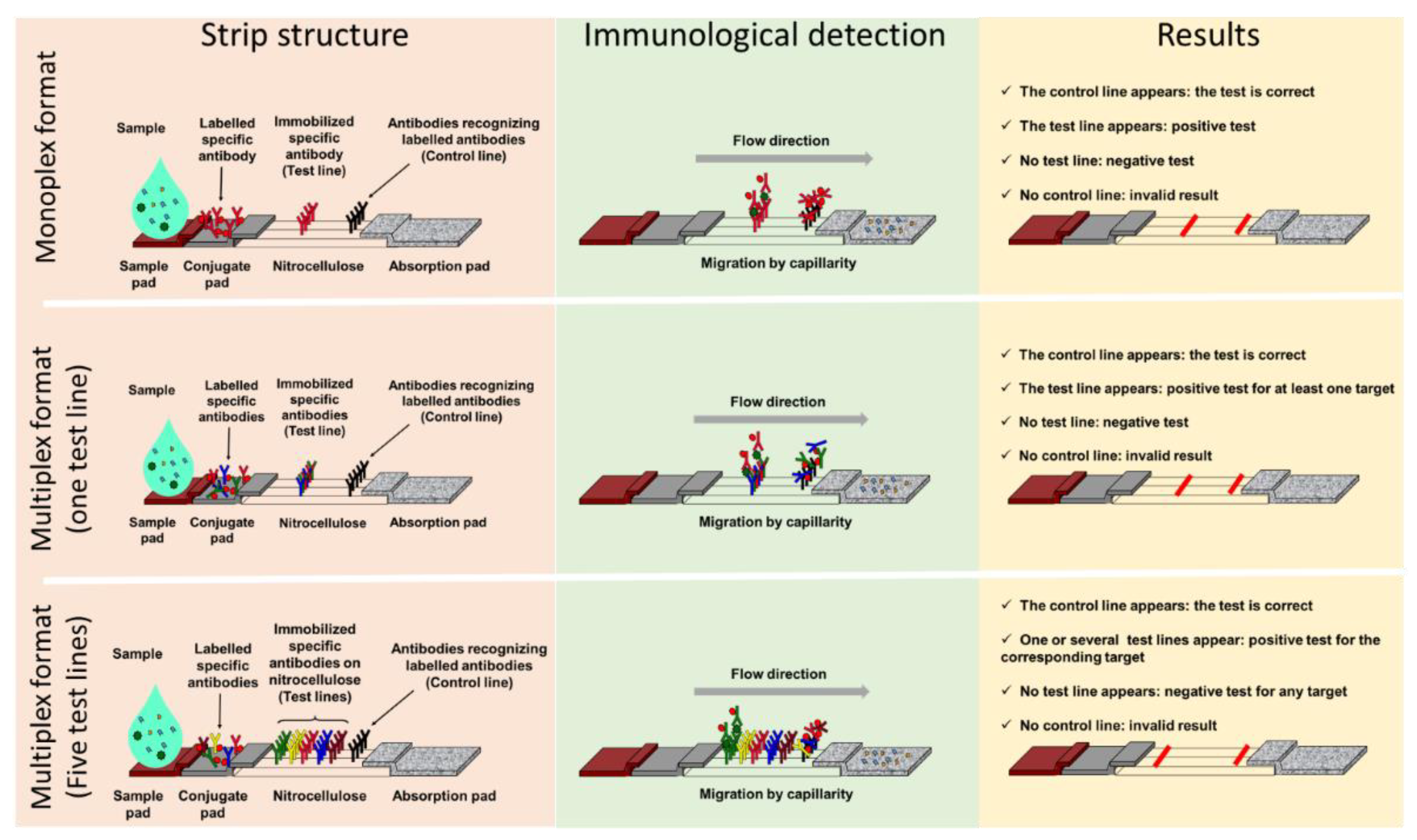
1.1. Components and Principle

Lateral Flow ImmunoAssay (LFIA) tests generally consist of a strip supporting different porous compounds on which liquids migrate by capillarity. The sample pad (SP), which is usually made of cellulose, receives the sample that successively drains onto the conjugate pad (CP). Made of glass fiber, the CP is the storage area for the conjugate, a labelled molecule that generates the signal on the strip. This signal is located on a nitrocellulose membrane printed with different compounds in order to form one test line and a control line (Figure 1). The test line captures the targets of interest while the control line serves as an inner control for confirmation of correct flow and compatible test conditions. Finally, the wicking pad acts as a pump as well as a reservoir for the liquid dispensed on the SP, and its capacity influences the volume of sample that can be analyzed. All components overlap one over the other and are generally enclosed in a plastic cassette. This cassette provides pressure points to maintain close contacts, providing efficient flow of the reagents through the strip, protection, easy handling, a localized sample dispensing area, and a reading frame.
Figure 1. Lateral flow assay formats: components and principle. Presentation of three lateral flow immunoassay formats with, from left to right, their structure (pink panel), immunological detection principle (green panel), and results interpretation (yellow panel). The monoplex format able to detect only one target is presented in the upper panel, a multiplex detection format with one test line and no possible identification of the target is in the central panel, and a multiplex detection format with spatial separation of the test lines and identification of the target(s) detected is presented in the lower panel.
Migration begins once the sample has been loaded on the SP that, depending on the nature of the samples, can be pre-treated in order to reduce matrix effects. The sample solution resuspends the conjugate that forms complexes with the analyte if present. Capillary pressure transports the complexes along the nitrocellulose membrane, and they accumulate on the test line and the excess of conjugate on the control line.
Most of the time, the test line and the conjugate involve antibodies specific for the target being detected, and two formats of LFIA are available. The first, known as non-competitive or sandwich immunoassay format, is for large molecular weight analytes such as proteins that provide several antigenic sites. In this case, a colored test line represents a positive result. The second format, known as competitive or inhibition immunoassay format, is dedicated to small molecular weight antigens, and an attenuated or absent test line indicates a positive result.

1.2. Advantages of Lateral Flow ImmunoAssay

LFIA are one-step assays that require no washing and only a small sample volume. The time to results, following an easy sample handling, is short (15–30 min), which positions LFIA as a good candidate for primary screening at PoC or PoN. They are inexpensive and do not have specific storage constraints, such as refrigeration, making them accessible to third-world countries [1][2]. The qualitative interpretation of the result can be visual, and no particular skills are required. Moreover, depending on the parameters of the test, results can also be semi-quantitative or even quantitative through the use of a reader [3][4]. In addition, if the reagents are available, the development time to market of an LFIA is relatively short, which can be useful to respond to an urgent sanitary crisis, as illustrated with the recent COVID-19 pandemic. The LFIA, due to its configuration, allows the detection of multiple analytes such as proteins, haptens, or nucleic acids [5].
Interest in LFIAs is best illustrated by the increase in publications describing their use in different fields of application over the last 10 years, especially in the clinical domain (Figure 2).The search query used was (Scopus format): TITLE-ABS-KEY (lateral AND flow AND immunoassay) OR TITLE (lateral AND flow AND assay) OR TITLE (immunochromatographic) AND PUBYEAR > 2009 AND PUBYEAR < 2020 AND (LIMIT-TO (DOCTYPE, “ar”)) AND (LIMIT-TO (LANGUAGE, “English”)). The research has been done in October 2020. Then, the articles were selected following the PRISMA guideline for systematic reviews [1]. It is likely that the ease of use, speed, and specificity of these tests are at the origin of their exponential use, including in the field of AMR.
Figure 2. Number of publications per year for different LFIA applications [1].

2. Classical Lateral Flow Assays and Antimicrobial Resistant 

Bacteria possess four main mechanisms that can confer resistance to antibiotics: (i) expression or overexpression of efflux pumps reducing antibiotic concentration within the bacteria; (ii) decreased permeability of the membrane or cell wall leading to ineffective drug concentration in the bacteria; (iii) changed target structure with affinity loss for antibiotics; and (iv) antibiotic degradation mediated by hydrolysis [6]. Any protein involved in a mechanism of AMR can then become a privileged target for LFIA. As a result, a number of LFIA tests have been developed, and some are commercially available.

2.1. Monoplex Lateral Flow ImmunoAssay to Address Antimicrobial Resistant (AMR) Detection

As the number of antibiotic-resistant isolates has increased, LFIA tests have been developed to target enzyme-mediated resistance traits in the clinically most important bacteria, with the primary objective of detecting enzymes involved in the resistance of most prevalent pathogens. A LFIA test detecting the expression of the Pseudomonas aeruginosa 6′-N-acetyltransferase AAC(6′)-Iae, which confers resistance to aminoglycosides, was described in 2010 and has a sensitivity of 105 cfu/test [7]. Another LFIA targets ArmA 16S rRNA methylase, which is one of the most prevalent 16S rRNA methylase reported and leads to pan-aminoglycoside resistance in Gram-negative bacteria (GNB) such as Acinetobacter baumannii and Escherichia coli [8].
Methicillin-resistant Staphylococcus aureus (MRSA) is another major pathogen responsible for severe morbidity and mortality in many hospitals worldwide, which also has an efficient capacity for spreading in the community [9]. The early detection of methicillin resistance, which confers resistance to all ß-lactams, is essential. It relies on the detection of the penicillin-binding-protein 2a (PBP2a), which has a reduced affinity for beta-lactam antibiotics. Antibody-based techniques for the detection of MRSA are challenging as S. aureus protein A binds to mammalian immunoglobulins. Nevertheless, the use of IgY anti-PBP2a antibodies has been described by Yamada et al. [10], and another study described an optimized LFIA with a detection limit of 104 cfu/mL [11].
Vancomycin-resistant enterococcus (VRE) is one of the most important nosocomial pathogens worldwide [12][13][14]. The vancomycin resistance mechanism in Enterococcus faecium and Enterococcus faecalis is mostly acquired and linked to the production of ligases. In Europe, the most prevalent are VanA and VanB [12][15]. In this context, a LFIA for the identification of VanA-VRE isolates was described with 100% sensitivity and 100% specificity and a limit of detection of 6.3 × 10⁶ cfu and 4.9 × 10⁵ cfu per test when the growth was performed on MH or ChromID® VRE plates, respectively [16]. Another LFIA for the detection of VanB-VRE isolates with a lower sensitivity and a mandatory pre-culture on vancomycin-containing media for induction of VanB ligase has also been reported [17].
Beta-lactams represent the major family of antibiotics to treat infections due to Gram-negative bacteria, but their use is currently challenged by the spread of beta-lactamases [18]. In particular, the spread of extended-spectrum beta-lactamases (ESBLs) among Enterobacterales represents a major threat as these enzymes are able to inactivate most beta-lactam molecules (including 3rd and 4th generation cephalosporins and aztreonam), sparing only carbapenems [19]. The most common family of the ESBL, the CTX-M, represented by five sub-groups: CTX-M-1, CTX-M-2, CTX-M-8, CTX-M-9, and CTX-M-25, has disseminated worldwide [20]. The increase in the prevalence of ESBL-producing Enterobacterales has led to an increased use of carbapenems, a last resort antibiotic, to treat infections with ESBL-producers. This has led to the selection and subsequent increase in bacteria resistant to these antibiotics. Carbapenem-resistant Enterobacterales (CREs) are usually resistant to most, if not all antibiotics, thus posing serious therapeutic issues in clinical practice. Among CREs, carbapenemase-producing isolates are the most worrisome, as they are capable of efficiently hydrolyzing carbapenems, and their genes are carried by plasmids that may be exchanged between bacteria. Early identification of carriers is essential in order to implement reinforced infection control measures, among which isolation of the patient is a prerequisite. There are five major carbapenemases, KPC, NDM, OXA-48, VIM, and IMP. LFIAs to detect KPC and OXA-48-like enzymes [21] (Coris Bioconcept, Gembloux Belgium), IMP (the most prevalent metallo-β-lactamases in Japan [22][23]), and NDM [24][25] have been developed, all with 100% sensitivity and specificity when used with isolated colony from agar plates.
Faced with infections resistant to carbapenems, the paucity of therapeutic options has led to the use of polymyxins such as colistin as last resort antibiotics [26]. Inexorably, bacteria acquired colistin resistance. While most colistin resistance is due to chromosomic mutations, plasmid-encoded mechanisms, such as MCR-1, have also been described. The latter are considered particularly threatening, as the mcr genes are plasmid encoded, and the resistance phenotype is difficult to detect. MCR-1, initially described in 2015, mediates the modification of the lipopolysaccharide by a phosphoethanolamine transferase activity [27]. More than eight different MCR-alleles have now been described [28]. Soon after the first description of MCR-1, a LFIA directed against this allele was commercialized (NG-Test MCR-1). This assay has been evaluated in a multicentric study against a collection of human and animal enterobacterial isolates. The results revealed 100% sensitivity for MCR-1 expressing isolates, but some MCR-2 carriers were missed, and the assay did not detect MCR-3, MCR-4, and MCR-5 carriers [29].

2.2. Multiplex Lateral Flow ImmunoAssay in the Antimicrobial Resistant Field

Monoplex LFIAs have proven efficacy for the detection and identification of resistance-determining markers. When targeting mechanisms involving a multitude of variants, such as the CTX-M family, a wider specificity can be relevant. Multiplex LFIAs, able to detect CTX-M enzymes but without discriminating the variant or subgroup to which they belong, have been developed. For example, the NG-Test CTX-M MULTI (NG Biotech, Guipry, France) is commercially available and relies on a cocktail of anti-CTX-M mouse antibodies, immobilized on a unique test line (Figure 1, central panel). This test allows the detection of the five CTX-M-subgroups. A recent study showed that the NG-Test CTX-M MULTI could detect 98% of ESBL-producers from a French clinical setting, either from colonies or positive blood cultures, missing only two SHV-ESBL producers [30]. A study, conducted in Italy, reported that the NG-Test CTX-M MULTI was a reliable assay for the detection of CTX-M-like ESBLs from bacterial pellets from blood culture broth, showing excellent sensitivity and specificity [31]. A further study recently described the detection of CTX-M-group-1, -2, and -9 producers using a monoclonal rabbit anti-CTX-M antibody, and showed 100% sensitivity and specificity with clinical isolates grown on agar plates [32]. Most resistance genes are carried by mobile genetic elements [33] and a single strain can harbor more than one resistance determinant. In this scenario, the detection and identification of more than one mechanism of resistance in a single test is relevant. To do so, several test lines are printed on the same strip (Figure 1, lower panel) and target identification is made through a spatial repartition [34]. Several multiplex assays have been described, so far only for the identification of carbapenemase-producing strains. Several versions of the RESIST LFIA test exist (from Coris Bioconcept), which are differentiated by the number of carbapenemases that can be detected. RESIST-3 [35] can detect NDM, KPC, and OXA-48 enzymes. This test has since been upgraded with the additional detection of VIM (RESIST-4 [36]), and IMP or OXA-163 (RESIST-5 [37][38][39]). All these multiplex assays consist of two-independent cassettes that are used in parallel with the same bacterial extract. Another assay, named NG CARBA-5 [40] (from NG Biotech, Guipry, France), also targets NDM, IMP, VIM, OXA-48, and KPC carpabenemases, but a major difference compared to the RESIST-LFIA test is that the sample has to be loaded onto one unique cassette. The NG Carba-5 has been evaluated by the Antimicrobial Resistance and Healthcare Associated Infections (AMRHAI) in London, with isolates covering the diversity of the carbapenemases. It showed 97.31% sensitivity and 99.75% specificity, missing IMP-13- and IMP-14-like enzymes [41], also not identified during a previous evaluation by the same team [42]. Missing such enzymes could be an issue in countries with a high IMP prevalence, and since the AMRHAI study, a new version has become available with improved detection of IMP variants [43]. In Europe, NG CARBA-5 shows high sensitivity (97.3% to 100%) and specificity (96.1% to 100%) according to recent studies [41][44][45][46][47]. NG CARBA-5 has also received U.S. Food and Drug Administration clearance and evaluation at three medical centers in the USA confirmed its accuracy for detecting and identifying the five most common carbapenemases [48]. NG Carba-5 is often compared to the molecular Xpert Carba-R test (Cepheid) and shows a very high correlation with Carba-R [48][49][50][51], but with the advantages of time efficiency and lower cost. LFIAs targeting carbapenemases using the CIM method and its various versions (mCIM, zCIM, and eCIM) make detection of most common and rare variants rapid, simple, and inexpensive [52][53]. Most studies have described the use of NG Carba-5 with Enterobacterales isolates, but in France, it is also valuable for the detection of carbapenemases produced by Pseudomonas spp., 89.4% detected compared with only 12.9% of carbapenemase-producing Acinetobacter spp. Indeed, the most prevalent carbapenemases in this organism are not targeted by this test [44].

2.3. Limitations of LFIA in the Context of AMR

Despite the many possibilities that LFIA offer, there are some limitations. For example, variations in sample volume loaded on the device can both reduce the accuracy of the result and impact the sensitivity of the test. Moreover, LFIA performance relies mostly on antibody affinity and specificity. However, even if the latter is determinant, researchers can only detect what researchers are looking for; thus, the specificity is a limitation in respect of the diversity of enzymes involved in bacterial resistance mechanisms. Implementation of an LFIA test in the clinical setting has to take into account the local epidemiological context in terms of prevalence of resistance mechanisms. The user should keep in mind that any new enzyme variant harboring a mutation in the epitope recognized by any of the antibodies involved in the test may give a reduced signal or a false negative result. Moreover, isolates with an AMR profile but which harbor a mechanism not targeted by the assay will also give a negative result. As for all diagnostic tests, interpretation of results must be made in light of the clinical data and viewed critically. Most LFIAs have been evaluated on colonies grown on agar-containing plates. The ability to use directly from clinical samples would therefore be an important improvement as it would increase turn-around time. For this to be achieved, the sensitivity of the assays has to be improved (on average 105–106 cfu), and interferences with the different biological matrixes needs to be evaluated. The sample or the matrix analyzed may require an additional sample pre-treatment to avoid interferences. Indeed, sample viscosity may prevent efficient migration on the nitrocellulose membrane leading to invalid results, or the matrix may generate interferences leading to false positive or negative results. Current LFIA systems must therefore be improved before they can be used directly with clinical samples [5][54]. Finally, LFIAs only detect the enzymes for which they have been developed. This is the case for the NG-Test CTX-M MULTI that detects the main ESBLs and CTX-Ms, but misses minor ESBLs, and plasmid-encoded cephalosporinase. Combining it with an LFIA that detects hydrolytic activity instead of the enzyme itself would therefore be a major improvement.

References

  1. Di Nardo, F.; Chiarello, M.; Cavalera, S.; Baggiani, C.; Anfossi, L. Ten Years of Lateral Flow Immunoassay Technique Applications: Trends, Challenges and Future Perspectives. Sensors 2021, 21, 5185.
  2. Posthuma-Trumpie, G.A.; Korf, J.; van Amerongen, A. Lateral Flow (Immuno)assay: Its Strengths, Weaknesses, Opportunities and Threats. A Literature Survey. Anal. Bioanal. Chem. 2009, 393, 569–582.
  3. Park, J. An Optimized Colorimetric Readout Method for Lateral Flow Immunoassays. Sensors 2018, 18, 4084.
  4. Xiao, R.; Lu, L.; Rong, Z.; Wang, C.; Peng, Y.; Wang, F.; Wang, J.; Sun, M.; Dong, J.; Wang, D.; et al. Portable and Multiplexed Lateral Flow Immunoassay Reader Based on SERS for Highly Sensitive Point-of-Care Testing. Biosens. Bioelectron. 2020, 168, 112524.
  5. Koczula, K.M.; Gallotta, A. Lateral Flow Assays. Essays Biochem. 2016, 60, 111–120.
  6. Mühlberg, E.; Umstätter, F.; Kleist, C.; Domhan, C.; Mier, W.; Uhl, P. Renaissance of Vancomycin: Approaches for Breaking Antibiotic Resistance in Multidrug-Resistant Bacteria. Can. J. Microbiol. 2020, 66, 11–16.
  7. Kitao, T.; Miyoshi-Akiyama, T.; Shimada, K.; Tanaka, M.; Narahara, K.; Saito, N.; Kirikae, T. Development of an Immunochromatographic Assay for the Rapid Detection of AAC(6′)-Iae-Producing Multidrug-Resistant Pseudomonas Aeruginosa. J. Antimicrob. Chemother. 2010, 65, 1382–1386.
  8. Oshiro, S.; Tada, T.; Kameoka, Y.; Suzuki, K.; Ohmagari, N.; Miyoshi-Akiyama, T.; Kirikae, T. Development and Evaluation of Immunochromatography to Detect Gram-Negative Bacteria Producing ArmA 16S rRNA Methylase Responsible for Aminoglycoside Resistance. J. Microbiol. Methods 2015, 118, 159–163.
  9. Turner, N.A.; Sharma-Kuinkel, B.K.; Maskarinec, S.A.; Eichenberger, E.M.; Shah, P.P.; Carugati, M.; Holland, T.L.; Fowler, V.G. Methicillin-Resistant Staphylococcus Aureus: An Overview of Basic and Clinical Research. Nat. Rev. Microbiol. 2019, 17, 203–218.
  10. Yamada, K.; Wanchun, J.; Ohkura, T.; Murai, A.; Hayakawa, R.; Kinoshita, K.; Mizutani, M.; Okamoto, A.; Namikawa, T.; Ohta, M. Detection of Methicillin-Resistant Staphylococcus Aureus Using a Specific Anti-PBP2a Chicken IgY Antibody. Jpn. J. Infect. Dis. 2013, 66, 103–108.
  11. Amini, M.; Pourmand, M.R.; Faridi-Majidi, R.; Heiat, M.; Mohammad Nezhady, M.A.; Safari, M.; Noorbakhsh, F.; Baharifar, H. Optimising Effective Parameters to Improve Performance Quality in Lateral Flow Immunoassay for Detection of PBP2a in Methicillin-Resistant Staphylococcus Aureus (MRSA). J. Exp. Nanosci. 2020, 15, 266–279.
  12. Cattoir, V.; Leclercq, R. Twenty-Five Years of Shared Life with Vancomycin-Resistant Enterococci: Is It Time to Divorce? J. Antimicrob. Chemother. 2013, 68, 731–742.
  13. Shadel, B.N.; Puzniak, L.A.; Gillespie, K.N.; Lawrence, S.J.; Kollef, M.; Mundy, L.M. Surveillance for Vancomycin-Resistant Enterococci: Type, Rates, Costs, and Implications. Infect. Control Hosp. Epidemiol. 2006, 27, 1068–1075.
  14. Prematunge, C.; MacDougall, C.; Johnstone, J.; Adomako, K.; Lam, F.; Robertson, J.; Garber, G. VRE and VSE Bacteremia Outcomes in the Era of Effective VRE Therapy: A Systematic Review and Meta-Analysis. Infect. Control Hosp. Epidemiol. 2016, 37, 26–35.
  15. Bender, J.K.; Cattoir, V.; Hegstad, K.; Sadowy, E.; Coque, T.M.; Westh, H.; Hammerum, A.M.; Schaffer, K.; Burns, K.; Murchan, S.; et al. Update on Prevalence and Mechanisms of Resistance to Linezolid, Tigecycline and Daptomycin in Enterococci in Europe: Towards a Common Nomenclature. Drug Resist. Updates 2018, 40, 25–39.
  16. Oueslati, S.; Volland, H.; Cattoir, V.; Bernabeu, S.; Girlich, D.; Dulac, D.; Plaisance, M.; Laroche, M.; Dortet, L.; Simon, S.; et al. Development and Validation of a Lateral Flow Immunoassay for Rapid Detection of VanA-Producing Enterococci. J. Antimicrob. Chemother. 2021, 76, 146–151.
  17. Oueslati, S.; Gonzalez, C.; Volland, H.; Cattoir, V.; Bernabeu, S.; Girlich, D.; Dulac, D.; Plaisance, M.; Boutigny, L.; Dortet, L.; et al. Rapid Detection of VanA/B-Producing Vancomycin-Resistant Enterococci Using Lateral Flow Immunoassay. Diagnosis 2021, 11, 1805.
  18. Jacoby, G.A.; Munoz-Price, L.S. The New β-Lactamases. N. Engl. J. Med. 2005, 352, 380–391.
  19. Bush, K.; Jacoby, G.A. Updated Functional Classification of β-Lactamases. Antimicrob. Agents Chemother. 2010, 54, 969–976.
  20. Woerther, P.-L.; Burdet, C.; Chachaty, E.; Andremont, A. Trends in Human Fecal Carriage of Extended-Spectrum β-Lactamases in the Community: Toward the Globalization of CTX-M. Clin. Microbiol. Rev. 2013, 26, 744–758.
  21. Wareham, D.W.; Shah, R.; Betts, J.W.; Phee, L.M.; Momin, M.H.F.A. Evaluation of an Immunochromatographic Lateral Flow Assay (OXA-48 K -SeT) for Rapid Detection of OXA-48-Like Carbapenemases in Enterobacteriaceae. J. Clin. Microbiol. 2016, 54, 471–473.
  22. Kitao, T.; Miyoshi-Akiyama, T.; Tanaka, M.; Narahara, K.; Shimojima, M.; Kirikae, T. Development of an Immunochromatographic Assay for Diagnosing the Production of IMP-Type Metallo-β-Lactamases That Mediate Carbapenem Resistance in Pseudomonas. J. Microbiol. Methods 2011, 87, 330–337.
  23. Notake, S.; Matsuda, M.; Tamai, K.; Yanagisawa, H.; Hiramatsu, K.; Kikuchi, K. Detection of IMP Metallo-β-Lactamase in Carbapenem-Nonsusceptible Enterobacteriaceae and Non-Glucose-Fermenting Gram-Negative Rods by Immunochromatography Assay. J. Clin. Microbiol. 2013, 51, 1762–1768.
  24. Tada, T.; Sekiguchi, J.-I.; Watanabe, S.; Kuwahara-Arai, K.; Mizutani, N.; Yanagisawa, I.; Hishinuma, T.; Zan, K.N.; Mya, S.; Tin, H.H.; et al. Assessment of a Newly Developed Immunochromatographic Assay for NDM-Type Metallo-β-Lactamase Producing Gram-Negative Pathogens in Myanmar. BMC Infect. Dis. 2019, 19, 565.
  25. Boutal, H.; Naas, T.; Devilliers, K.; Oueslati, S.; Dortet, L.; Bernabeu, S.; Simon, S.; Volland, H. Development and Validation of a Lateral Flow Immunoassay for Rapid Detection of NDM-Producing Enterobacteriaceae. J. Clin. Microbiol. 2017, 55, 2018–2029.
  26. Falagas, M.E.; Kasiakou, S.K.; Saravolatz, L.D. Colistin: The Revival of Polymyxins for the Management of Multidrug-Resistant Gram-Negative Bacterial Infections. Clin. Infect. Dis. 2005, 40, 1333–1341.
  27. Liu, Y.-Y.; Wang, Y.; Walsh, T.R.; Yi, L.-X.; Zhang, R.; Spencer, J.; Doi, Y.; Tian, G.; Dong, B.; Huang, X.; et al. Emergence of Plasmid-Mediated Colistin Resistance Mechanism MCR-1 in Animals and Human Beings in China: A Microbiological and Molecular Biological Study. Lancet Infect. Dis. 2016, 16, 161–168.
  28. Ling, Z.; Yin, W.; Shen, Z.; Wang, Y.; Shen, J.; Walsh, T.R. Epidemiology of Mobile Colistin Resistance Genes Mcr-1 to Mcr-9. J. Antimicrob. Chemother. 2020, 75, 3087–3095.
  29. Volland, H.; Dortet, L.; Bernabeu, S.; Boutal, H.; Haenni, M.; Madec, J.-Y.; Robin, F.; Beyrouthy, R.; Naas, T.; Simon, S. Development and Multicentric Validation of a Lateral Flow Immunoassay for Rapid Detection of MCR-1-Producing Enterobacteriaceae. J. Clin. Microbiol. 2019, 57, e01454-18.
  30. Bernabeu, S.; Ratnam, K.C.; Boutal, H.; Gonzalez, C.; Vogel, A.; Devilliers, K.; Plaisance, M.; Oueslati, S.; Malhotra-Kumar, S.; Dortet, L.; et al. A Lateral Flow Immunoassay for the Rapid Identification of CTX-M-Producing Enterobacterales from Culture Plates and Positive Blood Cultures. Diagnostics 2020, 10, 764.
  31. Bianco, G.; Boattini, M.; Iannaccone, M.; Cavallo, R.; Costa, C. Evaluation of the NG-Test CTX-M MULTI Immunochromatographic Assay for the Rapid Detection of CTX-M Extended-Spectrum-β-Lactamase Producers from Positive Blood Cultures. J. Hosp. Infect. 2020, 105, 341–343.
  32. Nishida, S.; Nakagawa, M.; Ouchi, Y.; Sakuma, C.; Nakajima, Y.; Shimizu, H.; Shibata, T.; Kurosawa, Y.; Maruyama, T.; Okumura, C.J.; et al. A Rabbit Monoclonal Antibody-Mediated Lateral Flow Immunoassay for Rapid Detection of CTX-M Extended-Spectrum β-Lactamase-Producing Enterobacterales. Int. J. Biol. Macromol. 2021, 185, 317–323.
  33. Partridge, S.R.; Kwong, S.M.; Firth, N.; Jensen, S.O. Mobile Genetic Elements Associated with Antimicrobial Resistance. Clin. Microbiol. Rev. 2018, 31, e00088-17.
  34. Anfossi, L.; Di Nardo, F.; Cavalera, S.; Giovannoli, C.; Baggiani, C. Multiplex Lateral Flow Immunoassay: An Overview of Strategies towards High-Throughput Point-of-Need Testing. Biosensors 2018, 9, 2.
  35. Hamprecht, A.; Vehreschild, J.J.; Seifert, H.; Saleh, A. Rapid Detection of NDM, KPC and OXA-48 Carbapenemases Directly from Positive Blood Cultures Using a New Multiplex Immunochromatographic Assay. PLoS ONE 2018, 13, e0204157.
  36. Glupczynski, Y.; Evrard, S.; Huang, T.-D.; Bogaerts, P. Evaluation of the RESIST-4 K-SeT Assay, a Multiplex Immunochromatographic Assay for the Rapid Detection of OXA-48-Like, KPC, VIM and NDM Carbapenemases. J. Antimicrob. Chemother. 2019, 74, 1284–1287.
  37. Hong, J.; Kang, D.; Kim, D. Performance Evaluation of the Newly Developed In Vitro Rapid Diagnostic Test for Detecting OXA-48-Like, KPC-, NDM-, VIM- and IMP-Type Carbapenemases: The RESIST-5 O.K.N.V.I. Multiplex Lateral Flow Assay. Antibiotics 2021, 10, 460.
  38. El Kettani, A.; Maaloum, F.; Nzoyikorera, N.; Khalis, M.; Katfy, K.; Belabbes, H.; Zerouali, K. Evaluation of the Performances of the Rapid Test RESIST-5 O.O.K.N.V Used for the Detection of Carbapenemases-Producing Enterobacterales. Antibiotics 2021, 10, 953.
  39. Han, R.; Guo, Y.; Peng, M.; Shi, Q.; Wu, S.; Yang, Y.; Zheng, Y.; Yin, D.; Hu, F. Evaluation of the Immunochromatographic NG-Test Carba 5, RESIST-5 O.O.K.N.V., and IMP K-SeT for Rapid Detection of KPC-, NDM-, IMP-, VIM-Type, and OXA-48-like Carbapenemase Among Enterobacterales. Front. Microbiol. 2021, 11, 3296.
  40. Boutal, H.; Vogel, A.; Bernabeu, S.; Devilliers, K.; Creton, E.; Cotellon, G.; Plaisance, M.; Oueslati, S.; Dortet, L.; Jousset, A.; et al. A Multiplex Lateral Flow Immunoassay for the Rapid Identification of NDM-, KPC-, IMP- and VIM-Type and OXA-48-like Carbapenemase-Producing Enterobacteriaceae. J Antimicrob Chemother 2018, 73, 909–915.
  41. Hopkins, K.L.; Meunier, D.; Naas, T.; Volland, H.; Woodford, N. Evaluation of the NG-Test CARBA 5 Multiplex Immunochromatographic Assay for the Detection of KPC, OXA-48-Like, NDM, VIM and IMP Carbapenemases. J. Antimicrob. Chemother. 2018, 73, 3523–3526.
  42. Findlay, J.; Hopkins, K.L.; Meunier, D.; Woodford, N. Evaluation of Three Commercial Assays for Rapid Detection of Genes Encoding Clinically Relevant Carbapenemases in Cultured Bacteria. J. Antimicrob. Chemother. 2015, 70, 1338–1342.
  43. Volland, H.; Girlich, D.; Laguide, M.; Gonzalez, C.; Paris, V.; Laroche, M.; Oueslati, S.; Dortet, L.; Simon, S.; Naas, T. Improvement of the Immunochromatographic NG-Test Carba 5 Assay for the Detection of IMP Variants Previously Undetected. Antimicrob. Agents Chemother. 2019, 64, e01940-19.
  44. Potron, A.; Fournier, D.; Emeraud, C.; Triponney, P.; Plésiat, P.; Naas, T.; Dortet, L. Evaluation of the Immunochromatographic NG-Test Carba 5 for Rapid Identification of Carbapenemase in Nonfermenters. Antimicrob. Agents Chemother. 2019, 63, e00968-19.
  45. Bodendoerfer, E.; Keller, P.M.; Mancini, S. Rapid Identification of NDM-, KPC-, IMP-, VIM- and OXA-48-like Carbapenemase-Producing Enterobacteriales from Blood Cultures by a Multiplex Lateral Flow Immunoassay. J. Antimicrob. Chemother. 2019, 74, 1749–1751.
  46. Giordano, L.; Fiori, B.; D’Inzeo, T.; Parisi, G.; Liotti, F.M.; Menchinelli, G.; De Angelis, G.; De Maio, F.; Luzzaro, F.; Sanguinetti, M.; et al. Simplified Testing Method for Direct Detection of Carbapenemase-Producing Organisms from Positive Blood Cultures Using the NG-Test Carba 5 Assay. Antimicrob. Agents Chemother. 2019, 63, e00550-19.
  47. Takissian, J.; Bonnin, R.A.; Naas, T.; Dortet, L. NG-Test Carba 5 for Rapid Detection of Carbapenemase-Producing Enterobacterales from Positive Blood Cultures. Antimicrob. Agents Chemother. 2019, 63, e00011-19.
  48. Jenkins, S.; Ledeboer, N.A.; Westblade, L.F.; Burnham, C.-A.D.; Faron, M.L.; Bergman, Y.; Yee, R.; Mesich, B.; Gerstbrein, D.; Wallace, M.A.; et al. Evaluation of NG-Test Carba 5 for Rapid Phenotypic Detection and Differentiation of Five Common Carbapenemase Families: Results of a Multicenter Clinical Evaluation. J. Clin. Microbiol. 2020, 58, e00344-20.
  49. Huang, Y.-T.; Kuo, Y.-W.; Lee, N.-Y.; Tien, N.; Liao, C.-H.; Teng, L.-J.; Ko, W.-C.; Hsueh, P.-R. Evaluating NG-Test CARBA 5 Multiplex Immunochromatographic and Cepheid Xpert CARBA-R Assays among Carbapenem-Resistant Enterobacterales Isolates Associated with Bloodstream Infection. Microbiol. Spectr. 2022, 10, e01728-21.
  50. Liu, Z.; Bai, L.; Liu, J.; Lei, J.; Gao, X.; Tenover, F.C.; Lei, K.; Tang, Y.-W.; Geng, Y.; He, A. Parallel Validation of the NG-Test Carba 5 and the Xpert Carba-R for Detection and Characterization of Carbapenem-Resistant Enterobacterales Causing Bloodstream Infections. J. Mol. Diagn. 2021, 23, 1007–1014.
  51. Yoon, J.; Kim, C.H.; Yoon, S.-Y.; Lim, C.S.; Lee, C.K. Application of a Multiplex Immunochromatographic Assay for Rapid Identification of Carbapenemases in a Clinical Microbiology Laboratory: Performance and Turn-around-Time Evaluation of NG-Test Carba 5. BMC Microbiol. 2021, 21, 260.
  52. Zhu, Y.; Jia, P.; Li, X.; Wang, T.; Zhang, J.; Zhang, G.; Duan, S.; Kang, W.; Xu, Y.; Yang, Q. Carbapenemase Detection by NG-Test CARBA 5—a Rapid Immunochromatographic Assay in Carbapenem-Resistant Enterobacterales Diagnosis. Ann. Transl. Med. 2021, 9, 769.
  53. Baeza, L.L.; Pfennigwerth, N.; Greissl, C.; Göttig, S.; Saleh, A.; Stelzer, Y.; Gatermann, S.G.; Hamprecht, A. Comparison of Five Methods for Detection of Carbapenemases in Enterobacterales with Proposal of a New Algorithm. Clin. Microbiol. Infect. 2019, 25, 1286.e9–1286.e15.
  54. Andryukov, B.G. Six Decades of Lateral Flow Immunoassay: From Determining Metabolic Markers to Diagnosing COVID-19. AIMS Microbiol. 2020, 6, 280–304.
More
Information
Contributors MDPI registered users' name will be linked to their SciProfiles pages. To register with us, please refer to https://encyclopedia.pub/register : , , , , ,
View Times: 472
Revisions: 2 times (View History)
Update Date: 15 Aug 2022
1000/1000