Submitted Successfully!
To reward your contribution, here is a gift for you: A free trial for our video production service.
Thank you for your contribution! You can also upload a video entry or images related to this topic.
Version Summary Created by Modification Content Size Created at Operation
1 -- 2200 2022-07-27 11:29:04 |
2 format correct + 1 word(s) 2201 2022-07-28 04:02:38 |

Video Upload Options

Do you have a full video?

Confirm

Are you sure to Delete?
Cite
If you have any further questions, please contact Encyclopedia Editorial Office.
Luo, H.;  Liu, S.;  Shi, L.;  Li, Z.;  Bai, Q.;  Du, X.;  Wang, L.;  Zha, H.;  Li, C. β-Adrenergic Agonist Residue Point-of-Care Testing. Encyclopedia. Available online: https://encyclopedia.pub/entry/25578 (accessed on 19 April 2024).
Luo H,  Liu S,  Shi L,  Li Z,  Bai Q,  Du X, et al. β-Adrenergic Agonist Residue Point-of-Care Testing. Encyclopedia. Available at: https://encyclopedia.pub/entry/25578. Accessed April 19, 2024.
Luo, Hongzhi, Shan Liu, Lina Shi, Zhu Li, Qianwen Bai, Xiaoxin Du, Lijun Wang, He Zha, Chenzhong Li. "β-Adrenergic Agonist Residue Point-of-Care Testing" Encyclopedia, https://encyclopedia.pub/entry/25578 (accessed April 19, 2024).
Luo, H.,  Liu, S.,  Shi, L.,  Li, Z.,  Bai, Q.,  Du, X.,  Wang, L.,  Zha, H., & Li, C. (2022, July 27). β-Adrenergic Agonist Residue Point-of-Care Testing. In Encyclopedia. https://encyclopedia.pub/entry/25578
Luo, Hongzhi, et al. "β-Adrenergic Agonist Residue Point-of-Care Testing." Encyclopedia. Web. 27 July, 2022.
β-Adrenergic Agonist Residue Point-of-Care Testing
Edit

The illegal use of β-adrenergic agonists during livestock growth poses a threat to public health; the long-term intake of this medication can cause serious physiological side effects and even death. Therefore, rapid detection methods for β-adrenergic agonist residues on-site are required. Traditional detection methods such as liquid chromatography have limitations in terms of expensive instruments and complex operations. In contrast, paper methods are low cost, ubiquitous, and portable, which has led to them becoming the preferred detection method in recent years. Various paper-based fluidic devices have been developed to detect β-adrenergic agonist residues, including lateral flow immunoassays (LFAs) and microfluidic paper-based analytical devices (μPADs).

β-adrenergic agonists paper-based device lateral flow immunoassay point-of-care testing biosensors microfluidics

1. Introduction

Food safety and food control are of increasing concern for food markets around the world. Animal-derived food provides a substantial protein source for humans and constitutes a notable proportion of many peoples’ diets. To maintain animal husbandry practices and help to develop the industry, veterinary drugs are widely used for the prevention and treatment of diseases, as well as for the promotion of growth [1]. Unfortunately, the uncontrolled use of veterinary drugs, especially synthesized β-adrenergic agonists known as “lean meat powder”, has caused many public health problems [2] and is now an issue of concern around the world.
The synthesized β-adrenergic agonist family (“lean meat powder”) includes clenbuterol (CLE), ractopamine (RAC), salbutamol (SAL), terbutaline, and clorprenaline, and they have been widely used to treat lung disease and asthma in humans. Furthermore, members of this family can also be used to improve fat decomposition and conversion and are therefore used as feed additives to accelerate growth and increase muscle in animals [3][4]. However, long-term or high-dose intake of residual β-adrenergic agonists through meat products can cause serious and harmful physiological side effects such as myalgia, dizziness, tachycardia, nervousness, and even death [5]. β-adrenergic agonists can accumulate in animal tissues as they do not readily decompose [6]. Due to this, β-adrenergic agonists are currently prohibited as feed additives in China and most European and American countries; however, several β-adrenergic agonists continue to be illegally added for economic benefits in many countries and regions [7][8]. For example, there was a clenbuterol (CLE) event in China in mid-March 2011 which sparked serious debate about food safety in relation to β-adrenergic agonists [9]. On 19 March 2021, the Ministry of Agriculture and Rural Affairs of the People’s Republic of China issued the “Notice on Launching a Special Rectification Action” for “lean meat powder” to severely crack down on its illegal use.
Due to the potential risks of β-adrenergic agonist residues on human health, and considering the need to monitor their illegal use, sensitive wide-ranged screening and convenient assays are required to allow detection on-site. In recent years, various analytical methods have been developed for the detection of β-adrenergic agonists, including liquid chromatography (LC) [10][11], gas chromatography-mass spectrometry (GC-MS) [12], LC-tandem mass spectrometry (LC-MS/MS) [13], GC-MS/MS [14], and capillary electrophoresis [10]. However, these methods are costly, laborious, and time-consuming, which hinders their use as on-site assays. In contrast, enzyme-linked immunosorbent assay (ELISA) is a sensitive and relatively simple procedure, yet it has a high false positive rate and is difficult to use on-site.
Paper has become a simple and reliable platform for analytical instruments, and one of the most well-known applications is the lateral flow immunoassay (LFA). Due to its unique advantages, including rapidity, portability, and simplicity, LFAs have been widely used as tests for clinical diagnosis [15][16], infectious diseases [17], and the detection of chemical contaminants [18]. LFAs combine immune-specific recognition and sensitive nano-signal characteristics, creating an excellent tool for use in point-of-care tests (POCT) for food safety. Microfluidic paper-based analytical devices (μPADs) are the newest generation of “lab-on-a-chip” devices. Since the first μPAD was released in 2007 [19], they have experienced rapid development. Using paper as a substrate for microfluidic devices has the advantages of low cost, wide availability, and simple fabrication; more importantly, externally powered equipment need not manipulate the fluid and conduct biochemical reactions. Although μPADs are still in the preliminary development stage for the detection of β-adrenergic agonists, satisfactory results have been achieved (Figure 1). Vertical flow immunoassays (VFAs) offer an improvement on the performance of LFAs due to their obvious advantages in comparison, such as shorter detection time and stronger multiplexing capability [20] and, consequently, they have the potential to be used for the high-throughput and ultra-sensitive on-site detection of “lean meat powder.”
Figure 1. Illegal addition of β-adrenergic agonists to livestock feed poses a huge threat to public health. Various paper-based fluidic devices have been developed to detect β-adrenergic agonist residues in animal-derived food, hair, and urine from livestock, including lateral flow immunoassays (LFAs) and microfluidic paper-based analytical devices (μPADs). During the past decades, researchers have improved the detection performance of devices by developing novel labeling and detection strategies.

2. LFA Formats and Principles

A typical LFA device contains four sections (sample pad, conjugate pad, nitrocellulose membrane, and absorbance pad) which are laminated in an orderly manner onto a sheet of plastic backing (Figure 2A). Various formats are possible depending on the type of target analyte [21], but sandwich and competitive assays are the two standard formats most frequently used.
Figure 2. Schematic illustration of the lateral flow immunoassay (LFA) test strip. (A) Typical composition of an LFA test strip, and (B) principles of competitive LFA testing.

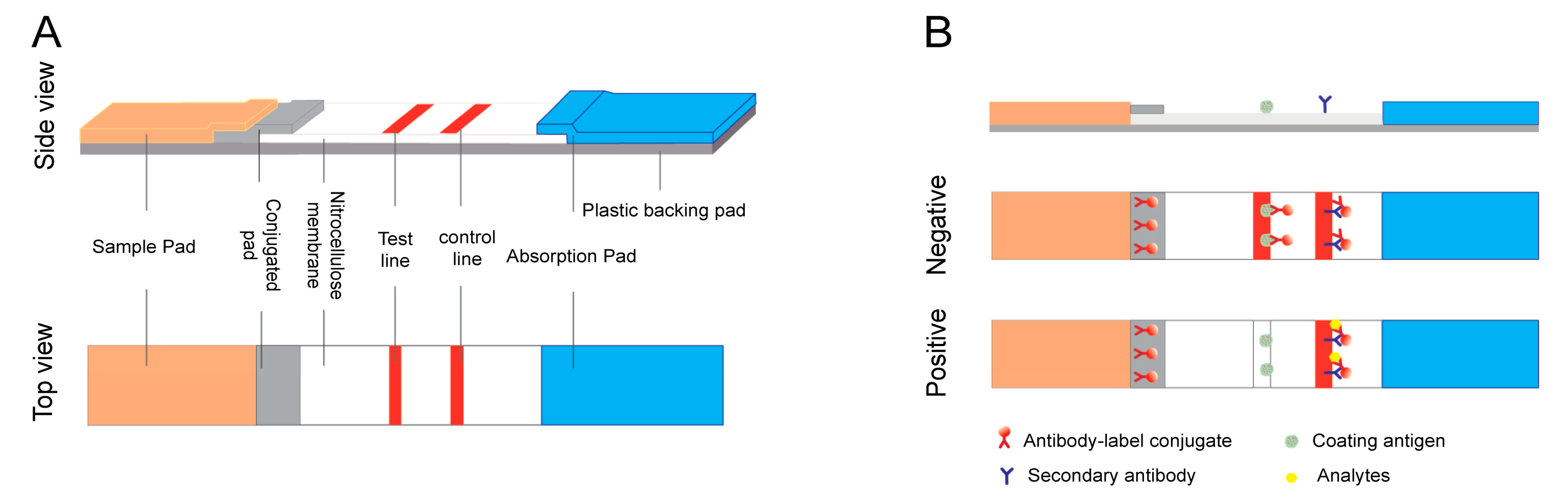
2.1. Sandwich Format

Sandwich assays are typically used for high molecular weight molecules or targets with multiple antigenic sites such as proteins and bacteria. Generally, this system uses a specific antibody pair, as the test line is coated with the unlabeled antibody, and the detected antibody is used to bind the signal label. When the sample is added to the test strip, the specific antibody reacts with the labeled antibody–target complex to form a clear test line through sample migration. The response detected in the test zone is directly proportional to the number of targets in the sample. The mixture then passes through the capture zone, where both the unbound and bound analytes bind to the capture antibody [22][23].

2.2. Competitive Format

Competitive assays are employed most often when testing low molecular weight analytes or when presenting a single antigenic determinant. For this format, the analyte–protein complex is immobilized on the test line, competing with the target in the sample to conjugate the labeled antibody. The response detected on the test line is negatively proportional to the amount of the target in the sample. Another specific antibody bound to the control line allows for the capture of excess antibody complex. Therefore, a band color appears in the control line regardless of the presence of the target analyte, confirming that the test was run correctly (Figure 2B) [22][23].
The test strip is a one-step procedure and the liquid sample to be analyzed is placed on the sample pad. All membrane pads are usually made with nitrocellulose. The reagent membrane comprises the immobilized specific antibodies and the labeled antibodies. With the addition of the sample, the reacting molecules are solubilized and combined with the expected metrics in the sample. Then, capillary action directs the fluid mixture to the reaction membrane [21].

3. Applications for Detecting β-Adrenergic Agonist Residues

β-adrenergic agonists are a group of synthetic phenethanolamine compounds, and according to their different aromatic ring structures they can be separated into three categories: aniline, phenol, and resorcinol [24]. LFA technology has been adequately developed to detect β-adrenergic agonists. As they are low molecular mass compounds, competitive assays should be used, the principles of which were described in Section 2 of this entry (2. LFA formats and principles). If the analyte is not present in the sample, then the labeled antibody will be caught by the bovine serum albumin (BSA)-analyte and immobilized on the membrane to form a clear test line with negative results; if the analyte is present in the sample exceeding the lower detectable concentration, it competes with the BSA-analyte immobilized on the test line and binds to the finite amount of labeled antibody, leading to an invisible test line with positive results. Published reports on LFA applications in this field are summarized in Table 1.
Table 1. Summary of the analytical performance of traditional paper-based LFAs for the detection of β-adrenergic agonists.
Analyte Label Assay Format Sample Assay Time LOD Reference
CLE 1 and RAC 3 AuNPs 2 Competitive LFA Swine urine 5 min 0.1 ± 0.01 ng/mL [25]
CLE AuNPs Competitive LFA Swine livers 10 min NA [26]
CLE AuNPs Competitive LFA Swine urine 10 min 3 ng/mL [27]
CLE AuNPs Competitive LFA Swine urine 10 min 0.1 ng/mL [28]
CLE, RAC, SAL 4 AuNPs Competitive LFA Swine urine 10 min 0.5 ng/mL [29]
CLE AuNPs Competitive LFA Swine urine 10 min 220 pg/mL [30]
SAL AuNPs Competitive LFA Swine urine 10 min 80 ng/mL [31]
RAC AuNPs Competitive LFA Swine urine 5 min 0.1 ng/mL [32]
SAL AuNPs Competitive LFA Meat and milk 10 min meat: 4.0 ng/g
milk: 3.0 ng/g
[33]
RAC AuNPs Competitive LFA Swine urine 45 min 0.13 ng/mL [34]
CLE SeNPs 5 Competitive LFA Swine urine / 3 ng/mL [35]
RAC and SAL SeNPs Competitive LFA Swine urine 5 min RAC: 1 ng/mL
SAL: 3 ng/mL
[36]
CLE and RAC SiNPs 6 Competitive LFA / 10 min CLE: 3 ng/mL
RAC: 2 ng/mL
[37]
CLE SiNPs Competitive LFA PBS, urine, and pork 10 min PBS: 3 ng/mL
urine: 6 ng/mL
pork: 5 ng/mL
[38]
Zilpaterol AuNPs Competitive LFA Feed 10 min 20 ng/g [39]
PA 7 AuNPs Competitive LFA Swine urine 10 min 0.188 ng/mL [40]
Clorprenaline AuNPs Competitive LFA Swine urine 3–5 min 0.104 ng/mL [41]
Clorprenaline AuNPs Competitive LFA Swine urine 9 min 0.15 ng/mL [42]
1 CLE: clenbuterol. 2 AuNPs: gold nanoparticles. 3 RAC: ractopamine. 4 SAL: salbutamol. 5 SeNPs: selenium nanoparticles. 6 SiNPs: silica nanoparticles. 7 PA: phenylethanolamine.
Various reagents can help visualize antigen–antibody interactions, but gold nanoparticles (AuNPs) are most frequently used in test strips. When many AuNPs aggregate, their color changes notably, and their application in colorimetric immunoassays is based on this principle [43]. AuNP-based LFAs have been used to detect β-adrenergic agonists. Zhang et al. [28] used AuNP-based LFA devices to detect clenbuterol in swine urine, as it was demonstrated that the limit of detection (LOD) for the device was 0.1 ng/mL when using the scanner and 1.0 ng/mL by eye, with the test completed within 10 min. In addition, Lai et al. [26] developed a lateral flow assay to detect CLE in swine livers. The results obtained when using this assay were compared to those of a commercial ELISA kit (the Ridascreen Clenbuterol Fast Kit [R1701, R-Biopharm, Darmstadt, Germany]), which is used to detect clenbuterol. Khamta et al. [31] developed a lateral flow assay to detect salbutamol. They used a salbutamol-BSA conjugate as the test line, goat antirabbit IgG as the control line, and an anti-SALB-colloidal gold conjugate (SALB-CGC) as the detector. The sensitivity to salbutamol in PBS is approximately 80 ng/mL. An AuNPs-based LFA was proposed by Liu et al. [32] to detect ractopamine (RAC) residues in swine urine. The LOD for the LFA strip, when using an optical density scanner, was 0.1 ± 0.013 ng/mL and the cut-off level was 1.0 ng/mL when observed with the naked eye. These data show that the lateral flow strip assay is a reliable screening method.
Zvereva et al. [33] proposed an indirect labeling method that could be applied with an LFA to detect SAL residues in food. The visual LOD was 4.0 ng/g for meat products and 3.0 ng/g for milk samples, and there was a high level of cross-reactivity with terbutaline (50%) and orciprenaline (10.5%). Xu et al. [44] prepared monoclonal antibodies against 12 β-adrenergic agonists (salbutamol, clenbuterol, brombuterol, clenpenterol, mabuterol, carbuterol, cimbuterol, mapenterol, pirbuterol, terbutaline, cimaterol, and clenproperol), which were applied to AuNPs-based LFA devices. The visual LOD values were 1, 1, 2.5, 2.5, 2.5, 2.5, 2.5, 5, 10, 10, 5, 10, and 10 ng/mL, respectively, and detection was completed within 5–10 min.
Quantitative detection is usually performed using the optical density (OD) of the signal detected in the test line [28][32]. However, the ODs in the test line also change with pH value, temperature, and reaction time. Therefore, test results determined solely by the strength of the test line will be inaccurate [30]. Li et al. [30] established a quantitative lateral flow immunoassay for the detection of CLE in porcine urine. The ODs of the test and control zones on the test strip were measured to compensate for the inherent factors of the strip and the influence of the sample matrix, with the LOD being as low as 220 pg/mL. Similarly, Ren et al. [34] used this method for the quantitative detection of ractopamine in swine urine.
LFA devices that use other colored NPs have been proposed for signal labeling. For example, an LFA device which used selenium nanoparticles (SeNPs) as labels was developed by Wang et al. [35] for the detection of CLE. The cut-off value for the visual detection of CLE in swine urine was 3 ng/mL. Wang et al. [36] also established a lateral flow immunoassay for the detection of RAC and SAL in swine urine using SeNPs as probes and simultaneously detected both on one test strip. The visual detection limits for RAC and SAL were 1 and 3 ng/mL, respectively. Yu et al. [37] used red and blue silica nanoparticles (SiNPs) as visible labels for the detection of CLE (red) and RAC (blue) for the first time, and their LODs were 3 and 2 ng/mL, respectively. Zhu et al. [38] proposed the use of a core-shell purple SiNP-based LFA for CLE screening via the naked eye, and the cut-off values were 3, 6, and 5 ng/mL in phosphate buffered saline, urine, and pork, respectively.
The sensitivity of these methods equals that of AuNPs-based methods. However, in recent years, researchers have developed more advanced LFA devices to detect β-adrenergic agonists on-site.

References

  1. Evans, T.; Chapple, N. The animal health market. Nat. Rev. Drug Discov. 2002, 1, 937–938.
  2. Cabello, F.C.; Godfrey, H.P. Even therapeutic antimicrobial use in animal husbandry may generate environmental hazards to human health. Environ. Microbiol. 2016, 18, 311–313.
  3. Mersmann, H.J. Overview of the effects of beta-adrenergic receptor agonists on animal growth including mechanisms of action. J. Anim. Sci. 1998, 76, 160–172.
  4. Lv, R.; Wu, E.; Wu, R.; Shen, W.; Ma, C.; Shi, R.; Guo, R.; Shao, M.; Liu, J. Sensitive detection of clenbuterol by hybrid iridium/silicon nanowire-enhanced laser desorption/ionization mass spectrometry. J. Mater. Chem. B 2020, 8, 7792–7800.
  5. Barbosa, J.; Cruz, C.; Martins, J.; Silva, J.M.; Neves, C.; Alves, C.; Ramos, F.; Noronha Da Silveira, M.I. MIN: Food poisoning by clenbuterol in Portugal. Food Addit. Contam. 2005, 22, 563–566.
  6. Kuiper, H.A.; Noordam, M.Y.; van Dooren-Flipsen, M.M.; Schilt, R.; Roos, A.H. Illegal use of beta-adrenergic agonists: European Community. J. Anim. Sci. 1998, 76, 195–207.
  7. Song, C.; Zhi, A.; Liu, Q.; Yang, J.; Jia, G.; Shervin, J.; Tang, L.; Hu, X.; Deng, R.; Xu, C.; et al. Rapid and sensitive detection of β-agonists using a portable fluorescence biosensor based on fluorescent nanosilica and a lateral flow test strip. Biosens. Bioelectron. 2013, 50, 62–65.
  8. Bulletins, M. The Catalogue of Drug Banned to Use in the Feed and Animal Drinking Water. 2002. Available online: https://www.fsis.usda.gov/sites/default/files/media_file/2021-03/China-176.pdf (accessed on 17 June 2022).
  9. Zhang, X.; Zhao, H.; Xue, Y.; Wu, Z.; Zhang, Y.; He, Y.; Li, X.; Yuan, Z. Colorimetric sensing of clenbuterol using gold nanoparticles in the presence of melamine. Biosens. Bioelectron. 2012, 34, 112–117.
  10. Wang, W.; Zhang, Y.; Wang, J.; Shi, X.; Ye, J. Determination of beta-agonists in pig feed, pig urine and pig liver using capillary electrophoresis with electrochemical detection. Meat Sci. 2010, 85, 302–305.
  11. Hu, Y.; Liu, R.; Li, Y.; Li, G. Investigation of ractopamine-imprinted polymer for dispersive solid-phase extraction of trace beta-agonists in pig tissues. J. Sep. Sci. 2010, 33, 2017–2025.
  12. Wang, P.-L.; Fan, L.; Su, X.-O.; Ye, Z.-H. Determination of four kinds of β-agonists in swine urine by molecularly imprinted solid phase extraction followed gas chromatography coupled mass spectrometry. Chin. J. Anal. Chem. 2012, 40, 470–473.
  13. Shao, B.; Jia, X.; Zhang, J.; Meng, J.; Wu, Y.; Duan, H.; Tu, X. Multi-residual analysis of 16 β-agonists in pig liver, kidney and muscle by ultra performance liquid chromatography tandem mass spectrometry. Food Chem. 2009, 114, 1115–1121.
  14. Bocca, B.; Fiori, M.; Cartoni, C.; Brambilla, G. Simultaneous determination of Zilpaterol and other beta agonists in calf eye by gas chromatography/tandem mass spectrometry. JAOAC Int. 2003, 86, 8–14.
  15. McPartlin, D.A.; O’Kennedy, R.J. Point-of-care diagnostics, a major opportunity for change in traditional diagnostic approaches: Potential and limitations. Expert Rev. Mol. Diagn. 2014, 14, 979–998.
  16. Zhu, X.; Sarwar, M.; Yue, Q.; Chen, C.; Li, C.-Z. Biosensing of DNA oxidative damage: A model of using glucose meter for non-glucose biomarker detection. Int. J. Nanomed. 2017, 12, 979–987.
  17. Wang, X.-L.; Wang, L.; Hasi, C.-L.; Wang, Y.-P.; Khan, A.; Ren, B.-Z.; Liu, Z.-Z.; Hou, S.-L.; Yang, L.-H.; Zhang, L.-Y.; et al. A rapid colloidal gold immunochromatographic assay for the diagnosis of coronavirus disease 2019. Chin. Med. J. (Engl.) 2020, 133, 1986–1988.
  18. Lin, L.-K.; Uzunoglu, A.; Stanciu, L.A. Aminolated and Thiolated PEG-Covered Gold Nanoparticles with High Stability and Antiaggregation for Lateral Flow Detection of Bisphenol A. Small 2018, 14, 1702828.
  19. Martinez, A.W.; Phillips, S.T.; Butte, M.J.; Whitesides, G.M. Patterned paper as a platform for inexpensive, low-volume, portable bioassays. Angew. Chem. Int. Ed. Engl. 2007, 46, 1318–1320.
  20. Clarke, O.; Goodall, B.; Hui, H.; Vats, N.; Brosseau, C. Development of a SERS-based rapid vertical flow assay for point-of-care diagnostics. Anal. Chem. 2017, 89, 1405–1410.
  21. Raeisossadati, M.J.; Danesh, N.M.; Borna, F.; Gholamzad, M.; Ramezani, M.; Abnous, K.; Taghdisi, S.M. Lateral flow based immunobiosensors for detection of food contaminants. Biosens. Bioelectron. 2016, 86, 235–246.
  22. Ngom, B.; Guo, Y.; Wang, X.; Bi, D. Development and application of lateral flow test strip technology for detection of infectious agents and chemical contaminants: A review. Anal. Bioanal. Chem. 2010, 397, 1113–1135.
  23. Chen, W.; Huang, Z.; Hu, S.; Peng, J.; Liu, D.; Xiong, Y.; Xu, H.; Wei, H.; Lai, W. Invited review: Advancements in lateral flow immunoassays for screening hazardous substances in milk and milk powder. J. Dairy Sci. 2019, 102, 1887–1900.
  24. Wang, R.; Zhang, W.; Wang, P.; Su, X. A paper-based competitive lateral flow immunoassay for multi β-agonist residues by using a single monoclonal antibody labelled with red fluorescent nanoparticles. Mikrochim. Acta 2018, 185, 191.
  25. Zhang, M.-Z.; Wang, M.-Z.; Chen, Z.-L.; Fang, J.-H.; Fang, M.-M.; Liu, J.; Yu, X.-P. Development of a colloidal gold-based lateral-flow immunoassay for the rapid simultaneous detection of clenbuterol and ractopamine in swine urine. Anal. Bioanal. Chem. 2009, 395, 2591–2599.
  26. Lai, W.H.; Fung, D.Y.; Xu, Y.; Xiong, Y.H. Screening procedures for clenbuterol residue determination in raw swine livers using lateral-flow assay and enzyme-linked immunosorbent assay. J. Food Prot. 2008, 71, 865–869.
  27. Lai, W.; Xu, Y.; Fung, D.Y.; Xiong, Y. Development of a lateral-flow assay for rapid screening of the performance-enhancing sympathomimetic drug clenbuterol used in animal production; food safety assessments. Asia Pac. J. Clin. Nutr. 2007, 16, 106–110.
  28. Zhang, G.P.; Wang, X.N.; Yang, J.F.; Yang, Y.Y.; Xing, G.X.; Li, Q.M.; Zhao, D.; Chai, S.J.; Guo, J.Q. Development of an immunochromatographic lateral flow test strip for detection of beta-adrenergic agonist Clenbuterol residues. J. Immunol. Methods 2006, 312, 27–33.
  29. Wu, Q.; Song, Q.; Wang, X.; Yao, L.; Xu, J.; Lu, J.; Liu, G.; Chen, W. Simultaneous Detection of Multiple β-Adrenergic Agonists with 2-Directional Lateral Flow Strip Platform. Anal. Sci. 2020, 36, 653–658.
  30. Li, C.; Luo, W.; Xu, H.; Zhang, Q.; Xu, H.; Aguilar, Z.P.; Lai, W.; Wei, H.; Xiong, Y. Development of an immunochromatographic assay for rapid and quantitative detection of clenbuterol in swine urine. Food Control. 2013, 34, 725–732.
  31. Khamta, Y.; Pattarawarapan, M.; Nangola, S.; Tayapiwatana, C. Development of immunochromatographic assay for the on-site detection of salbutamol. J. Immunoass. Immunochem. 2009, 30, 441–456.
  32. Liu, A.; Lin, J.; Dai, M.; Wu, Y.; Fang, J.; Zhang, M. Development of a monoclonal antibody-based immunochromatographic assay detecting ractopamine residues in swine urine. Food Analytical. Methods 2016, 9, 2016–2025.
  33. Zvereva, E.; Zherdev, A.; Xu, C.; Dzantiev, B. Highly sensitive immunochromatographic assay for qualitative and quantitative control of beta-agonist salbutamol and its structural analogs in foods. Food Control. 2018, 86, 50–58.
  34. Ren, M.L.; Chen, X.L.; Li, C.H.; Xu, B.; Liu, W.J.; Xu, H.Y.; Xiong, Y.H. Lateral flow immunoassay for quantitative detection of ractopamine in swine urine. Biomed. Environ. Sci. 2014, 27, 134–137.
  35. Wang, Z.; Jing, J.; Ren, Y.; Guo, Y.; Tao, N.; Zhou, Q.; Zhang, H.; Ma, Y.; Wang, Y. Preparation and application of selenium nanoparticles in a lateral flow immunoassay for clenbuterol detection. Mater. Lett. 2019, 234, 212–215.
  36. Wang, Z.; Zhou, Q.; Guo, Y.; Hu, H.; Zheng, Z.; Li, S.; Wang, Y.; Ma, Y. Rapid Detection of Ractopamine and Salbutamol in Swine Urine by Immunochromatography Based on Selenium Nanoparticles. Int. J. Nanomed. 2021, 16, 2059–2070.
  37. Chen, Y.; Huang, Z.; Hu, S.; Zhang, G.; Peng, J.; Xia, J.; Lai, W. Integrated immunochromatographic assay for qualitative and quantitative detection of clenbuterol. Anal. Biochem. 2019, 577, 45–51.
  38. Zhu, C.; Zhao, G.; Dou, W. Immunochromatographic assay using brightly colored silica nanoparticles as visible label for point-of-care detection of clenbuterol. Sens. Actuators B Chemical. 2018, 266, 392–399.
  39. Shelver, W.L.; Smith, D.J. Development of an immunochromatographic assay for the β-adrenergic agonist feed additive zilpaterol. Food Addit. Contam. Part A 2018, 35, 1519–1529.
  40. Dai, M.; Gong, Y.; Liu, A.; Zhang, L.; Lin, J.; Zhang, M.; Yu, X. Development of a colloidal gold-based lateral-flow immunoassay for the rapid detection of phenylethanolamine A in swine urine. Anal. Methods 2015, 7, 4130–4137.
  41. Jiang, W.; Zeng, L.; Liu, L.; Song, S.; Kuang, H. Development of an immunochromatographic assay for rapid detection of clorprenaline in pig urine. Food Agric. Immunol. 2018, 29, 536–547.
  42. Peng, T.; Zhang, F.S.; Yang, W.C.; Li, D.X.; Chen, Y.; Xiong, Y.H.; Wei, H.; Lai, W.H. Lateral-flow assay for rapid quantitative detection of clorprenaline residue in swine urine. J. Food Prot. 2014, 77, 1824–1829.
  43. Wang, Z.; Zhao, J.; Xu, X.; Guo, L.; Xu, L.; Sun, M.; Hu, S.; Kuang, H.; Xu, C.; Li, A. An Overview for the Nanoparticles-Based Quantitative Lateral Flow Assay. Small Methods 2022, 6, e2101143.
  44. Xu, X.; Xu, X.; Sun, L.; Wu, A.; Song, S.; Kuang, H.; Xu, C. An ultrasensitive colloidal gold immunosensor to simultaneously detect 12 beta (2)-adrenergic agonists. J. Chromatogr. B 2022, 1191, 123119.
More
Information
Contributors MDPI registered users' name will be linked to their SciProfiles pages. To register with us, please refer to https://encyclopedia.pub/register : , , , , , , , ,
View Times: 961
Revisions: 2 times (View History)
Update Date: 28 Jul 2022
1000/1000