Submitted Successfully!
To reward your contribution, here is a gift for you: A free trial for our video production service.
Thank you for your contribution! You can also upload a video entry or images related to this topic.
Version Summary Created by Modification Content Size Created at Operation
1 -- 4377 2022-07-14 02:13:37 |
2 format correct -15 word(s) 4362 2022-07-14 03:11:18 |

Video Upload Options

Do you have a full video?

Confirm

Are you sure to Delete?
Cite
If you have any further questions, please contact Encyclopedia Editorial Office.
Balistreri, C.R. Preserving Adult Endothelium Health and Reversing Its Dysfunction. Encyclopedia. Available online: https://encyclopedia.pub/entry/25118 (accessed on 26 June 2024).
Balistreri CR. Preserving Adult Endothelium Health and Reversing Its Dysfunction. Encyclopedia. Available at: https://encyclopedia.pub/entry/25118. Accessed June 26, 2024.
Balistreri, Carmela Rita. "Preserving Adult Endothelium Health and Reversing Its Dysfunction" Encyclopedia, https://encyclopedia.pub/entry/25118 (accessed June 26, 2024).
Balistreri, C.R. (2022, July 14). Preserving Adult Endothelium Health and Reversing Its Dysfunction. In Encyclopedia. https://encyclopedia.pub/entry/25118
Balistreri, Carmela Rita. "Preserving Adult Endothelium Health and Reversing Its Dysfunction." Encyclopedia. Web. 14 July, 2022.
Preserving Adult Endothelium Health and Reversing Its Dysfunction
Edit

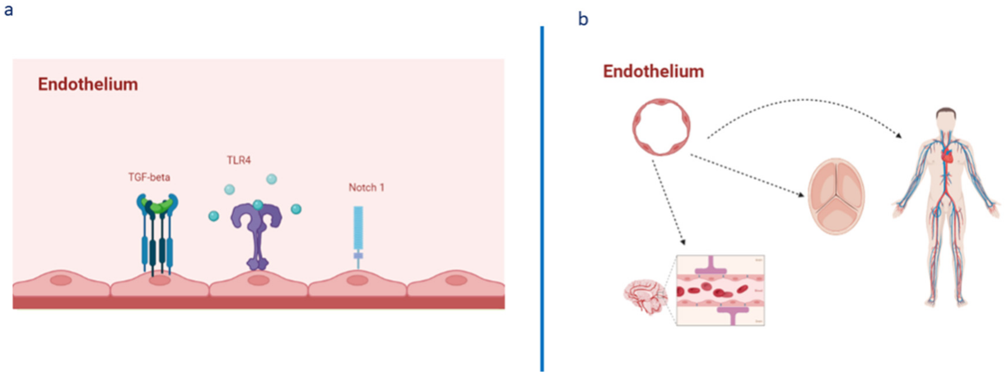
The endothelium has multiple functions, ranging from maintaining vascular homeostasis and providing nutrition and oxygen to tissues to evocating inflammation under adverse conditions and determining endothelial barrier disruption, resulting in dysfunction. Endothelial dysfunction represents a common condition associated with the pathogenesis of all diseases of the cardiovascular system, as well as of diseases of all of the other systems of the human body, including sepsis, acute respiratory distress syndrome, and COVID-19 respiratory distress. Such evidence is leading to the identification of potential biomarkers and therapeutic targets for preserving, reverting, or restoring endothelium integrity and functionality by promptly treating its dysfunction.

endothelium endothelium dysfunction newer omics technologies

1. Introduction

The term “endothelium” indicates the cellular layer that acts as barrier between blood flow and the inner lining of the vessel wall. Considered for a number of years as an inert vascular component, the endothelium is recognized today as the hub of the homeostasis of the entire cardiovascular system, including the heart valves, where endothelial cells (EC), defined as valvular ECs (VECs), can control the proliferation, differentiation, apoptosis, and remodeling of valvular interstitial cells (VICs) [1]. Thus, the endothelium represents a vital organ in the cardiovascular system and the principal element of the stroma of all body tissues, executing diverse regulatory activities thanks to the production and release of several endothelial-derived vasoactive factors, acting via endocrine, paracrine, or autocrine signaling. Accordingly, endothelial control includes not only the regulation of vascular tone, but also angiogenesis, inflammation, platelet aggregation, leukocyte adhesion, vascular cell proliferation, and oxidative stress [2][3][4][5][6] (see Figure 1). Such essential functions of the endothelium are already marked during fetal life. Such essential functions of the endothelium are already marked during fetal life. In the fetus, the endothelium has the fundamental role of ensuring the correct ontogenesis of immune cells (i.e., tissue-resident macrophages, microglia cells, and epidermal γδ T cells) and cells of the hematopoietic system, as well as the development of other systems, such as the nervous and cardiovascular systems [6][7][8].
Figure 1. Architecture, EC pathways, and their heterogeneous presence and complexity in various body tissues. (a) Endothelium architecture and expression of some crucial pathways involved in its homeostasis, as well as in its activation, determining changes in the expression of specific genes and in the consequent phenotypic EC changes related to the evocation of dysfunction, caused by inflammation and endothelial barrier disruption (increased vascular permeability, edema formation, release of proinflammatory cytokines, and leukocyte extravasation). (b) The endothelium as an essential element of not only of the entire cardiovascular system, but also as the “heart” of all the tissues of the body, consequently contributing to their physiological homeostasis and function.
Consistent with these observations, the current evidence reports a close relationship between biochemical or structural injury, altered genesis, homeostasis, and function of the endothelium, and the onset and progression of pathological conditions correlated to inflammation, thrombosis, and atherosclerosis, as well as alterations in the vascular resistance and blood pressure [2][4][9][10][11]. Consequently, endothelial dysfunction is assumed as the axis of activation of all pathophysiological mechanisms and related disease pathways linked to excessive morbidity and mortality, such as hypertension, myocardial infarction, aneurysm, stroke, diabetes, metabolic syndrome, rheumatic diseases, renal failure, neurodegenerative diseases, cancer, and inflammatory diseases [2][4][5][8][9][11][12][13][14][15][16][17][18][19][20] (see Figure 2). For instance, tumor neovascularization is characterized by a highly disordered vascular endothelium network and the generation of new blood vessels. In addition, the abovementioned pathologies are characterized as coexisting in patients with a median age of about 65 years old and over by increasing the risk of adverse events [21][22]. Furthermore, a significant association between typical EC plasma proteins and individual risk of cardiovascular diseases (CVD), detected using the Framingham risk score, has also been evidenced in recent studies confirming the close relationship between CVD onset and endothelium dysfunction, as well as corresponding biomarker profiles [23]. Interestingly, alterations in the vascular endothelium have also been associated with organ dysfunction in patients affected by severe SARS-CoV-2 infection, belonging to the 70–89 age group [24][25][26][27] (see Figure 2).
Figure 2. Endothelium dysfunction and its relationship with human diseases.
Endothelial dysfunction constitutes a common pathological condition, which accompanies the aging process of all the tissues, organs, and systems, and the development of cardiovascular diseases including atherosclerosis, thrombosis, hypertension, aneurysms, valve diseases, but also of cancer, onco-haematology diseases, diabetes, and neurodegenerative disorders, as well as SARS-CoV-2 infection and related complications.
Considering these observations, it is of crucial importance to develop strategies with the aim of not only warding off aggressive agents or risk factors for preventing endothelium dysfunction but also identifying therapies which can reverse or mitigate the damaging effects generated by such conditions. Certainly, regenerative strategies via induced pluripotent stem cells (IPSCs) and direct reprogramming targeting ECs via transcription factors (TFs) involved in endothelial cell fate are optimal examples. TFs, such as OCT4, SOX2, KLF4, c-MYC, ETV2, and ERG, have recently become the subject of many studies, and the direct reprogramming strategy appears to be promising for the treatment of diseases such as CVD. However, limitations in their efficiency linked principally to epigenetic resistance and the methods used have been evidenced [28] (see Figure 3). Thus, the clinical applicability and translational nature of such strategies do not appear be evident. Here, attention is focused on new promising methodologies and biological matrices for use in achieving the suggested objectives. Accordingly, the use of blood liquid biopsy and omics technologies is described in the next section, in addition to their application in studies on endothelial dysfunction aimed at identifying biomarkers and emerging therapeutic targets.
Figure 3. Some strategies for obtaining mature EC.

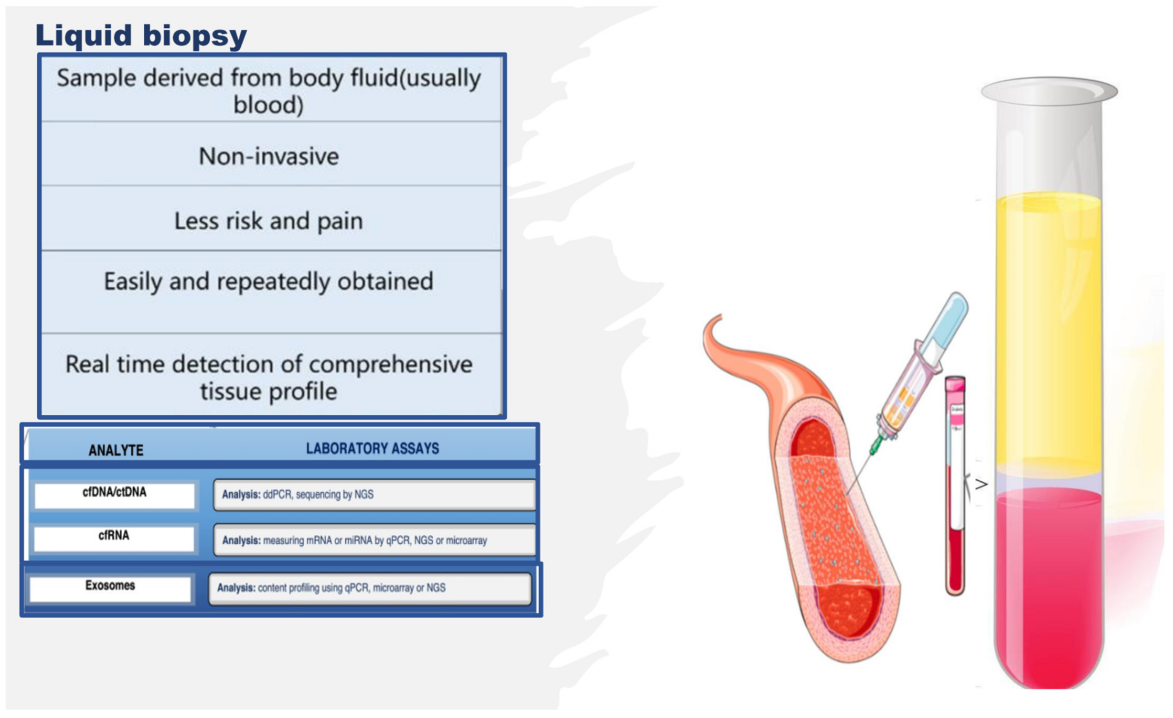
2. Omics Technologies and Liquid Biopsy as Emerging Tools for Identifying Endothelial Biomarkers and Therapeutic Targets

In proposing new strategies for preserving the functionality and efficiency of adult endothelium tissue, it is imperative to identify molecules as optimal biomarkers and therapeutic targets. Biomarkers represent crucial indicators of both physiological and pathological processes, such as endothelium dysfunction. Specific changes in molecular and cellular mechanisms of physiological processes result in biochemical alterations at both the tissue and the systemic level, which can provide comprehensive information regarding the health status of tissues (the endothelium), as well as the nature of any pathological tissue dysfunction and disease. In addition, any disease biomarker should be specific and reliable, able to distinguish between the physiological condition of a tissue, organ, or system and a dysfunctional condition or disease, and diverse across various diseases or their subgroups and phenotypes. Accordingly, biomarkers can predict the risk of disease, facilitate early diagnosis, and enable setting guidelines for the development of new therapies for treating diseases. New-generation omics technologies and their platforms, including next-generation sequencing (NGS), epigenomics, proteomics, metabolomics, and microbiomics [29][30] (see Figure 4), can detect molecules involved in the close relationship between genotype and the environment, nutritional habits, and endothelial health or dysfunction, at an unprecedented grade of importance and resolution [31][32][33]. They also allow for obtaining an enormous quantity of data, albeit accompanied by consequent computational difficulties in their interpretation [31][32][33]. Today, such a challenge has been resolved thanks to potent bioinformatics algorithms, which can easily interpret and combine big data, by offering the possibility of both analyzing them at diverse multilevel networks and identifying genes, proteins, metabolites, and microbiomes, as well as their interactions in the complex human interactome, which can be used as emerging biomarkers and drug targets [31][32][33] (see Figure 4). Such a strategy exemplifies network medicine with the feature of combining molecular and bioinformatic data, which can improve current medical practices in the management of endothelial dysfunction-related pathologies by overcoming the limits of the obsolete points of view regarding endothelial dysfunction, considered the direct consequence of a single molecular alteration. An example of such an approach is machine learning (see Figure 4). The Abasht group [34] used this approach to evaluate vascular endothelial dysfunction in wooden breast syndrome, while the Treder group [35] used this approach to detect graft detachment in Descemet membrane endothelial keratoplasty. Furthermore, it is currently being applied to develop algorithms for vascular endothelial senescence, as recently evidenced by Sun and Feinberg [36].
Figure 4. Network medicine and its features in endothelium studies.
Furthermore, omics technologies enable evaluating changes at a single-cell level, i.e., the evaluation of gene expression profiles of the endothelium at the transcriptome and epigenome level, when exposed to one or more stressors, such as disturbed blood flow, high-glucose status, or inflammation. Specifically, it is possible to evaluate the transcriptomic and chromatin accessibility variations at the resolution of a single-EC. This allows comparing the effects of stressors in healthy ECs vs. stressed ECs in the same vessel or in diverse vessels, thereby following the changes in physiological status of the endothelium as a result of dysfunction or other conditions, e.g., hyperplasia (see below). A similar study design was recently adopted by Kumar and coworkers [37] to evaluate the gene expression profile in ECs isolated, via an innovative protocol, from the lumen of carotid arteries from mice subjected to a partial carotid ligation (PCL), which induces disturbed blood flow (d-flow) in the left carotid artery, while maintaining stable laminar flow in the right carotid artery, used as a control. Such a mouse PCL model allows comparing the effects induced in ECs by blood flow under disturbed conditions or with stable laminar flow (s-flow). ECs were obtained from the carotid arteries by dissection 2 days or 2 weeks post PCL surgery, before subjecting their lumen to collagenase digestion to acquire endothelial-enriched single cells or single nuclei. These single-cell and single-nuclei preparations were utilized for RNA preparation, library generation, single-cell RNA sequencing (scRNA-seq), and single-cell transposase-accessible chromatin sequencing (scATACseq), which were used to determine the transcriptomic and chromatin accessibility changes. The data obtained consequently allowed evaluating the genes expressed under the effects induced by d-flow or s-flow at a single-cell level, resulting in the activation of proatherogenic or physiological responses. These proatherogenic genes and their molecules were then suggested as targets for developing therapies. Specifically, it was confirmed that Krüppel-like factors 4 and 2 (KLF4/KLF2) act as s-flow-sensitive transcription factor (TF)-binding sites. The identified TFs sensitive to d-flow were RELA, AP1, STAT1, and TEAD1, which were suggested to induce d-flow EC reprogramming from an athero-protective to a proatherogenic phenotype, including endothelial–mesenchymal transition [37][38]. Furthermore, thanks to the integration of diverse omics assays, including the acetylation of histone 3 lysine 27 (H3K27ac) ChIP-seq (chromatin immunoprecipitation followed by high-throughput sequencing), ATAC-seq (an assay for transposase-accessible chromatin sequencing), and RNA-seq (RNA-sequencing), it was possible to detect the genome-wide epigenetic regulations in ECs in response to athero-protective pulsatile shear stress (PS) [39]. The combined data obtained showed that inositol 1,4,5-trisphosphate receptor 3 (ITPR3) was upregulated under PS conditions via KLF4 or statins, in ECs isolated from mouse aorta, lung ECs isolated from EC-KLF4-transgenic versus EC-KLF4-knoukout mice, and atorvastatin-treated ECs. The use of KLF4 ATAC-qPCR (quantitative polymerase chain reaction) and ChIP-qPCR also led to the identification of a specific locus in the promoter region of the ITPR3 gene indispensable for KLF4 binding, H3K27ac enrichment, chromatin accessibility, RNA polymerase II recruitment, and ITPR3 transcriptional activation [39]. By evocating the deletion of KLF4 binding locus in EC thanks to the use of a clustered regularly interspaced short palindromic repeats-associated protein 9 (CRISPR/Cas9) system, a blunted calcium influx, reduced expression of endothelial nitric oxide synthase, and diminished nitric oxide bioavailability were evidenced. In the complex, such interesting results obtained thanks to novel multi-omics investigations suggested that KLF4 is crucial for PS-modulated H3K27ac, which allows the transcriptional activation of ITPR3. Such a mechanism contributes to Ca2+-dependent endothelial nitric oxide synthase (eNOS) activation and EC homeostasis, and these molecules are potential candidates for biomarkers and therapeutic targets [39].
Another group integrated investigations of in vivo epigenomic mapping with conditional knockout, gene transfer, and pharmacology with the aim of studying, in rodent models, the endothelial hyperplasia/proliferation (IH), which represents the primary etiology of vascular stenosis. The data from injured (IH-prone) rat arteries demonstrated a rise in genome-wide occupancy by histone 3 lysine 27 trimethylation (H3K27me3), a gene-repression indicator. This result was unexpected when considering the traditional point of view on prevailing post-injury gene activation rather than repression. Additional evaluation has also demonstrated a shift in H3K27me3 enrichment to anti-proliferative genes from pro-proliferative genes, characterized by gene activation via H3K27ac (acetylation) accumulation. H3K27ac and its reader BRD4 (bromodomain protein) showed co-enrichment of Ezh2, while conditional BRD4 knockout in injured mouse arteries resulted in a decrease in H3K27me3 and its writer EZH2, which positively regulates another pro-IH chromatin modulator, UHRF1. Thus, these results reveal an injury-induced locus-specific H3K27me3 redistribution in the epigenomic landscape involving BRD4 → EZH2 → UHRF1 hierarchical regulation, while suggesting that these players may be pharmaceutical targets [40].
Another interesting study was conducted in 2021 by a group in Napoli examining the glucose-dependent and dose-responsive alterations in endothelial DNA methylation to detect a putative epigenetic mechanism underlying diabetic vasculopathy. Specifically, they discovered the disproportionate glucose-dependent methylation and gene expression of VEGF and NO signaling cascades, constituting a physiological imbalance known to evocate endothelial dysfunction in diabetes. Consequently, they assumed that epigenetic mechanisms encode glycemic memory within EC, and the pathways involved can be used as therapeutic targets [41]. Specifically, their novel evidence demonstrated that hyperglycemia triggers dose-responsive variations in DNA methylation dynamics, affecting key physiological processes involved in the maintenance of endothelial function, including a glucose-dependent physiologic uncoupling of VEGF and NO signaling, which causes endothelial dysfunction.
Additionally, recent advances in molecular genetics and omics technologies also showed significant molecular heterogeneity within brain endothelial and perivascular cell types [42]. The blend of these conventional and modern approaches has enabled detecting phenotypical variations between healthy and abnormal conditions at the single-cell level. Accordingly, a deep understanding of brain vascular cell states during physiological, pathological, and aging processes has rapidly emerged, and therapeutic approaches have been developed, including vascularization models of human brain organoids on a chip (widely quoted in [42]), self-assembling multicellular blood–brain barrier (BBB) spheroids (widely quoted in [42]), and improved endothelial BBB differentiation protocols using human pluripotent or induced pluripotent stem cells (widely quoted in [8][42]). Human organoid and cell reprogramming technologies have rapidly improved, allowing the simulation of human brain development and disease modeling with the construction of a functional vasculature. These in vitro models will become potent instruments for exploring human-specific vascular traits absent in animal models, for investigating drug delivery across the BBB, and for screening drugs targeting BBB dysfunction in neurological diseases. Consequently, this research recently led researchers to hypothesize a close relationship between endothelial BBB conditions and neurodegenerative diseases (widely quoted in [8][42]).
Therefore, the literature is seeing an increase in the number of multi-omics investigations on endothelium, and the studies reported above constitute only some examples. However, many of these studies were complex with regard to their design, often invasive or necessitating the sacrifice of animals, along with an intricate approach to collecting the biological study samples and the related costs. Alternatively, studies on other types of samples, whose collection attracts a minor cost and a simple technique, are growing in number. Indeed, studies aimed at identifying noninvasive indicators of endothelial tissue status, simply using liquid biopsy, are increasing. Liquid biopsy is attractive as it facilitates the collection of an optimal biological sample, thereby enabling the extrapolation of big data via multi-omics technologies [43][44][45]. Liquid biopsy consists of an easy sampling of diverse bodily fluids, principally including blood, saliva, and urine, for the evaluation of various potential circulating noninvasive biomarkers [43][44][45]. As a biopsy, it also enables obtaining circulating intact cells, cell-free nucleic acids, circulating epigenetic-sensitive molecules (methylated DNA, modified histones, and noncoding RNAs), circulating metabolites, and other cell products, such as micro-vesicles and exosomes [43][44][45] (see Figure 5). However, unlike tissue biopsy, liquid biopsy offers the advantage of colleting heterogeneous cellular phenotypes by giving information on many circulating cell types and their products at specific times, thereby allowing the real-time monitoring of the evolution of a pathological condition or of the switching of the endothelium from physiological to dysfunctional [46]. In addition, emerging NGS platforms executed on body fluid samples, principally blood, can detect novel circulating noninvasive biomarkers, which may further the development of precision medicine and personalized therapy in the field of endothelium dysfunction-related pathologies, by preserving the health of the endothelium or reverting its alterations [43][44][45][46]. Furthermore, liquid biopsy shows several benefits compared to tissue biopsy; it is (a) noninvasive with negligible pain and risk, (b) more quickly executed, (c) easier to collect, (d) able to provide a real-time systemic profile, (e) able to give spatiotemporal information, and (f) able to monitor physiological or pathological conditions over time.
Figure 5. Liquid biopsy, describing some components and laboratory analysis techniques. Liquid biopsy involves the collection and analysis of diverse components from peripheral blood samples, including cell-free nucleic acids (cfDNA/ctDNA and cfRNA) and exosomes. cfDNA: circulating free DNA, ctDNA: circulating tumor DNA, cfRNA: cell-free RNA, CTCs: circulating tumor cells, TEPs: tumor-educated platelets, NGS: next-generation sequencing, qPCR: quantitative polymerase chain reaction.
As mentioned above, fluid-based assays may provide noninvasive indicators that indicate the endothelium status or allow sequential monitoring of the evolution of dysfunction. Interesting evidence can be gained by assessing the number, function, and senescence grade of endothelial progenitor cells (EPCs), as well as by evaluating the effects induced by stem-cell therapies (i.e., human umbilical cord-derived mesenchymal stem cells (MSCs)) or by detecting the quantity and quality of circulating metabolites and other cell products, such as micro-vesicles and exosomes or molecules able to modulate the gene expression at transcriptional, post-transcriptional, and translational levels, including circular RNAs (circRNAs) and ADAR enzymes. The next section reports the description of some of these metabolites and cell products, stressing their emerging potential as biomarkers and therapeutic targets of endothelium dysfunction, as well as their related limitations.

3. Endothelial Progenitor Cells (EPCs) as Potential Biomarkers of Endothelium Dysfunction and Therapeutic Agents

Circulating EPCs represent the progenitors of ECs, and they originate from bone marrow (BM)-derived hematopoietic stem cells (HSCs) [47][48][49]. EPCs constitute a real resource of ECs, thus maintaining vascular and tissue homeostasis and appropriate oxygen regulation and transport. Accordingly, EPCs represent a reservoir of circulating cells able to target injury sites, restore endothelium integrity, and enable physiological activities [47][48][49]. The impact of EPCs in the vascularization process has been proven in both animal models and humans. Such evidence has also led to the assumption that reductions in EPC circulating number and/or alterations in their functions related to different causes could impact endothelium function and architecture, as well as the onset and complications of endothelium dysfunction and, consequently, the survival of affected persons. Increased or decreased circulating EPC levels, as well as alterations in their function (for a detailed description, see reference [48]), have indeed been associated with vascular endothelium aging and diverse endothelium dysfunction pathologies, including coronary artery disease, stroke, diabetes, systemic sclerosis, autoimmune disorders, and aneurysms [50][51][52][53][54][55][56][57][58]. A group, for instance, recently evidenced that subjects affected by bicuspid aorta valve syndrome show a significant decrease in both the tissue and circulating levels of the Notch pathway, as well as a decrease in blood EPC number, compared to subjects with a tricuspid physiological valve, whether in the presence or absence of aorta aneurysm (AAA) [51][59]. In addition, using both blood and human tissue aorta samples, that the unique independent risk predictors for vascular aging are age and a reduced EPC number, as well as reduced EPC migratory activity and senescence, mediated by high SA-β-Gal activity and high levels of TP53, p21, p16, and inflammatory genes [59]. Furthermore, it has been reported in another study [60] that a decreased circulating number of EPCs, associated with increased RDW values, augmented blood levels of high-sensitivity C-reactive protein, and reduced mean values of both leukocyte telomere length and telomerase activity, can predict both vascular aging and ascending aorta aneurysm (AAA) onset and prognosis. Consequently, it can be affirmed that these factors might be used as an ideal biomarker profile for vascular aging, as well as for the diagnosis and outcome of sporadic AAA.
Such evidence might be of clinical relevance, and possible new recommendations and preventive measures might be applied. Accordingly, Hill and colleagues [61] already showed that the number of circulating EPCs represents a better predictor of vascular reactivity than conventional cardiovascular risk factors. Furthermore, a significant correlation between in vitro EPC senescence and endothelium dysfunction pathologies risk was also detected in blood donors [59]. Consequently, EPCs might be considered as an optimal predictive biomarker, as well as a diagnostic and prognostic biomarker of endothelium dysfunction and related pathologies. Certainly, ulterior studies are needed. However, such evidence has also led to the use of EPCs as therapeutic agents in the autologous or heterologous treatment of several endothelium dysfunction diseases, as well as in clinical trials, despite contradictory results being obtained. In particular, a favorable improvement in left-ventricular (LV) function in a rat model of myocardial infarction (MI) after intravenous injection of ex vivo expanded human CD34+ cells was reported [47][48][49]. Another study examined the effect of catheter-based intramyocardial transplantation in a swine model of MI, providing encouraging outcomes in favoring the application of EPCs as a potential cell therapy in clinical trials [47][48][49]. Naruse and colleagues conducted a study to assess the therapeutic treatment of diabetic neuropathy using in vivo expanded human EPC and streptozotocin-induced diabetic nude rats [62]. They observed numerous micro-vessels at the site of EPC injection [63]. Another group evidenced an improvement in neurological functions in chronic cerebral ischemic rats injected with CD34+ HSC cells, including EPCs [63]. The ability of EPCs to expand in culture under in vitro conditions represents another barrier for their therapeutic use. Genetically modified and ex vivo expanded EPCs may become promising agents able to appropriately rescue the impaired neovascularization process under disease conditions. In a rhesus model, ex vivo CD34+ cell transfection with recombinant non-replicative herpes virus vector and subsequent cell transplantation resulted in the expression of vector genes in angiogenic areas of skin autografts of rhesus macaques. Since CD34+ cells possess a natural angiogenic tropism to the injured endothelium, they may serve as ideal candidates for the delivery of genes into areas of angiogenesis [64]. These results encouraged the execution of clinical trials for evaluating the potential of EPCs to enhance endothelial integrity and vascularization at ischemia sites in patients with CVDs. Three different strategies were mainly applied: (a) the administration of granulocyte-colony stimulating factor (G-CSF) for verifying the recruitment of the patient’s own bone morrow resident progenitors, with two preliminary studies demonstrating increased LV function [65]; (b) the intracoronary infusion of BM progenitor cells in patients with MI, which demonstrated positive effects on LV function in three smaller studies [47][48][49]. Subsequently, two prospective large trials assessed significant LV function after 4–6 months of administration of BM progenitor cells. Ten recent and large trials confirmed the success and safety of this procedure with a follow-up over 1.5 years (widely quoted in [47][48][49]). In addition, intramyocardial and intracoronary administration has recently been suggested as a suitable strategy for the treatment of patients with refractory angina (widely quoted in [47][48][49]); (c) a more invasive strategy involving the direct injection of cells into target tissues [59]. This treatment (specifically, transepicardial or transendocardial injection of unfractioned BM cells) has been performed in patients with diffuse coronary artery disease and intractable angina with no option of recanalization. Ventricular function and physical capacity were observed to increase, but the small sample size of these studies necessitates confirmation in larger studies (widely quoted in [47][48][49]).
Studies using autologous cell therapy are also interesting. Accordingly, Yamamoto’s group executed an intramuscular injection of autologous BM-derived mononuclear cells containing 1% CD34+ cells in patients with chronic limb ischemia [66]. They quantitatively assessed the expression of EPCs and endothelial markers (i.e., CD133 and VE-cadherin) before the experiment and after the injection. Before investigation, the transcription of these molecules was undetectable. Autologous injection determined an elevation of EPC marker transcription. Thus, they established that autologous BM cells may be used in the therapy of patients with arterial diseases. A replication of these results was obtained by Lenk and colleagues [67]. Erbs and colleagues also used this autologous treatment in patients who underwent recanalization of chronic coronary total occlusion [68]. Autologous treatment with EPCs, expanded for 4 days in endothelium growth medium, improves coronary endothelium function and wall motion abnormalities, and it has a beneficial effect on metabolism in the target area in patients with symptomatic coronary atherosclerosis.
Despite these promising data, the clinical application of EPCs in exogenous or autologous cell therapy remains unclear for different reasons. Accordingly, the validity of their results is influenced by different factors: (a) the insignificant number of patients in these studies; (b) their missing randomization; (c) participation of a limited number of centers; (d) the imprecise phenotypic features of EPC utilized in the treatments, (e) the different methods of administration applied, and (f) the security and viability of the treatments [47][48][49]. Teratoma formation, immunoreactivity, or arrhythmias constitute the major unfavorable effects which have been detected after such treatments. In addition, other limitations have not been considered, including the limited number of EPCs in circulation, which usually requires their cultured expansion in a sufficient number of subpopulations from peripheral blood. The in vitro quantification of progenitor cells to obtain a quantity sufficient to utilize in therapeutic treatment can be modulated by phenotypic changes during their differentiation in vitro with the risk of obtaining senescent cells, requiring artificial cell pre-activation or stimulation for this approach. In addition to such evidenced limitations, another aspect influencing the results of this study is the lack of standardized criteria and a consensus for defining, characterizing, and identifying EPCs with well-established surface biomarkers, protocols, and methodologies. Consequently, other investigations are imperative [47][48][49].

Other Candidates for Therapeutic Agents of Endothelium Dysfunction

Promising evidence has recently been reported on the great potential of human umbilical cord-derived MSCs (hucMSCs) in repairing diabetic vascular endothelial damage, by applying assays of resazurin staining, MTT cell viability, wound healing, transwell migration, and Matrigel tube formation on human umbilical vein endothelial cells (HUVECs), and by evaluating how hucMSCs work through the assessment of RNA sequencing (RNA-seq) and molecular experiments [69]. Specifically, the use of a conditioned medium of hucMSCs (MSC-CM) revealed that hucMSCs improved the cell viability, wound healing, migration, and angiogenesis of HUVECs damaged by high glucose via paracrine signaling, and the altered gene expressions of IL-6, TNF-α, ICAM-1, VCAM-1, BAX, P16, P53, and ET-1 were significantly restored by MSC-CM. RNA-seq incorporated with real-time PCR and Western blot clarified that high glucose activates MAPK/ERK signaling in HUVECs, while MSC-CM reverses the abnormal phosphorylation of ERK and overexpression of MKNK2, ERBB3, MYC, and DUSP5 in the MAPK/ERK signaling pathway [69]. Certainly, other studies are needed.

References

  1. Inai, K. Chapter 17: Atrioventricular Valve Abnormalities: From Molecular Mechanisms Underlying Morphogenesis to Clinical Perspective. In Etiology and Morphogenesis of Congenital Heart Disease: From Gene Function and Cellular Interaction to Morphology ; Nakanishi, T., Markwald, R.R., Baldwin, H.S., Keller, B.B., Srivastava, D., Yamagishi, H., Eds.; Springer: Tokyo, Japan, 2016.
  2. Vita, J.A. Endothelial Function. Circulation 2011, 124, e906–e912.
  3. Sabe, S.A.; Feng, J.; Sellke, F.W.; Abid, M.R. Mechanisms and clinical implications of endothelium-dependent vasomotor dysfunction in coronary microvasculature. Am. J. Physiol. Circ. Physiol. 2022, 322, H819–H841.
  4. Boulanger, C.M. Endothelium. Arterioscler. Thromb. Vasc. Biol. 2016, 36, e26–e31.
  5. Balistreri, C.R.; Madonna, R.; Melino, G.; Caruso, C. The emerging role of Notch pathway in ageing: Focus on the related mechanisms in age-related diseases. Ageing Res. Rev. 2016, 29, 50–65.
  6. Balistreri, C.R.; Garagnani, P.; Madonna, R.; Vaiserman, A.; Melino, G. Developmental programming of adult haematopoiesis system. Ageing Res. Rev. 2019, 54, 100918.
  7. Allegra, A.; Giarratana, R.M.; Scola, L.; Balistreri, C.R. The close link between the fetal programming imprinting and neurodegeneration in adulthood: The key role of “hemogenic endothelium” programming. Mech. Ageing Dev. 2021, 195, 111461.
  8. Schirò, G.; Balistreri, C.R. The close link between brain vascular pathological conditions and neurodegenerative diseases: Focus on some examples and potential treatments. Vasc. Pharmacol. 2021, 142, 106951.
  9. Pober, J.S.; Min, W.; Bradley, J.R. Mechanisms of Endothelial Dysfunction, Injury, and Death. Annu. Rev. Pathol. Mech. Dis. 2009, 4, 71–95.
  10. Regina, C.; Panatta, E.; Candi, E.; Melino, G.; Amelio, I.; Balistreri, C.R.; Annicchiarico-Petruzzelli, M.; Di Daniele, N.; Ruvolo, G. Vascular ageing and endothelial cell senescence: Molecular mechanisms of physiology and diseases. Mech. Ageing Dev. 2016, 159, 14–21.
  11. Madonna, R.; Novo, G.; Balistreri, C.R. Cellular and molecular basis of the imbalance between vascular damage and repair in ageing and age-related diseases: As biomarkers and targets for new treatments. Mech. Ageing Dev. 2016, 159, 22–30.
  12. Wang, Y.-F.; Hsu, Y.-J.; Wu, H.-F.; Lee, G.-L.; Yang, Y.-S.; Wu, J.-Y.; Yet, S.-F.; Wu, K.K.; Kuo, C.-C. Endothelium-Derived 5-Methoxytryptophan Is a Circulating Anti-Inflammatory Molecule That Blocks Systemic Inflammation. Circ. Res. 2016, 119, 222–236.
  13. Miura, S.; Morimoto, Y.; Furihata, T.; Takeuchi, S. Functional analysis of human brain endothelium using a microfluidic device integrating a cell culture insert. APL Bioeng. 2022, 6, 016103.
  14. Jalnapurkar, S.; Landes, S.; Wei, J.; Mehta, P.K.; Shufelt, C.; Minissian, M.; Pepine, C.J.; Handberg, E.; Cook-Wiens, G.; Sopko, G.; et al. Coronary endothelial dysfunction appears to be a manifestation of a systemic process: A report from the Women’s Ischemia Syndrome Evaluation—Coronary Vascular Dysfunction (WISE-CVD) study. PLoS ONE 2021, 16, e0257184.
  15. Meilhac, A.; Cautela, J.; Thuny, F. Cancer Therapies and Vascular Toxicities. Curr. Treat. Options Oncol. 2022, 23, 333–347.
  16. Camilli, M.; La Vecchia, G.; Lillo, R.; Iannaccone, G.; Lamendola, P.; Montone, R.A.; Hohaus, S.; Aspromonte, N.; Massetti, M.; Lanza, G.A.; et al. Cardiovascular involvement in patients affected by multiple myeloma: A comprehensive review of recent advances. Expert Rev. Hematol. 2021, 14, 1115–1128.
  17. Love, K.M.; Barrett, E.J.; Malin, S.K.; Reusch, J.E.B.; Regensteiner, J.G.; Liu, Z. Diabetes pathogenesis and management: The endothelium comes of age. J. Mol. Cell Biol. 2021, 13, 500–512.
  18. Trejo-Lopez, J.A.; Yachnis, A.T.; Prokop, S. Neuropathology of Alzheimer’s Disease. Neurotherapeutics 2021, 19, 173–185.
  19. Olivieri, F.; Prattichizzo, F.; Grillari, J.; Balistreri, C.R. Cellular Senescence and Inflammaging in Age-Related Diseases. Mediat. Inflamm. 2018, 2018, 9076485.
  20. Peng, H.; Wang, X.; Du, J.; Cui, Q.; Huang, Y.; Jin, H. Metabolic Reprogramming of Vascular Endothelial Cells: Basic Research and Clinical Applications. Front. Cell Dev. Biol. 2021, 9, 626047.
  21. Jaul, E.; Barron, J. Characterizing the Heterogeneity of Aging: A Vision for a Staging System for Aging. Front. Public Health 2021, 9, 513557.
  22. Pisano, C.; Polisano, D.; Balistreri, C.; Altieri, C.; Nardi, P.; Bertoldo, F.; Trombetti, D.; Asta, L.; Ferrante, M.; Buioni, D.; et al. Role of Cachexia and Fragility in the Patient Candidate for Cardiac Surgery. Nutrients 2021, 13, 517.
  23. Iglesias, M.J.; Kruse, L.D.; Sanchez-Rivera, L.; Enge, L.; Dusart, P.; Hong, M.-G.; Uhlén, M.; Renné, T.; Schwenk, J.M.; Bergstrom, G.; et al. Identification of Endothelial Proteins in Plasma Associated with Cardiovascular Risk Factors. Arterioscler. Thromb. Vasc. Biol. 2021, 41, 2990–3004.
  24. Amraei, R.; Rahimi, N. COVID-19, Renin-Angiotensin System and Endothelial Dysfunction. Cells 2020, 9, 1652.
  25. Pons, S.; Fodil, S.; Azoulay, E.; Zafrani, L. The vascular endothelium: The cornerstone of organ dysfunction in severe SARS-CoV-2 infection. Crit. Care 2020, 24, 353.
  26. Zhang, J.; Tecson, K.M.; McCullough, P.A. Endothelial dysfunction contributes to COVID-19-associated vascular inflammation and coagulopathy. Rev. Cardiovasc. Med. 2020, 21, 315–319.
  27. Lio, D.; Scola, L.; Giarratana, R.M.; Candore, G.; Colonna-Romano, G.; Caruso, C.; Balistreri, C.R. SARS CoV2 infection _The longevity study perspectives. Ageing Res. Rev. 2021, 67, 101299.
  28. Wingo, M.; Rafii, S. Endothelial reprogramming for vascular regeneration: Past milestones and future directions. Semin. Cell Dev. Biol. 2021, 122, 50–55.
  29. León, P.; Wang, J.; Huertas-Vazquez, A. Relevance of Multi-Omics Studies in Cardiovascular Diseases. Front. Cardiovasc. Med. 2019, 6, 91.
  30. Scola, L.; Giarratana, R.M.; Torre, S.; Argano, V.; Lio, D.; Balistreri, C.R. On the Road to Accurate Biomarkers for Cardiometabolic Diseases by Integrating Precision and Gender Medicine Approaches. Int. J. Mol. Sci. 2019, 20, 6015.
  31. Greene, J.A.; Loscalzo, J. Putting the Patient Back Together—Social Medicine, Network Medicine, and the Limits of Reductionism. N. Engl. J. Med. 2017, 377, 2493–2499.
  32. Cheng, F.; Lu, W.; Liu, C.; Fang, J.; Hou, Y.; Handy, D.E.; Wang, R.; Zhao, Y.; Yang, Y.; Huang, J.; et al. A genome-wide positioning systems network algorithm for in silico drug repurposing. Nat. Commun. 2019, 10, 3476.
  33. Lee, L.Y.-H.; Loscalzo, J. Network Medicine in Pathobiology. Am. J. Pathol. 2019, 189, 1311–1326.
  34. Abasht, B.; Papah, M.B.; Qiu, J. Evidence of vascular endothelial dysfunction in Wooden Breast disorder in chickens: Insights through gene expression analysis, ultra-structural evaluation and supervised machine learning methods. PLoS ONE 2021, 16, e0243983.
  35. Treder, M.; Lauermann, J.L.; Alnawaiseh, M.; Eter, N. Using Deep Learning in Automated Detection of Graft Detachment in Descemet Membrane Endothelial Keratoplasty: A Pilot Study. Cornea 2018, 38, 157–161.
  36. Sun, X.; Feinberg, M.W. Vascular Endothelial Senescence: Pathobiological Insights, Emerging Long Noncoding RNA Targets, Challenges and Therapeutic Opportunities. Front. Physiol. 2021, 12, 693067.
  37. Kumar, S.; Andueza, A.; Kim, J.; Kang, D.-W.; Jo, H. Isolation of Endothelial Cells from the Lumen of Mouse Carotid Arteries for Single-cell Multi-omics Experiments. J. Vis. Exp. 2021, 176, e63128.
  38. Andueza, A.; Kumar, S.; Kim, J.; Kang, D.-W.; Mumme, H.L.; Perez, J.I.; Villa-Roel, N.; Jo, H. Endothelial Reprogramming by Disturbed Flow Revealed by Single-Cell RNA and Chromatin Accessibility Study. Cell Rep. 2020, 33, 108491.
  39. He, M.; Huang, T.-S.; Li, S.; Hong, H.-C.; Chen, Z.; Martin, M.; Zhou, X.; Huang, H.-Y.; Su, S.-H.; Zhang, J.; et al. Atheroprotective Flow Upregulates ITPR3 (Inositol 1,4,5-Trisphosphate Receptor 3) in Vascular Endothelium via KLF4 (Krüppel-Like Factor 4)-Mediated Histone Modifications. Arterioscler. Thromb. Vasc. Biol. 2019, 39, 902–914.
  40. Zhang, M.; Urabe, G.; Ozer, H.G.; Xie, X.; Webb, A.; Shirasu, T.; Li, J.; Han, R.; Kent, K.C.; Wang, B.; et al. Angioplasty induces epigenomic remodeling in injured arteries. Life Sci. Alliance 2022, 5, e202101114.
  41. Pepin, M.E.; Schiano, C.; Miceli, M.; Benincasa, G.; Mansueto, G.; Grimaldi, V.; Soricelli, A.; Wende, A.R.; Napoli, C. The human aortic endothelium undergoes dose-dependent DNA methylation in response to transient hyperglycemia. Exp. Cell Res. 2021, 400, 112485.
  42. Matsuoka, R.L.; Buck, L.D.; Vajrala, K.P.; Quick, R.E.; Card, O.A. Historical and current perspectives on blood endothelial cell heterogeneity in the brain. Experientia 2022, 79, 372.
  43. Benincasa, G.; Mansueto, G.; Napoli, C. Fluid-based assays and precision medicine of cardiovascular diseases: The ‘hope’ for Pandora’s box? J. Clin. Pathol. 2019, 72, 785–799.
  44. Mattox, A.K.; Bettegowda, C.; Zhou, S.; Papadopoulos, N.; Kinzler, K.W.; Vogelstein, B. Applications of liquid biopsies for cancer. Sci. Transl. Med. 2019, 11, eaay1984.
  45. Rubis, G.; Krishnan, S.; Bebawy, M. Liquid biopsies in cancer diagnosis, monitoring, and prognosis. Trends Pharmacol. Sci. 2019, 40, 172–186.
  46. Perakis, S.; Speicher, M.R. Emerging concepts in liquid biopsies. BMC Med. 2017, 15, 75.
  47. Balistreri, C.R.; Buffa, S.; Pisano, C.; Lio, D.; Ruvolo, G.; Mazzesi, G. Are Endothelial Progenitor Cells the Real Solution for Cardiovascular Diseases? Focus on Controversies and Perspectives. BioMed Res. Int. 2015, 2015, 835934.
  48. Balistrieri, C. Endothelial Progenitor Cells: A New Real Hope? Springer International Publishing: Dordrecht, The Netherlands, 2017; pp. 1–80.
  49. Olivieri, F.; Pompilio, G.; Balistreri, C.R. Endothelial progenitor cells in ageing. Mech. Ageing Dev. 2016, 159, 1–3.
  50. Altabas, V.; Altabas, K.; Kirigin, L. Endothelial progenitor cells (EPCs) in ageing and age-related diseases: How currently available treatment modalities affect EPC biology, atherosclerosis, and cardiovascular outcomes. Mech. Ageing Dev. 2016, 159, 49–62.
  51. Balistreri, C.R.; Crapanzano, F.; Schirone, L.; Allegra, A.; Pisano, C.; Ruvolo, G.; Forte, M.; Greco, E.; Cavarretta, E.; Marullo, A.G.M.; et al. Deregulation of Notch1 pathway and circulating endothelial progenitor cell (EPC) number in patients with bicuspid aortic valve with and without ascending aorta aneurysm. Sci. Rep. 2018, 8, 13834.
  52. Balistreri, C.R.; Pisano, C.; Bertoldo, F.; Massoud, R.; Dolci, S.; Ruvolo, G. Red Blood Cell Distribution Width, Vascular Aging Biomarkers, and Endothelial Progenitor Cells for Predicting Vascular Aging and Diagnosing/Prognosing Age-Related Degenerative Arterial Diseases. Rejuvenation Res. 2019, 22, 399–408.
  53. Ferrante, A.; Guggino, G.; Di Liberto, D.; Ciccia, F.; Cipriani, P.; Balistreri, C.R.; Sireci, G.; Giacomelli, R.; Triolo, G. Endothelial progenitor cells: Are they displaying a function in autoimmune disorders? Mech. Ageing Dev. 2016, 159, 44–48.
  54. Loiola, R.A.; García-Gabilondo, M.; Grayston, A.; Bugno, P.; Kowalska, A.; Duban-Deweer, S.; Rizzi, E.; Hachani, J.; Sano, Y.; Shimizu, F.; et al. Secretome of endothelial progenitor cells from stroke patients promotes endothelial barrier tightness and protects against hypoxia-induced vascular leakage. Stem Cell Res. Ther. 2021, 12, 552.
  55. Morrone, D.; Picoi, M.E.L.; Felice, F.; De Martino, A.; Scatena, C.; Spontoni, P.; Naccarato, A.G.; Di Stefano, R.; Bortolotti, U.; Monte, M.D.; et al. Endothelial Progenitor Cells: An Appraisal of Relevant Data from Bench to Bedside. Int. J. Mol. Sci. 2021, 22, 12874.
  56. Ota, Y.; Kuwana, M. Endothelial cells and endothelial progenitor cells in the pathogenesis of systemic sclerosis. Eur. J. Rheumatol. 2020, 7, 139–146.
  57. Pulito-Cueto, V.; Remuzgo-Martínez, S.; Genre, F.; Atienza-Mateo, B.; Mora-Cuesta, V.; Iturbe-Fernández, D.; Lera-Gómez, L.; Pérez-Fernández, R.; Prieto-Peña, D.; Portilla, V.; et al. Endothelial Progenitor Cells: Relevant Players in the Vasculopathy and Lung Fibrosis Associated with the Presence of Interstitial Lung Disease in Systemic Sclerosis Patients. Biomedicines 2021, 9, 847.
  58. Endothelial progenitor cells and coronary artery disease: Current concepts and future research directions. World J. Clin. Cases 2021, 9, 8953–8966.
  59. Buffa, S.; Borzì, D.; Chiarelli, R.; Crapanzano, F.; Lena, A.M.; Nania, M.; Candi, E.; Triolo, F.; Ruvolo, G.; Melino, G.; et al. Biomarkers for vascular ageing in aorta tissues and blood samples. Exp. Gerontol. 2019, 128, 110741.
  60. Poz, D.; De Falco, E.; Pisano, C.; Madonna, R.; Ferdinandy, P.; Balistreri, C.R. Diagnostic and Prognostic Relevance of Red Blood Cell Distribution Width for Vascular Aging and Cardiovascular Diseases. Rejuvenation Res. 2019, 22, 146–162.
  61. Hill, J.M.; Zalos, G.; Halcox, J.P.J.; Schenke, W.H.; Waclawiw, M.A.; Quyyumi, A.A.; Finkel, T. Circulating Endothelial Progenitor Cells, Vascular Function, and Cardiovascular Risk. N. Engl. J. Med. 2003, 348, 593–600.
  62. Naruse, K.; Hamada, Y.; Nakashima, E.; Kato, K.; Mizubayashi, R.; Kamiya, H.; Yuzawa, Y.; Matsuo, S.; Murohara, T.; Matsubara, T.; et al. Therapeutic Neovascularization Using Cord Blood–Derived Endothelial Progenitor Cells for Diabetic Neuropathy. Diabetes 2005, 54, 1823–1828, Erratum in Diabetes 2006, 55, 1534.
  63. Shyu, W.-C.; Lin, S.-Z.; Chiang, M.-F.; Su, C.-Y.; Li, H. Intracerebral Peripheral Blood Stem Cell (CD34+) Implantation Induces Neuroplasticity by Enhancing beta1 Integrin-Mediated Angiogenesis in Chronic Stroke Rats. J. Neurosci. 2006, 26, 3444–3453.
  64. Gómez-Navarro, J.; Contreras, J.L.; Arafat, W.; Jiang, X.L.; Krisky, D.; Oligino, T.; Marconi, P.; Hubbard, B.; Glorioso, J.C.; Curiel, D.T.; et al. Genetically modified CD34+ cells as cellular vehicles for gene delivery into areas of angiogenesis in a rhesus model. Gene Ther. 2000, 7, 43–52.
  65. Ohtsuka, M.; Takano, H.; Zou, Y.; Toko, H.; Akazawa, H.; Qin, Y.; Suzuki, M.; Hasegawa, H.; Nakaya, H.; Komuro, I. Cytokine therapy prevents left ventricular remodeling and dysfunction after myocardial infarction through neovascularization. FASEB J. 2004, 18, 851–853.
  66. Yamamoto, K.; Kondo, T.; Suzuki, S.; Izawa, H.; Kobayashi, M.; Emi, N.; Komori, K.; Naoe, T.; Takamatsu, J.; Murohara, T. Molecular Evaluation of Endothelial Progenitor Cells in Patients with Ischemic Limbs. Arterioscler. Thromb. Vasc. Biol. 2004, 24, e192–e196.
  67. Lenk, K.; Adams, V.; Lurz, P.; Erbs, S.; Linke, A.; Gielen, S.; Schmidt, A.; Scheinert, D.; Biamino, G.; Emmrich, F.; et al. Therapeutical potential of blood-derived progenitor cells in patients with peripheral arterial occlusive disease and critical limb ischaemia. Eur. Heart J. 2005, 26, 1903–1909.
  68. Erbs, S.; Linke, A.; Adams, V.; Lenk, K.; Thiele, H.; Diederich, K.-W.; Emmrich, F.; Kluge, R.; Kendziorra, K.; Sabri, O.; et al. Transplantation of Blood-Derived Progenitor Cells After Recanalization of Chronic Coronary Artery Occlusion. Circ. Res. 2005, 97, 756–762.
  69. Liu, Y.; Chen, J.; Liang, H.; Cai, Y.; Li, X.; Yan, L.; Zhou, L.; Shan, L.; Wang, H. Human umbilical cord-derived mesenchymal stem cells not only ameliorate blood glucose but also protect vascular endothelium from diabetic damage through a paracrine mechanism mediated by MAPK/ERK signaling. Stem Cell Res. Ther. 2022, 13, 258.
More
Information
Contributor MDPI registered users' name will be linked to their SciProfiles pages. To register with us, please refer to https://encyclopedia.pub/register :
View Times: 375
Revisions: 2 times (View History)
Update Date: 14 Jul 2022
1000/1000
Video Production Service