Submitted Successfully!
To reward your contribution, here is a gift for you: A free trial for our video production service.
Thank you for your contribution! You can also upload a video entry or images related to this topic.
Version Summary Created by Modification Content Size Created at Operation
1 -- 2023 2022-07-08 10:07:03 |
2 format change Meta information modification 2023 2022-07-08 13:08:09 |

Video Upload Options

Do you have a full video?

Confirm

Are you sure to Delete?
Cite
If you have any further questions, please contact Encyclopedia Editorial Office.
Mourouzis, I.;  Apostolaki, V.;  Trikas, A.;  Kokkinos, L.;  Alexandrou, N.;  Avdikou, M.;  Giannoulopoulou, M.;  Vassi, A.;  Tseti, I.;  Pantos, C. Role of Thyroid Hormone in COVID-19. Encyclopedia. Available online: https://encyclopedia.pub/entry/24935 (accessed on 03 July 2024).
Mourouzis I,  Apostolaki V,  Trikas A,  Kokkinos L,  Alexandrou N,  Avdikou M, et al. Role of Thyroid Hormone in COVID-19. Encyclopedia. Available at: https://encyclopedia.pub/entry/24935. Accessed July 03, 2024.
Mourouzis, Iordanis, Vassiliki Apostolaki, Athanasios Trikas, Leonidas Kokkinos, Natassa Alexandrou, Maria Avdikou, Myrto Giannoulopoulou, Aimilia Vassi, Ioulia Tseti, Constantinos Pantos. "Role of Thyroid Hormone in COVID-19" Encyclopedia, https://encyclopedia.pub/entry/24935 (accessed July 03, 2024).
Mourouzis, I.,  Apostolaki, V.,  Trikas, A.,  Kokkinos, L.,  Alexandrou, N.,  Avdikou, M.,  Giannoulopoulou, M.,  Vassi, A.,  Tseti, I., & Pantos, C. (2022, July 08). Role of Thyroid Hormone in COVID-19. In Encyclopedia. https://encyclopedia.pub/entry/24935
Mourouzis, Iordanis, et al. "Role of Thyroid Hormone in COVID-19." Encyclopedia. Web. 08 July, 2022.
Role of Thyroid Hormone in COVID-19
Edit

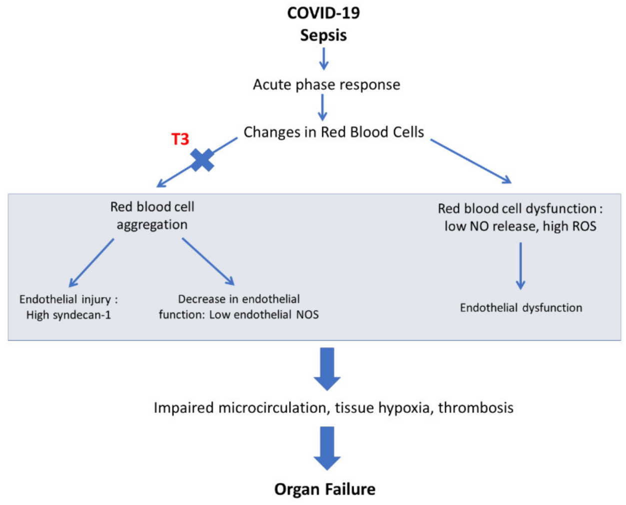
Tissue hypoxia is one of the main pathophysiologic mechanisms in sepsis and particularly in COVID-19. Microvascular dysfunction, endothelialitis and alterations in red blood cell hemorheology are all implicated in severe COVID-19 hypoxia and multiorgan dysfunction. Tissue hypoxia results in tissue injury and remodeling with re-emergence of fetal programming via hypoxia-inducible factor-1α (HIF-1a)-dependent and -independent pathways. In this context, thyroid hormone (TH), a critical regulator of organ maturation, may be of relevance in preventing fetal-like hypoxia-induced remodeling in COVID-19 sepsis. Acute triiodothyronine (T3) treatment can prevent cardiac remodeling and improve recovery of function in clinical settings of hypoxic injury as acute myocardial infarction and by-pass cardiac surgery. Furthermore, T3 administration prevents tissue hypoxia in experimental sepsis. On the basis of this evidence, the use of T3 treatment was proposed for ICU (Intensive Care Unit) COVID-19 patients (Thy-Support, NCT04348513).

thyroid hormone COVID-19 erythrocyte sepsis right ventricle hypoxia

1. Introduction

It is well-recognized that changes in Thyroid Hormone (TH) metabolism occur in response to acute illness, known as non-thyroidal illness syndrome (NTIS). In this syndrome, an initial drop of circulating triiodothyronine (T3) levels is observed, and at later stages a decrease in both circulating L-thyroxine (T4) and T3 levels occurs. Numerous studies have reported that NTIS in acute illness is associated with poor outcomes and high mortality [1]. Similarly, changes in TH metabolism have been observed in COVID-19 patients with an impact on disease progression and outcome [2]. The management of this condition still remains controversial.
Tissue hypoxia due to disturbed microcirculation seems to be a unifying pathophysiologic mechanism in a spectrum of clinical conditions such as myocardial infarction, sepsis and trauma [3]. In this context, TH signaling is shown to be implicated in hypoxia- induced cardiac remodeling [4] and T3 therapy has favorable effects on postischemic cardiac function and infarct healing both in experimental studies and in humans [5][6]. Along this line, T3 has recently been shown to prevent tissue hypoxia in experimental sepsis [7].

2. Tissue Hypoxia: A Unifying Pathophysiologic Mechanism in Myocardial Infarction, Sepsis and Severe COVID-19

Impairment of microcirculation remains one of the main causes of tissue hypoxia and organ dysfunction in acute critical illness. Despite hemodynamic restoration and oxygen availability, there is a lack of improvement in organ perfusion both in sepsis and ischemia–reperfusion, such as myocardial infarction and by-pass surgery, due to the impaired microcirculation [8][9]. Persistent microcirculatory changes are associated with organ failure and death in patients with septic shock [10]. Similarly, microvascular obstruction after acute myocardial infarction is associated with heart failure and high mortality [11][12]. Along this line, changes in microcirculation were found in patients with severe COVID-19 [13].
Endothelial injury and altered blood cell hemorheology are considered important pathogenetic mechanisms which may lead to impaired microcirculation [14]. Endothelial injury appears to be common in sepsis and myocardial ischemia–reperfusion injury as in myocardial infarction and by-pass surgery [3]. Syndecan-1 and thrombomodulin are the most studied biomarkers of endothelial integrity and function. Syndecan is a heparin sulfate proteoglycan expressed in endothelial cells and the main marker of endothelial glycocalyx degradation [15]. Thrombomodulin is a type I transmembrane glycoprotein that is present on the luminal surfaces of endothelial cells. Plasma levels of syndecan-1 and thrombomodulin are higher in patients with sepsis, and both biomarkers predicted the risk of circulatory failure or death [16]. Similarly, in by-pass surgery, a clinical setting of ischemia–reperfusion injury, acute microcirculatory perfusion changes persisted in the first three postoperative days and were associated with increased heparin sulphate and Syndecan-1 levels [8]. Interestingly, COVID-19 patients with high levels of Syndecan-1 had significantly higher levels of thrombomodulin, interleukin-6 and TNF-α and higher mortality [17]. Furthermore, pulmonary vascular endothelialitis was reported from autopsies of COVID-19 patients [18].
Erythrocyte abnormalities are common in sepsis as well as in ischemia–reperfusion injury and have an impact on microcirculation. Erythrocyte aggregation initially occurs in areas of slow flow and impairs microcirculation. Impaired vascular flow enhances erythrocyte aggregation (a vicious circle), and this in turn causes sludging of blood and thrombosis [14]. Moreover, endothelial nitric oxide synthase (NOS) expression is decreased due to lower shear stress resulting from the axial accumulation of red blood cells (RBCs) [19]. RBC aggregation can disrupt the endothelial glycocalyx as the aggregates rub against and induce endothelial injury [20]. Erythrocyte aggregation occurs in experimental sepsis [21]. The erythrocyte sedimentation rate (ESR), an indirect index of erythrocyte aggregation, is elevated in sepsis. In acute myocardial infarction, patients with a high ESR have a greater risk for cardiovascular death and major adverse cardiovascular events (MACEs) [22]. Erythrocyte aggregation may also occur in COVID-19 [23]. In vitro studies showed a strong contribution of plasma fibrinogen in RBC hyperaggregation. RBC aggregation correlated positively with clot firmness, negatively with clot formation time and positively with the length of hospitalization. Patients receiving oxygen supplementation had higher RBC aggregation and blood viscosity. In addition, patients with pulmonary lesions had higher RBC aggregation and enhanced coagulation [23]. More recently, erythrocyte reactive oxygen species (ROS) production was shown to be increased and NO release to be decreased in COVID-19 vasculopathy [24][25]. Collectively, it appears that erythrocytes may serve an important role in microcirculation and tissue hypoxia in critical illness and particularly in severe COVID-19. Figure 1.
Figure 1. Schematic of the potential role of erythrocyte in COVID-19 vasculopathy and the potential effect of T3 in this response. T3: triiodothyronine, ROS: reactive oxygen species, NO: nitric oxide, NOS: NO synthase.

3. TH and Tissue Hypoxia

3.1. Differential Effects of TH on Healthy and Injured Tissue

The use of TH therapy in clinical practice has been limited due to the long-standing belief that TH can aggravate myocardial ischemia by increasing the metabolic rate and oxygen demand. However, over the past years, landmark experimental studies have challenged this long-standing belief, showing that the effects of TH are different in normal and diseased states (recently reviewed in [26]).
Similar observations have been reported for other organs, such as the kidney and liver, in different experimental settings. TH treatment appears to induce protection against ischemic injury in the kidney [27][28]. T3 treatment 24 h prior to renal ischemia–reperfusion resulted in a marked decrease in proteinuria [27]. In addition, T3 treatment prevented acute tubular necrosis after renal ischemia–reperfusion injury [29]. T3 treatment in experimental sepsis prevented cardiac and liver tissue hypoxia (pO2 less than 10 mmHg, a threshold below which HIF-1α-regulated mechanisms are activated) [7]. On the contrary, high-dose T3 administration in normal animals for 10 days or 7 weeks resulted in increased oxygen consumption, intrarenal tissue hypoxia and proteinuria [30][31]. These data clearly indicate that TH effects are different in stressed tissue than in normal.

3.2. Potential Underlying Mechanisms of TH Effects on Tissue Hypoxia

TH regulates the microvascular function both directly and indirectly. T3, acting through thyroid hormone receptor α (TRα), induces endothelium-dependent vasodilation via endothelial nitric oxide synthase [32]. The increased metabolic rate induced by TH may also lead to vasodilation indirectly. Furthermore, TH can induce physiologic angiogenesis, increasing new small vessels with normal permeability and function via direct regulation of angiogenic factors such as Vascular Endothelial Growth Factor-A, Fibroblast Growth Factor-2, angiopoietin-2 and Platelet-derived Growth Factor [26]. TH may also increase tolerance to hypoxic injury via induction of adaptive molecules such as the Heat Shock Proteins [33]. Additional evidence suggests that TH interacts with HIF-α and the hypoxia response pathway. This action may have profound effects on the HIF-induced organ remodeling and dysfunction [26][34].

3.3. TH as Potential Therapy for Tissue Hypoxia

The therapeutic effect of TH on cardiac tissue hypoxia has been tested in experimental models and recently in clinical trials. TH pre-treatment can protect against myocardial ischemia–reperfusion injury in a pattern similar to ischemic pre-conditioning [35]. High-dose T3 and not T4 administration at reperfusion improved cardiac function and limited apoptosis, particularly in the mid-layer of the myocardium, an area where microvascular circulation prevails [36]. This effect required the TRα1 receptor [37] and was mediated by up-regulation of pro-survival Akt and suppression of p38 MAPK [36].
Post-ischemic cardiac remodeling appears to be associated with distinct temporal changes in TRs [38], and an interaction between (TRs) and adrenergic or inflammatory signaling was shown to occur in cell-based experimental models [33]. Accordingly, TH administration in a dose-dependent manner prevented cardiac remodeling in experimental models of myocardial infarction [39]. This experimental evidence was recently translated into a pilot randomized clinical trial (Thy-Repair) which investigated the effects of early high-dose L-triiodothyronine (LT3) in patients with anterior MI undergoing primary angioplasty [6]. In accordance with the experimental evidence, this study showed that LT3 can prevent dilatation of the left ventricle (LV), electrical remodeling and can also improve infarct healing, without any major adverse effects. A favorable effect on microvascular obstruction (MVO) was also observed [6]. In addition, early T3 administration in by-pass surgery was shown to improve cardiac function and limit troponin release, too [40].
Along this line, T3 therapy has favorable effects on cardiac dysfunction due to excessive inflammation driven by TNFα overexpression [41]. TNFα overexpression resulted in down-regulation of TRβ1 as previously found in cell-based experimental models [42]. Interestingly, in this model, a gender-specific reduction in T3 levels could cause the worst cardiac phenotype observed in female mice, and T3 administration improved cardiac function and calcium handling via controlled Akt activation [41].

4. The Role of TH in COVID-19

There is accumulating evidence showing that alterations in TH metabolism frequently occur in COVID-19 patients. In spite of the relatively high heterogeneity between studies, low TH levels on admission were associated with COVID-19 disease severity and mortality. Furthermore, the probability of finding low Free T3 (FT3) and low Thyroid-Stimulating Hormone (TSH) levels was related to disease severity in patients without known thyroid disease. It should be noted that blood samples, in most of the studies reported in the literature, were collected on patients’ admission or during the first three days of hospitalization either in the ward or in ICU.
A retrospective study of 46 patients evaluated the thyroid function in two different groups of consecutive patients affected by COVID-19, one with pneumonia and the other with acute respiratory syndrome (ARDS) requiring ICU admission in comparison with euthyroid patients. COVID-19 patients showed a statistically significant reduction in FT3 and TSH levels measured one day after admission, and as far as ICU patients were concerned, a further statistically significant reduction in FT3 and TSH was found. These findings probably indicate a negative association between FT3 and TSH levels with disease severity at initial presentation [43].
Another cohort study by Lui et al. [44], after a follow-up of 191 patients with COVID-19, reported that around 15% of patients with mild-to-moderate disease had abnormal thyroid function. A decreasing trend of FT3 with increasing COVID-19 severity was found, and patients with low FT3 had more adverse COVID-19-related outcomes.
Along this line, a retrospective observational study of 127 COVID-19 patients showed that serum FT3 levels on admission were lower in non-survivors compared to survivors among moderate-to-critical patients, and the low FT3 state on admission was associated with an increased risk of all-cause in-hospital mortality in these patients [45]. Furthermore, in a retrospective study of 78 critically ill COVID-19 patients, the levels of FT3 on ICU admission were lower in non-survivors compared to survivors, and patients who survived had higher levels of FT3, FT4 and TSH than non-survivors by day 5 [46]. However, in a recent meta-analysis including 3609 COVID-19 patients, the relation between FT3 or TSH levels and survival could not be reliably assessed due to high heterogeneity between studies. This analysis further showed that survivors had higher FT4 levels than non-survivors [47].
The potential mechanisms implicated in TH abnormalities in COVID-19 are not fully understood. It is likely that changes in the Hypothalamic–Pituitary–Thyroid (HPT) axis and iodothyronine deiodinase activity due to abnormal systemic inflammatory response may occur. Furthermore, SARS-CoV-2, via its direct action, can result in destruction of the thyroid gland [47]. Thyroid follicular cells have been shown to express ACE2 (the target of the spike protein), while typical histopathological features of thyroid injury have been found in patients who died of COVID-19 [48][49].
Concomitant medications used in COVID-19 patients may also affect thyroid function. Systemic corticosteroids, mainly dexamethasone, are recommended for patients with severe and critical COVID-19 based on the guidelines of the World Health Organization (WHO) to control the exacerbated inflammatory response [2], though glucocorticoids are known to affect serum TSH levels in humans. A physiological dose of hydrocortisone appears to play a role in the daily variation in serum TSH levels [50]. Acute inhibition of TSH secretion has also been reported after administration of pharmacological doses of glucocorticoids [51].

References

  1. Padhi, R.; Kabi, S.; Panda, B.N.; Jagati, S. Prognostic significance of nonthyroidal illness syndrome in critically ill adult patients with sepsis. Int. J. Crit. Illn. Inj. Sci. 2018, 8, 165–172.
  2. Chen, W.; Tian, Y.; Li, Z.; Zhu, J.; Wei, T.; Lei, J. Potential Interaction Between SARS-CoV-2 and Thyroid: A Review. Endocrinology 2021, 162, bqab004.
  3. Johansson, P.I.; Stensballe, J.; Ostrowski, S.R. Shock induced endotheliopathy (SHINE) in acute critical illness—A unifying pathophysiologic mechanism. Crit. Care 2017, 21, 25.
  4. Pantos, C.; Mourouzis, I.; Cokkinos, D.V. Rebuilding the post-infarcted myocardium by activating ‘physiologic’ hypertrophic signaling pathways: The thyroid hormone paradigm. Heart Fail. Rev. 2010, 15, 143–154.
  5. Pantos, C.; Mourouzis, I. Thyroid hormone receptor α1 as a novel therapeutic target for tissue repair. Ann. Transl. Med. 2018, 6, 254.
  6. Pantos, C.I.; Trikas, A.G.; Pisimisis, E.G.; Grigoriou, K.P.; Stougiannos, P.N.; Dimopoulos, A.K.; Linardakis, S.I.; Alexopoulos, N.A.; Evdoridis, C.G.; Gavrielatos, G.D.; et al. Effects of acute triiodothyronine treatment in patients with anterior myocardial infarction undergoing primary angioplasty: Evidence from a pilot randomized clinical trial (ThyRepair study). Thyroid 2022, 32, 714–724.
  7. Mourouzis, I.S.; Lourbopoulos, A.I.; Trikas, A.G.; Tseti, I.K.; Pantos, C.I. Triiodothyronine prevents tissue hypoxia in experimental sepsis: Potential therapeutic implications. Intensive Care Med. Exp. 2021, 9, 17.
  8. Dekker, N.A.M.; Veerhoek, D.; Koning, N.J.; van Leeuwen, A.L.I.; Elbers, P.W.G.; van den Brom, C.E.; Vonk, A.B.A.; Boer, C. Postoperative microcirculatory perfusion and endothelial glycocalyx shedding following cardiac surgery with cardiopulmonary bypass. Anaesthesia 2019, 74, 609–618.
  9. Vincent, J.L.; De Backer, D. Microvascular dysfunction as a cause of organdysfunction in severe sepsis. Crit. Care 2005, 9 (Suppl. S4), S9–S12.
  10. Sakr, Y.; Dubois, M.J.; De Backer, D.; Creteur, J.; Vincent, J.L. Persistent microcirculatory alterations are associated with organ failure and death in patients with septic shock. Crit. Care Med. 2004, 32, 1825–1831.
  11. Konijnenberg, L.S.F.; Damman, P.; Duncker, D.J.; Kloner, R.A.; Nijveldt, R.; van Geuns, R.M.; Berry, C.; Riksen, N.P.; Escaned, J.; van Royen, N. Pathophysiology and diagnosis of coronary microvascular dysfunction in ST-elevation myocardial infarction. Cardiovasc. Res. 2020, 116, 787–805.
  12. van Kranenburg, M.; Magro, M.; Thiele, H.; de Waha, S.; Eitel, I.; Cochet, A.; Cottin, Y.; Atar, D.; Buser, P.; Wu, E.; et al. Prognostic value of microvascular obstruction and infarct size, as measured by CMR in STEMI patients. JACC Cardiovasc. Imaging 2014, 7, 930–939.
  13. Kanoore Edul, V.S.; Caminos Eguillor, J.F.; Ferrara, G.; Estenssoro, E.; Siles, D.S.P.; Cesio, C.E.; Dubin, A. Microcirculation alterations in severe COVID-19 pneumonia. J. Crit. Care 2021, 61, 73–75.
  14. Sloop, G.D.; Pop, G.A.; Weidman, J.J.; Cyr, J.A. The Detrimental Role of Elevated Blood Viscosity in Patients With COVID-19. J. Cardiol. Ther. 2021, 8, 976–980.
  15. Johansson, P.I.; Stensballe, J.; Rasmussen, L.S.; Ostrowski, S.R. A high admission syndecan-1 level, a marker of endothelial glycocalyx degradation, is associated with inflammation, protein C depletion, fibrinolysis, and increased mortality in trauma patients. Ann. Surg. 2011, 254, 194–200.
  16. Johansen, M.E.; Johansson, P.I.; Ostrowski, S.R.; Bestle, M.H.; Hein, L.; Jensen, A.L.; Soe-Jensen, P.; Andersen, M.H.; Steensen, M.; Mohr, T.; et al. Profound endothelial damage predicts impending organ failure and death in sepsis. Semin. Thromb. Hemost. 2015, 41, 16–25.
  17. Zhang, D.; Li, L.; Chen, Y.; Ma, J.; Yang, Y.; Aodeng, S.; Cui, Q.; Wen, K.; Xiao, M.; Xie, J.; et al. Syndecan-1, an indicator of endothelial glycocalyx degradation, predicts outcome of patients admitted to an ICU with COVID-19. Mol. Med. 2021, 27, 151.
  18. Ackermann, M.; Verleden, S.E.; Kuehnel, M.; Haverich, A.; Welte, T.; Laenger, F.; Vanstapel, A.; Werlein, C.; Stark, H.; Tzankov, A.; et al. Pulmonary Vascular Endothelialitis, Thrombosis, and Angiogenesis in COVID-19. N. Engl. J. Med. 2020, 383, 120–128.
  19. Baskurt, O.K.; Yalcin, O.; Ozdem, S.; Armstrong, J.K.; Meiselman, H.J. Modulation of endothelial nitric oxide synthase expression by red blood cell aggregation. Am. J. Physiol. Heart Circ. Physiol. 2004, 286, H222–H229.
  20. Iffrig, E.; Oakley, J.; Fibben, K.; Sakurai, Y.; Esper, A.M.; Lam, W. Severe COVID-19 Is an Acquired Red Cell Disease- Potential Mechanisms Underlying Endothelialitis. Am. J. Respir. Crit. Care Med. 2021, 203, A4470.
  21. Baskurt, O.K.; Temiz, A.; Meiselman, H.J. Red blood cell aggregation in experimental sepsis. J. Lab. Clin. Med. 1997, 130, 183–190.
  22. Li, C.; Wang, Y.; Zhang, Q.; Wang, L.; Li, K.; Yang, X. Incorporating the erythrocyte sedimentation rate for enhanced accuracy of the global registry of acute coronary event score in patients with ST-segment elevated myocardial infarction: A retrospective cohort study. Medicine 2020, 99, e22523.
  23. Nader, E.; Nougier, C.; Boisson, C.; Poutrel, S.; Catella, J.; Martin, F.; Charvet, J.; Girard, S.; Havard-Guibert, S.; Martin, M.; et al. Increased blood viscosity and red blood cell aggregation in patients with COVID-19. Am. J. Hematol. 2022, 97, 283–292.
  24. Brugnara, C. The Erythrocyte, a Novel Disease-Mediator for COVID-19 Vasculopathy? JACC Basic Transl. Sci. 2022, 7 Pt 1, 205–206.
  25. Mahdi, A.; Collado, A.; Tengbom, J.; Jiao, T.; Wodaje, T.; Johansson, N.; Farnebo, F.; Färnert, A.; Yang, J.; Lundberg, J.O.; et al. Erythrocytes Induce Vascular Dysfunction in COVID-19. JACC Basic Transl. Sci. 2022, 7 Pt 1, 193–204.
  26. Lourbopoulos, A.I.; Mourouzis, I.S.; Trikas, A.G.; Tseti, I.K.; Pantos, C.I. Effects of Thyroid Hormone on Tissue Hypoxia: Relevance to Sepsis Therapy. J. Clin. Med. 2021, 10, 5855.
  27. Ferreyra, C.; O’Valle, F.; Osorio, J.M.; Moreno, J.M.; Rodriguez, I.; Vargas, F.; Osuna, A. Effect of preconditioning with triiodothyronine on renal ischemia/reperfusion injury and poly(ADP-ribose) polymerase expression in rats. Transpl. Proc. 2009, 41, 2073–2075.
  28. Sutter, P.M.; Thulin, G.; Stromski, M.; Ardito, T.; Gaudio, K.M.; Kashgarian, M.; Siegel, N.J. Beneficial effect of thyroxin in the treatment of ischemic acute renal failure. Pediatr. Nephrol. 1988, 2, 1–7.
  29. Ferreyra, C.; Vargas, F.; Rodriguez-Gomez, I.; Perez-Abud, R.; O’Valle, F.; Osuna, A. Preconditioning with triiodothyronine improves the clinical signs and acute tubular necrosis induced by ischemia/reperfusion in rats. PLoS ONE 2013, 8, e74960.
  30. Friederich-Persson, M.; Persson, P.; Fasching, A.; Hansell, P.; Nordquist, L.; Palm, F. Increased kidney metabolism as a pathway to kidney tissue hypoxia and damage: Effects of triiodothyronine and dinitrophenol in normoglycemic rats. Adv. Exp. Med. Biol. 2013, 789, 9–14.
  31. Sivertsson, E.; Friederich-Persson, M.; Persson, P.; Nangaku, M.; Hansell, P.; Palm, F. Thyroid hormone increases oxygen metabolism causing intrarenal tissue hypoxia; a pathway to kidney disease. PLoS ONE 2022, 17, e0264524.
  32. Geist, D.; Hones, G.S.; Gassen, J.; Kerp, H.; Kleinbongard, P.; Heusch, G.; Fuhrer, D.; Moeller, L.C. Noncanonical Thyroid Hormone Receptor alpha Action Mediates Arterial Vasodilation. Endocrinology 2021, 162, bqab099.
  33. Pantos, C.; Mourouzis, I.; Cokkinos, D.V. Thyroid hormone and cardiac repair/regeneration: From Prometheus myth to reality? Can. J. Physiol. Pharmacol. 2012, 90, 977–987.
  34. Forini, F.; Nicolini, G.; Iervasi, G. Mitochondria as key targets of cardioprotection in cardiac ischemic disease: Role of thyroid hormone triiodothyronine. Int. J. Mol. Sci. 2015, 16, 6312–6336.
  35. Pantos, C.I.; Malliopoulou, V.A.; Mourouzis, I.S.; Karamanoli, E.P.; Paizis, I.A.; Steimberg, N.; Varonos, D.D.; Cokkinos, D.V. Long-term thyroxine administration protects the heart in a pattern similar to ischemic preconditioning. Thyroid 2002, 12, 325–329.
  36. Pantos, C.; Mourouzis, I.; Saranteas, T.; Clave, G.; Ligeret, H.; Noack-Fraissignes, P.; Renard, P.Y.; Massonneau, M.; Perimenis, P.; Spanou, D.; et al. Thyroid hormone improves postischaemic recovery of function while limiting apoptosis: A new therapeutic approach to support hemodynamics in the setting of ischaemia-reperfusion? Basic Res. Cardiol. 2009, 104, 69–77.
  37. Pantos, C.; Mourouzis, I.; Saranteas, T.; Brozou, V.; Galanopoulos, G.; Kostopanagiotou, G.; Cokkinos, D.V. Acute T3 treatment protects the heart against ischemia-reperfusion injury via TRalpha1 receptor. Mol. Cell. Biochem. 2011, 353, 235–241.
  38. Pantos, C.; Mourouzis, I.; Galanopoulos, G.; Gavra, M.; Perimenis, P.; Spanou, D.; Cokkinos, D.V. Thyroid hormone receptor alpha1 downregulation in postischemic heart failure progression: The potential role of tissue hypothyroidism. Horm. Metab. Res. 2010, 42, 718–724.
  39. Mourouzis, I.; Mantzouratou, P.; Galanopoulos, G.; Kostakou, E.; Roukounakis, N.; Kokkinos, A.D.; Cokkinos, D.V.; Pantos, C. Dose-dependent effects of thyroid hormone on post-ischemic cardiac performance: Potential involvement of Akt and ERK signalings. Mol. Cell. Biochem. 2012, 363, 235–243.
  40. Ranasinghe, A.M.; Quinn, D.W.; Pagano, D.; Edwards, N.; Faroqui, M.; Graham, T.R.; Keogh, B.E.; Mascaro, J.; Riddington, D.W.; Rooney, S.J.; et al. Glucose-insulin-potassium and tri-iodothyronine individually improve hemodynamic performance and are associated with reduced troponin I release after on-pump coronary artery bypass grafting. Circulation 2006, 114 (Suppl. S1), I245–I250.
  41. Ntari, L.; Mantzouratou, P.; Katsaouni, A.; Pantos, C.; Kollias, G.; Mourouzis, I. Changes in Thyroid Hormone Signaling Mediate Cardiac Dysfunction in the Tg197 Mouse Model of Arthritis: Potential Therapeutic Implications. J. Clin. Med. 2021, 10, 5512.
  42. Pantos, C.; Xinaris, C.; Mourouzis, I.; Kokkinos, A.D.; Cokkinos, D.V. TNF-alpha administration in neonatal cardiomyocytes is associated with differential expression of thyroid hormone receptors: A response prevented by T3. Horm. Metab. Res. 2008, 40, 731–734.
  43. Baldelli, R.; Nicastri, E.; Petrosillo, N.; Marchioni, L.; Gubbiotti, A.; Sperduti, I.; Di Giacinto, P.; Rizza, L.; Rota, F.; Franco, M.; et al. Thyroid dysfunction in COVID-19 patients. J. Endocrinol. Investig. 2021, 44, 2735–2739.
  44. Lui, D.T.W.; Lee, C.H.; Chow, W.S.; Lee, A.C.H.; Tam, A.R.; Fong, C.H.Y.; Law, C.Y.; Leung, E.K.H.; To, K.K.W.; Tan, K.C.B.; et al. Thyroid Dysfunction in Relation to Immune Profile, Disease Status, and Outcome in 191 Patients with COVID-19. J. Clin. Endocrinol. Metab. 2021, 106, e926–e935.
  45. Lang, S.; Liu, Y.; Qu, X.; Lu, R.; Fu, W.; Zhang, W.; Wang, H.; Hong, T. Association between Thyroid Function and Prognosis of COVID-19: A Retrospective Observational Study. Endocr. Res. 2021, 46, 170–177.
  46. Ballesteros Vizoso, M.A.; Castilla, A.F.; Barceló, A.; Raurich, J.M.; Argente del Castillo, P.; Morell-García, D.; Velasco, J.; Pérez-Bárcena, J.; Llompart-Pou, J.A. Thyroid Disfunction in Critically Ill COVID-19 Patients. Relationship with In-Hospital Mortality. J. Clin. Med. 2021, 10, 5057.
  47. Chen, Y.; Li, X.; Dai, Y.; Zhang, J. The Association Between COVID-19 and Thyroxine Levels: A Meta-Analysis. Front. Endocrinol. 2021, 12, 779692.
  48. Muller, I.; Cannavaro, D.; Dazzi, D.; Covelli, D.; Mantovani, G.; Muscatello, A.; Ferrante, E.; Orsi, E.; Resi, V.; Longari, V.; et al. SARS-CoV-2-related atypical thyroiditis. Lancet Diabetes Endocrinol. 2020, 8, 739–741.
  49. Rotondi, M.; Coperchini, F.; Ricci, G.; Denegri, M.; Croce, L.; Ngnitejeu, S.T.; Villani, L.; Magri, F.; Latrofa, F.; Chiovato, L. Detection of SARS-CoV-2 receptor ACE-2 mRNA in thyroid cells: A clue for COVID-19-related subacute thyroiditis. J. Endocrinol. Investig. 2021, 44, 1085–1090.
  50. Samuels, M.H.; McDaniel, P.A. Thyrotropin levels during hydrocortisone infusions that mimic fasting-induced cortisol elevations: A clinical research center study. J. Clin. Endocrinol. Metab. 1997, 82, 3700–3704.
  51. Nicoloff, J.T.; Fisher, D.A.; Appleman, M.D., Jr. The role of glucocorticoids in the regulation of thyroid function in man. J. Clin. Investig. 1970, 49, 1922–1929.
More
Information
Contributors MDPI registered users' name will be linked to their SciProfiles pages. To register with us, please refer to https://encyclopedia.pub/register : , , , , , , , , ,
View Times: 413
Entry Collection: COVID-19
Revisions: 2 times (View History)
Update Date: 08 Jul 2022
1000/1000
Video Production Service