Submitted Successfully!
To reward your contribution, here is a gift for you: A free trial for our video production service.
Thank you for your contribution! You can also upload a video entry or images related to this topic.
Version Summary Created by Modification Content Size Created at Operation
1 + 5047 word(s) 5047 2022-02-14 04:05:05 |
2 corrected the format and inserted the references -2239 word(s) 2808 2022-03-16 02:14:59 | |
3 Update figure 1 -2239 word(s) 2808 2022-03-16 02:19:52 |

Video Upload Options

Do you have a full video?

Confirm

Are you sure to Delete?
Cite
If you have any further questions, please contact Encyclopedia Editorial Office.
Odun-Ayo, F. Gastrointestinal Microbiota Dysbiosis with SARS-CoV-2 in Colorectal: Probiotic. Encyclopedia. Available online: https://encyclopedia.pub/entry/20592 (accessed on 06 July 2024).
Odun-Ayo F. Gastrointestinal Microbiota Dysbiosis with SARS-CoV-2 in Colorectal: Probiotic. Encyclopedia. Available at: https://encyclopedia.pub/entry/20592. Accessed July 06, 2024.
Odun-Ayo, Frederick. "Gastrointestinal Microbiota Dysbiosis with SARS-CoV-2 in Colorectal: Probiotic" Encyclopedia, https://encyclopedia.pub/entry/20592 (accessed July 06, 2024).
Odun-Ayo, F. (2022, March 15). Gastrointestinal Microbiota Dysbiosis with SARS-CoV-2 in Colorectal: Probiotic. In Encyclopedia. https://encyclopedia.pub/entry/20592
Odun-Ayo, Frederick. "Gastrointestinal Microbiota Dysbiosis with SARS-CoV-2 in Colorectal: Probiotic." Encyclopedia. Web. 15 March, 2022.
Gastrointestinal Microbiota Dysbiosis with SARS-CoV-2 in Colorectal: Probiotic
Edit

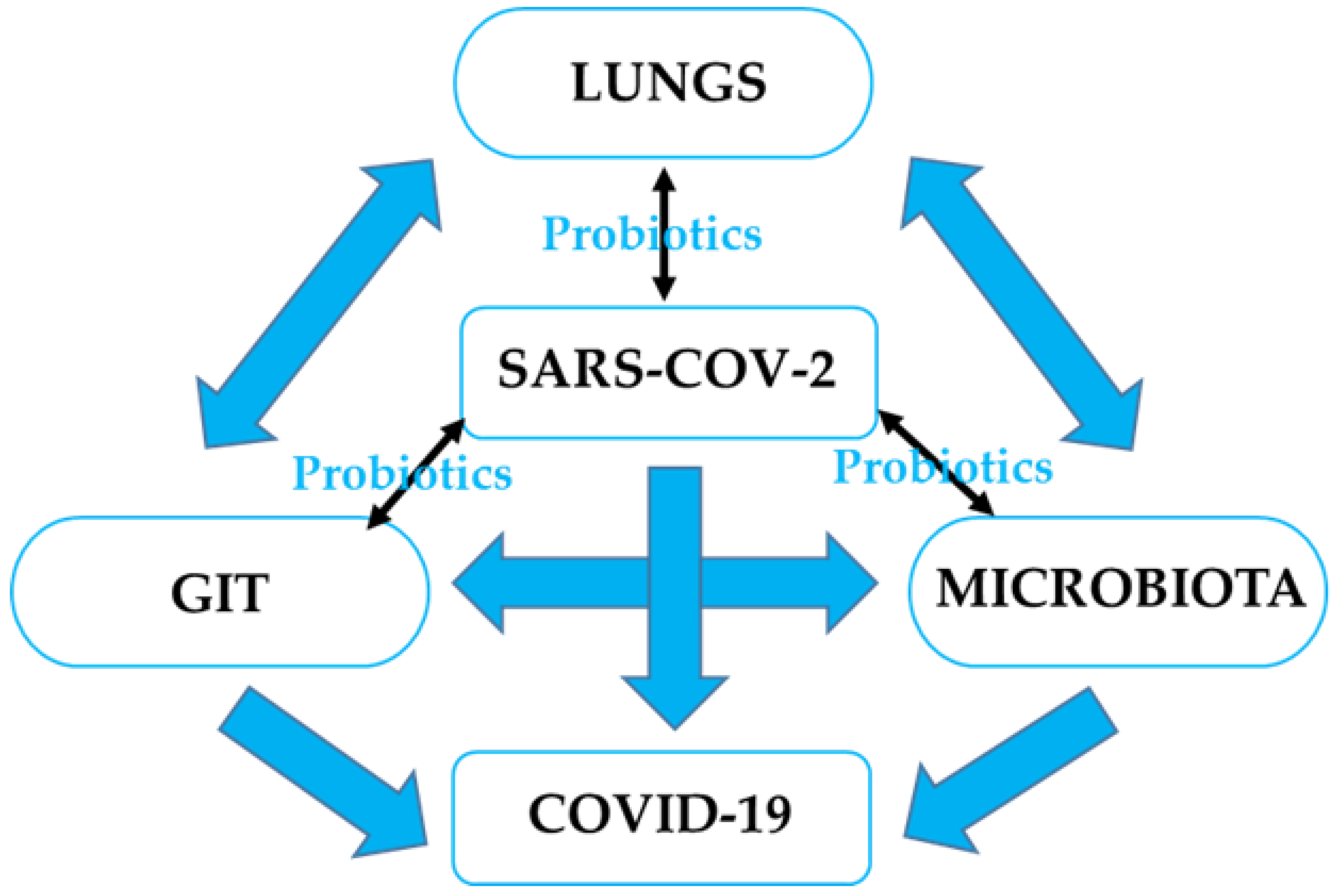
GI microbiota dysbiosis has been associated with respiratory disorders, including COVID-19, as well as sporadic colorectal cancer (CRC) through imbalanced microbiota and compromised immune response. It is pertinent to understand the possible role of probiotics in stabilizing the microbial environment and maintaining the integrity of the respiratory and GI tracts in SARS-CoV-2 induced dysbiosis and colorectal carcinogenesis. 

gut microbiota colorectal cancer respiratory tract infection SARS-CoV-2 Dysbiosis Virus Probiotic Lung

1. Introduction

Probiotics are defined as “live microorganisms which when administered orally in adequate amount confer a health benefit on the host” [1][2]. They are described as a live microbial feed and food supplement that beneficially affects the host’s intestinal tract [3]. Probiotics are non-pathogenic microbes that exert a variety of beneficial effects, such as antipathogenic effects, immunomodulatory factors, the production of key nutrients, and the development of mucosal epithelia. Products derived from bacteria or their end products cannot be considered probiotic because they are not alive when administered or during consumption [4]. One important point common to all these definitions is the ability of the probiotic to confer a beneficial effect on the health of the host. The implantation or colonization of these viable microorganisms improves the microbial balance of the intestinal tract. Viruses are the cause of nearly 90% of upper respiratory tract infections [5]. However, certain probiotic strains may prevent bacterial and viral diseases, such as gastroenteritis [6][7] and respiratory tract infections (RTIs), including COVID-19 [5][8][9][10][11]. It is worth noting that not all probiotics, even those that offer GI advantages, help to reduce the risk of respiratory infection in every way. For example, Lactobacillus Rhamnosus GG and Bifidobacterium animalis ssp. lactis may help the GIT, but they do not diminish the number of viruses in the nasopharynx [12]. Many in vivo and in vitro studies reveal an association between these beneficial bacteria and human immune-modulatory responses. This has led to a shift in the focus of research towards the beneficial use of probiotics in the treatment of various diseases in recent years. It is vital, therefore, to understand some of the areas regarding GIT and RTI diseases to which probiotics have been applied extensively in recent years, as well as to perform meaningful estimates for future applications, particularly in the treatment of COVID-19. 

2. GI Microbiota and CRC

The gut microbiota is linked to the occurrence and progression of CRC. Alterations in the immunological response, epithelial hemostasis, metabolic profile and activity, DNA damage, and abnormal cellular and molecular activities in colonocytes can all contribute to carcinogenesis [13][14][15]. The whole microbial composition of an organ or system is referred to as the human microbiome, which includes bacteria, fungi, viruses, their surrounding environmental circumstances, genomes, and host relationships [16]. The human GI microbiota consists of hundreds of types of microorganism, with an estimated value of over 1013–1014 bacteria acting as a natural infection-defeating barrier. Furthermore, the microbiota plays an important role in gut homeostasis by performing a variety of defensive, structural, and metabolic functions in the intestinal epithelium, as well as the development of a healthy immune system [17]. Some of these bacteria grow and colonize the intestinal region of the host, becoming the Gl microbiota, which acts as a line of defense against pathogenic organisms. Microorganisms are spread unevenly throughout the digestive tract, including the stomach (<103), duodenum (<103), small intestine (102–103), and large intestine (1010–1012) [18]. The human colon consists of a complex microbial composition mostly of bacteria, which consist of more than 50 genera [19][20]. The bacterial composition of the colon is estimated to be as high as 1014 [21][22]. The colon mostly comprises anaerobes, such as Bacteroides, Porphyromonas, Bifidobacterium, Lactobacillus, and Clostridium, which outgrow aerobes by a factor of 102–103: 1 [23]. Some bacteria, including Bacteroides fragilis and Eubacterium rectale, inhabit discrete zones within the intestinal lumen of the human colon, while some become adherent to the mucosal surface [24]. Microorganisms may occasionally find themselves in a favorable environment for proliferation, but this is not the same habitat as their typical flora, resulting in the overgrowth and, eventually, the suppression of the normal flora [25]. The GI microbiota can exert both positive and harmful effects by modulating epithelial proliferation and differentiation, in addition to impacting host nutrition via the metabolism [26].
Several bacterial species appear to be involved in the pathogenesis of CRC [27][28][29]. The loss of bacterial diversity and dysbiosis are common observations in CRC. However, despite the existence of conflicting evidence, several studies have found significant changes in the mucosal and fecal microbiota of CRC patients and controls. Streptococcus gallolyticus (formerly Streptococcus bovis) is found in around 20–50% of CRC and less than 5% of healthy people. CRC patients were reported to have lower levels of Bifidobacterium longumClostridium clostridioforme, and Ruminococcus bromii than healthy people [30]. However, upon further study, Bacteroides were shown to be more prevalent in CRC tissues than in normal tissues; they were associated with an increase in IL 17 immunoreactive cells in the mucosa of CRC patients [30]. The presence of Fusobacterium nucleatum sequences was detected in CRC tumors and linked to lymph node metastasis [31]. In addition, another taxonomy-based comparison study was undertaken to assess the differences between the microbiota of cancerous and neighboring non-cancerous colorectal tissues [27]Firmicutes were the most prevalent phyla, accounting for 63.46% and 39.54% of the GI microbiota in malignant cancerous and adjacent non-cancerous tissues, respectively. This was followed by 12.77% and 19% of Bacteroidetes in the cancerous and adjacent non-cancerous colorectal tissues. The study further confirms that the genera Lactococcus, Bacteroides, Fusobacterium, Prevotella, and Streptococcus were found in greater abundance in cancerous cells than in non-cancerous cells [27]. Even though FirmicutesBacteroides, and lactic acid bacteria are frequently reduced, Fusobacterium and Porphyromonas are often increased [15]. It was demonstrated that the concentration of Fusobacterium within the tumor microenvironment is the most notable and consistent finding. This suggests that Fusobacterium is linked to inflammatory bowel diseases, such as ulcerative colitis and Crohn’s disease, which are known to increase the risk of CRC [27][32]Fusobacterium sp have virulence properties that promote their adhesion to host epithelial cells and their ability to infiltrate epithelial cells, as well as the ability to trigger host pro-inflammatory responses [28]Fusobacterium nucleatum, a typical driving bacteria, promotes CRC carcinogenesis in APCmin mice. However, the F. nucleatum cannot colonize the colon on its own. This requires the help of a few other species to form colonies, which then support the growth of Peptostreptococcus and Porphyromonas [33][34]Lactococcus, which are commonly known to be GIT commensals with probiotic properties, were found to be over-represented in CRC patients. This implies that the microbial shifts are induced by the quite severe physiological and metabolic changes that occur as a result of colon carcinogenesis [35]. These species could be considered CRC bacterial passengers, according to the driver–passenger concept in CRC [27][36]. The “driver-passenger” paradigm proposes that a microbial leader assembles a group of disease-facilitating microorganisms to start the biological mechanisms that cause CRC. First, “driver” bacteria cause DNA damage and the malignant transformation of epithelial stem cells, resulting in a pro-oncogenic environment. After cancer begins, “passenger” bacteria that are better adapted to the tumor environment appear, such as F. nucleatum and S. gallolyticus [36][37].
In an animal study performed under germ-free conditions, it was noted that mutant mice genetically prone to CRC produce considerably fewer tumors than when they have typical microbiota [38]Enterococcus faecalis produces extracellular genotoxins and DNA-damaging superoxide, causing the acute induction of chromosomal instability, which can contribute to the development of CRC [39]. CRC is caused by the activation of oncogenes in combination with the inactivation of tumor-suppressor genes due to mutations. In total, 85% of CRC cases involve gene mutations in APC or other tumor suppressor genes that activate the Wnt pathway, leading to chromosomal instability [40]. In the majority of CRC patients, the hyperactivation of Wnt/β-catenin signaling is a typical characteristic. The neural cell adhesion receptor L1CAM (L1) is a target gene of β-catenin signaling activated in CRC patients’ carcinoma cells, where it plays a significant role in CRC metastasis [41]. By acting as a co-transcriptional activator of Wnt target genes in the nucleus, together with the T-cell factor, β-catenin aids in the transmission of the Wnt signal to the nucleus [42]. The loss of DNA mismatch repair affects 15% of patients, resulting in a high level of microsatellite instability [43]. As demonstrated by Escherichia coli NC101, the inflammatory environment can change microbial gene functions and boost the cancer-promoting activities of specific bacterium strains [44].

3. GI Microbiota Dysbiosis Associated with SARS-CoV-2 Infection and CRC

The role of the digestive system and GI microbiota in SARS-CoV-2 infection evokes the idea of the gut–lung axis. This refers to the bi-directional interplay between the GI microbiota and the lungs, which can affect immune responses and influence the course of respiratory disorders [45]. Based on previous studies and findings, a unique idea of tight connections within the microbiota-gut-lung triad as related to SARS-CoV-2 infection has emerged [46]. It is believed that the interactions within this triangle could have a direct impact on the development of COVID-19, as well as its clinical symptoms and therapy, as shown in Figure 1.
Figure 1. The interaction within the microbiota-GIT-lung could affect the development of COVID-19; hence, potential therapy through probiotic intervention could maintain microbial balance and immune modulation.
One of the key aspects of the interactions within microbiota-GIT-lung cross-talk is the expression of ACE-2, which serves as an entry receptor for SARS-CoV-2 and an important regulator of inflammation that is higher in the GIT than in the lungs [47][48]. ACE-2 regulates amino acid transport in the intestinal epithelium, which has been linked to the synthesis of antimicrobial peptides affecting the makeup and function of the GI microbiota [49]. However, some gut microorganisms may influence ACE-2 expression in the opposite direction [47]. In recent studies, it has been reported that COVID-19 patients acquire dysbiotic GI microbiota [50][51][48][52][53][54]. This implies that disturbed GI microbiota may play a specific role in COVID-19′s pathogenesis. It is important to note that various hypotheses were investigated to understand the association of dysbiotic GI microbiota with SARS-CoV-2 infection. Certain GI microbiota compositions have been hypothesized to be either positively or negatively linked by inflammatory cytokine levels and to be predictive of severe COVID-19, potentially through the modulation of fecal metabolites and host immunity [55]. The further reveals that the GI microbiota may play a role in typical individuals’ susceptibility to severe COVID-19 through positively associated pro-inflammatory cytokines.
SARS-CoV-2 and COVID-19 severity were linked to alterations in the fecal microbiota. During or throughout hospitalization, patients with COVID-19 had significant abnormalities in their fecal microbiota compared to controls, characterized by the enrichment of opportunistic pathogens and the loss of beneficial commensals [47]. It was further reported that Bacteroides sp. reduces the expression of ACE-2 in the mouse gut, which is inversely related to SARS-CoV-2 burden in the patient’s fecal material. Even after SARS-CoV-2 clearance (as indicated by throat swabs) and the resolution of respiratory symptoms, depleted symbionts, and gut dysbiosis persisted [47]Bacteroides and Streptococcus genus were negatively associated with most pro-inflammatory factors. The fecal metabolomics analysis of the gut microbial from a COVID-19 patient was linked to amino acid metabolism, particularly the aminoacyl-tRNA, arginine, valine, leucine, and isoleucine biosynthesis pathways [55]. A deficiency in or insufficiency of amino acids results in the depletion of the available aminoacylated tRNA, which is essential for host immune response [56][57]; hence, the pro-inflammatory response induced by cytokines was significantly reduced [58]. As these amino acids play a key role in immunoregulation and enhancing intestinal development [59], they may invariably affect GI microbiota in COVID-19 patients and CRC.
ACE-2 and TMPRSS2 expressions have been linked to a variety of bacterial genera such as Chlamydia, which is the microbiota that has been found to be the most strongly positively correlated with ACE-2 expression in CRC patients [60]. Given that ACE-2 is highly expressed in the ileum and colon, the importance of ACE-2 is key to maintaining dietary amino acid balance and innate immunity [61]. In SARS-CoV-2 infection, the virus is attached to the host’s ACE-2 receptor, with the upper airway and lungs being the predominant sites of infection. On the other hand, studies have shown that the intestinal enterocytes at the epithelial layer and the colon epithelial cells have the highest expression of ACE-2 in the human body. SARS-CoV-2 replication is aided by their support, culminating in GI barrier disruption [62][63][64][65], and possibly in CRC. Although the influence of the gut microbiota on COVID-19 risk in CRC patients remains poorly understood, the possible mechanisms targeting microbial dysbiosis should be further investigated [66].
During influenza infection, the inducement of interferons type I promotes the reduction of obligatory anaerobic bacteria and the enrichment of Proteobacteria in the gut, resulting in a “dysbiotic” milieu [67]. Interferons have been demonstrated to decrease antimicrobial and inflammatory responses in the GIT during Salmonella-induced colitis. This has been linked to increased Salmonella intestinal colonization and dissemination, a risk factor for CRC [68]Salmonella can lead to protracted intestinal infection, dysbiotic gut microbiota, and chronic inflammation, all of which can lead to DNA damage and chromosome instability or epigenetic change. Salmonella effector proteins activate cancer-related signaling pathways. They promote the Wnt/-catenin signaling pathway during persistent infection, causing host cell change. Bacterial proteins cause leaky gut, microbiota imbalance, and inflammation, all of which contribute to the development of CRC [68]. Furthermore, in HINI influenza infection, interleukin 17A (IL-17A) signaling enhanced fast viral infiltration of the lungs by pleural cavity B-1a cells via the increase in Blimp-1 expression and NF-kB activation in B-1a cells. IL-17A deficit resulted in highly diminished B-1a-derived antibody production in the respiratory tract, leading to viral clearance deficiencies [69][70]. In CRC patients’ tumor tissue samples, IL-17 immune cells were discovered in the majority of samples and the lamina propria of homologous normal mucosa, whereas they were rarely or not observed in normal mucosa in typical individuals. In addition, the gene amplification of Bacteroides was substantially detected at a higher level in tumor tissue compared to normal homologous tissue [30]. This implies an association between the IL-17 immune cells and Bacteroides in the mucosa cells causing dysbiosis in CRC patients. It was further reported that it was unclear why there was a link between Bacteroides density elevation and malignant CRC, as measured by qPCR [30]. It is noteworthy that the activation of pro-inflammatory and immunological cells in the colon mucosa is crucial in the development of cancer, as well as in the development of severe cases of COVID-19. Some GIT microbiota members may influence host mucosal regulatory T-cell responses involving Th17 cells. As a result, T-cell activation may be linked to a shift in mucosal IL-17 caused by Bacteroides, as seen in animal models [71][72]. These findings support the presence of a skewed immune response in CRC tissues, with IL-17 overproduction aggravating the disease, which was most likely caused by Bacteroides [73][74].
The interaction between the host immune environment and CRC or SARS-CoV-2 infection uses similar mechanisms, such as hypercoagulability, dysregulated immune response, elevated cytokine levels, altered expression of ACE-2 and TMPRSS2, and prothrombotic states. This throws the human body into disarray and may exacerbate the effects of SARS-CoV-2 in some cancer patients [75]. Numerous infiltrating plasma cells and lymphocytes with interstitial edema were detected in the lamina propria of the stomach, duodenum, and rectum [76]. Most patients infected with SARS-CoV-2 have mild GI symptoms and a good prognosis after infection, indicating that the immune function is a strong defense against this virus. Seven to fourteen days after the onset of symptoms, lymphopenia (changes in T lymphocytes) is commonly observed, with an increase in IL-6 and other inflammatory cytokines (pneumonia phase). Lymphopenia and cytokine storm syndrome lead to disease development and a poor prognosis. Lymphocytes are principally responsible for immunological responses to viral infections. However, within the first few days of infection, which is a critical stage, the rate of immune response and the level of lymphocytes produced may not be sufficient to combat or immunosuppress the rate of replication of the virus, especially in the case of SARS-CoV-2, which is new to the host body. This implies that a higher number of lymphocytes is required to compete against the virus [9], irrespective of how quickly the virus or T cell replicates. Although there are various ways for the immune defense function to eradicate infections, it is noteworthy that if the immune response is effective, viral suppression occurs. However, this may not occur if the patient has other co-morbidities, including cancer [77]. The immune dysregulation produced by SARS-CoV-2 could result in even more serious problems for an already fragile population [78].
The gut microbiota has been linked to the development of CRC. The SARS-CoV-2 infection causes changes in the gut microbiota, including the enrichment of opportunistic pathogens, the depletion of beneficial commensals, an overall drop in microbial diversity, and a loss of butyrate-producing bacteria. The increased expression of CRC carcinogenesis markers, tumor immunosuppression, and inflammation induction produced by SARS-CoV-2 infection may exacerbate CRC progression, resulting in gut barrier breakdown and the worsening of CRC advancement [78]. Regardless of the clinical stage of the disease, patients with CRC may be at a high risk of contracting COVID-19 and are crucial protection targets in epidemic prevention. Although further validation of clinical data is needed, these findings are of practical importance. Patients with clinically mild or moderate COVID-19 with a diagnosis of CRC should be paid special attention because of a possible longer course of the disease or a higher risk of severe infection. Although further studies are required,  researchers lays the groundwork for the influence and impact of SARS-CoV-2 on the progression of CRC.

References

  1. Hill, C.; Guarner, F.; Reid, G.; Gibson, G.R.; Merenstein, D.J.; Pot, B.; Morelli, L.; Canani, R.B.; Flint, H.J.; Salminen, S. Expert consensus document: The International Scientific Association for Probiotics and Prebiotics consensus statement on the scope and appropriate use of the term probiotic. Nat. Rev. Gastroenterol. Hepatol. 2014, 11, 506–514.
  2. Hotel, A.C.P.; Cordoba, A. Health and nutritional properties of probiotics in food including powder milk with live lactic acid bacteria. Prevention 2001, 5, 1–10.
  3. Vasiljevic, T.; Shah, N.P. Probiotics—from Metchnikoff to bioactive. Int. Dairy J. 2008, 18, 714–728.
  4. Sanders, M.E.; Gibson, G.; Gill, H.S.; Guarner, F. Probiotics: Their potential to impact human health. Counc. Agric. Sci. Technol. Issue Pap. 2007, 36, 1–20.
  5. Baud, D.; Dimopoulou Agri, V.; Gibson, G.R.; Reid, G.; Giannoni, E. Using probiotics to flatten the curve of coronavirus disease COVID-2019 pandemic. Front. Public Health 2020, 8, 186.
  6. Lenoir-Wijnkoop, I.; Gerlier, L.; Roy, D.; Reid, G. The clinical and economic impact of probiotics consumption on respiratory tract infections: Projections for Canada. PLoS ONE 2016, 11, e0166232.
  7. Szajewska, H.; Kołodziej, M.; Gieruszczak-Białek, D.; Skórka, A.; Ruszczyński, M.; Shamir, R. Systematic review with meta-analysis: Lactobacillus rhamnosus GG for treating acute gastroenteritis in children—A 2019 update. Aliment. Pharmacol. Ther. 2019, 49, 1376–1384.
  8. Guillemard, E.; Tondu, F.; Lacoin, F.; Schrezenmeir, J. Consumption of a fermented dairy product containing the probiotic Lactobacillus casei DN-114 001 reduces the duration of respiratory infections in the elderly in a randomized controlled trial. Br. J. Nutr. 2010, 103, 58–68.
  9. Manna, S.; Chowdhury, T.; Chakraborty, R.; Mandal, S.M. Probiotics-derived peptides and their immunomodulatory molecules can play a preventive role against viral diseases including COVID-19. Probiotics Antimicrob. Proteins 2021, 13, 611–623.
  10. Power, D.; Burton, J.; Chilcott, C.; Dawes, P.; Tagg, J. Preliminary investigations of the colonization of upper respiratory tract tissues of infants using a pediatric formulation of the oral probiotic Streptococcus salivarius K12. Eur. J. Clin. Microbiol. Infect. Dis. 2008, 27, 1261–1263.
  11. Weiss, G.; Rasmussen, S.; Zeuthen, L.H.; Nielsen, B.N.; Jarmer, H.; Jespersen, L.; Frøkiær, H. Lactobacillus acidophilus induces virus immune defense genes in murine dendritic cells by a Toll-like receptor-2-dependent mechanism. Immunology 2010, 131, 268–281.
  12. Lehtoranta, L.; Kalima, K.; He, L.; Lappalainen, M.; Roivainen, M.; Närkiö, M.; Mäkelä, M.; Siitonen, S.; Korpela, R.; Pitkäranta, A. Specific probiotics and virological findings in symptomatic conscripts attending military service in Finland. J. Clin. Virol. 2014, 60, 276–281.
  13. Hendler, R.; Zhang, Y. Probiotics in the treatment of colorectal cancer. Medicines 2018, 5, 101.
  14. Meng, C.; Bai, C.; Brown, T.D.; Hood, L.E.; Tian, Q. Human gut microbiota and gastrointestinal cancer. Genom. Proteom. Bioinform. 2018, 16, 33–49.
  15. Raskov, H.; Burcharth, J.; Pommergaard, H.-C. Linking gut microbiota to colorectal cancer. J. Cancer 2017, 8, 3378.
  16. Sender, R.; Fuchs, S.; Milo, R. Are we really vastly outnumbered? Revisiting the ratio of bacterial to host cells in humans. Cell 2016, 164, 337–340.
  17. Boleij, A.; Tjalsma, H. Gut bacteria in health and disease: A survey on the interface between intestinal microbiology and colorectal cancer. Biol. Rev. 2012, 87, 701–730.
  18. Aureli, P.; Capurso, L.; Castellazzi, A.M.; Clerici, M.; Giovannini, M.; Morelli, L.; Poli, A.; Pregliasco, F.; Salvini, F.; Zuccotti, G.V. Probiotics and health: An evidence-based review. Pharmacol. Res. 2011, 63, 366–376.
  19. Claesson, M.J.; Cusack, S.; O′Sullivan, O.; Greene-Diniz, R.; de Weerd, H.; Flannery, E.; Marchesi, J.R.; Falush, D.; Dinan, T.; Fitzgerald, G. Composition, variability, and temporal stability of the intestinal microbiota of the elderly. Proc. Natl. Acad. Sci. USA 2011, 108 (Suppl. S1), 4586–4591.
  20. Marchesi, J.R. Human distal gut microbiome. Environ. Microbiol. 2011, 13, 3088–3102.
  21. Hakansson, A.; Molin, G. Gut microbiota and inflammation. Nutrients 2011, 3, 637–682.
  22. Sekirov, I.; Russell, S.L.; Antunes, L.C.M.; Finlay, B.B. Gut microbiota in health and disease. Physiol. Rev. 2010, 90, 859–904.
  23. Quigley, E.M.M. Gut microbiota and the role of probiotics in therapy. Curr. Opin. Pharmacol. 2011, 11, 593–603.
  24. Swidsinski, A.; Weber, J.; Loening-Baucke, V.; Hale, L.P.; Lochs, H. Spatial organization, and composition of the mucosal flora in patients with inflammatory bowel disease. J. Clin. Microbiol. 2005, 43, 3380–3389.
  25. Quigley, E.M.; Abu-Shanab, A. Small intestinal bacterial overgrowth. Infect. Dis. Clin. 2010, 24, 943–959.
  26. Srikanth, C.; McCormick, B.A. Interactions of the intestinal epithelium with the pathogen and the indigenous microbiota: A three-way crosstalk. Interdiscip. Perspect. Infect. Dis. 2008, 2008, 626827.
  27. Gao, Z.; Guo, B.; Gao, R.; Zhu, Q.; Qin, H. Microbiota dysbiosis is associated with colorectal cancer. Front. Microbiol. 2015, 6, 20.
  28. Castellarin, M.; Warren, R.L.; Freeman, J.D.; Dreolini, L.; Krzywinski, M.; Strauss, J.; Barnes, R.; Watson, P.; Allen-Vercoe, E.; Moore, R.A. Fusobacterium nucleatum infection is prevalent in human colorectal carcinoma. Genome Res. 2012, 22, 299–306.
  29. Marchesi, J.R.; Dutilh, B.E.; Hall, N.; Peters, W.H.; Roelofs, R.; Boleij, A.; Tjalsma, H. Towards the human colorectal cancer microbiome. PLoS ONE 2011, 6, e20447.
  30. Sobhani, I.; Tap, J.; Roudot-Thoraval, F.; Roperch, J.P.; Letulle, S.; Langella, P.; Corthier, G.; Van Nhieu, J.T.; Furet, J.P. Microbial dysbiosis in colorectal cancer (CRC) patients. PLoS ONE 2011, 6, e16393.
  31. Ray, K. Fusobacterium nucleatum found in colon cancer tissue—Could an infection cause colorectal cancer? Nat. Rev. Gastroenterol. Hepatol. 2011, 8, 662.
  32. Strauss, J.; Kaplan, G.G.; Beck, P.L.; Rioux, K.; Panaccione, R.; DeVinney, R.; Lynch, T.; Allen-Vercoe, E. Invasive potential of gut mucosa-derived Fusobacterium nucleatum positively correlates with IBD status of the host. Inflamm. Bowel Dis. 2011, 17, 1971–1978.
  33. Flynn, K.J.; Baxter, N.T.; Schloss, P.D. Metabolic and community synergy of oral bacteria in colorectal cancer. Msphere 2016, 1, e00102–e00116.
  34. Kostic, A.D.; Chun, E.; Robertson, L.; Glickman, J.N.; Gallini, C.A.; Michaud, M.; Clancy, T.E.; Chung, D.C.; Lochhead, P.; Hold, G.L. Fusobacterium nucleatum potentiates intestinal tumorigenesis and modulates the tumor-immune microenvironment. Cell Host Microbe 2013, 14, 207–215.
  35. Hirayama, A.; Kami, K.; Sugimoto, M.; Sugawara, M.; Toki, N.; Onozuka, H.; Kinoshita, T.; Saito, N.; Ochiai, A.; Tomita, M. Quantitative metabolome profiling of colon and stomach cancer microenvironment by capillary electrophoresis time-of-flight mass spectrometry. Cancer Res. 2009, 69, 4918–4925.
  36. Tjalsma, H.; Boleij, A.; Marchesi, J.R.; Dutilh, B.E. A bacterial driver–passenger model for colorectal cancer: Beyond the usual suspects. Nat. Rev. Microbiol. 2012, 10, 575–582.
  37. Sears, C.L.; Garrett, W.S. Microbes, microbiota, and colon cancer. Cell Host Microbe 2014, 15, 317–328.
  38. Uronis, J.M.; Mühlbauer, M.; Herfarth, H.H.; Rubinas, T.C.; Jones, G.S.; Jobin, C. Modulation of the intestinal microbiota alters colitis-associated colorectal cancer susceptibility. PLoS ONE 2009, 4, e6026.
  39. Wang, X.; Allen, T.D.; May, R.J.; Lightfoot, S.; Houchen, C.W.; Huycke, M.M. Enterococcus faecalis induces aneuploidy and tetraploidy in colonic epithelial cells through a bystander effect. Cancer Res. 2008, 68, 9909–9917.
  40. Advani, S.M.; Advani, P.S.; Brown, D.W.; DeSantis, S.M.; Korphaisarn, K.; VonVille, H.M.; Bressler, J.; Lopez, D.S.; Davis, J.S.; Daniel, C.R. Global differences in the prevalence of the CpG island methylator phenotype of colorectal cancer. BMC Cancer 2019, 19, 964.
  41. Cheriyamundath, S.; Ben-Ze’ev, A. Wnt/β-Catenin target genes in colon cancer metastasis: The special case of L1cam. Cancers 2020, 12, 3444.
  42. McCrea, P.D.; Gottardi, C.J. Beyond β-catenin: Prospects for a larger catenin network in the nucleus. Nat. Rev. Mol. Cell Biol. 2016, 17, 55–64.
  43. Boland, C.R.; Goel, A. Microsatellite instability in colorectal cancer. Gastroenterology 2010, 138, 2073–2087.
  44. Arthur, J.C.; Gharaibeh, R.Z.; Mühlbauer, M.; Perez-Chanona, E.; Uronis, J.M.; McCafferty, J.; Fodor, A.A.; Jobin, C. Microbial genomic analysis reveals the essential role of inflammation in bacteria-induced colorectal cancer. Nat. Commun. 2014, 5, 4724.
  45. Enaud, R.; Prevel, R.; Ciarlo, E.; Beaufils, F.; Wieërs, G.; Guery, B.; Delhaes, L. The gut-lung axis in health and respiratory diseases: A place for inter-organ and inter-kingdom crosstalks. Front. Cell. Infect. Microbiol. 2020, 10, 9.
  46. Mulak, A. The impact of probiotics on interactions within the microbiota-gut-lung triad in COVID-19. Int. J. Food Sci. Nutr. 2021, 72, 577–578.
  47. Zuo, T.; Zhang, F.; Lui, G.C.; Yeoh, Y.K.; Li, A.Y.; Zhan, H.; Wan, Y.; Chung, A.C.; Cheung, C.P.; Chen, N. Alterations in the gut microbiota of patients with COVID-19 during the time of hospitalization. Gastroenterology 2020, 159, 944–955.
  48. Zuo, T.; Liu, Q.; Zhang, F.; Lui, G.C.-Y.; Tso, E.Y.; Yeoh, Y.K.; Chen, Z.; Boon, S.S.; Chan, F.K.; Chan, P.K. Depicting SARS-CoV-2 fecal viral activity in association with gut microbiota composition in patients with COVID-19. Gut 2021, 70, 276–284.
  49. Viana, S.D.; Nunes, S.; Reis, F. ACE2 imbalance as a key player for the poor outcomes in COVID-19 patients with age-related comorbidities–Role of gut microbiota dysbiosis. Ageing Res. Rev. 2020, 62, 101123.
  50. Gu, S.; Chen, Y.; Wu, Z.; Chen, Y.; Gao, H.; Lv, L.; Guo, F.; Zhang, X.; Luo, R.; Huang, C. Alterations of the gut microbiota in patients with coronavirus disease 2019 or H1N1 influenza. Clin. Infect. Dis. 2020, 71, 2669–2678.
  51. Zuo, T.; Zhan, H.; Zhang, F.; Liu, Q.; Tso, E.Y.; Lui, G.C.; Chen, N.; Li, A.; Lu, W.; Chan, F.K. Alterations in the fecal fungal microbiome of patients with COVID-19 during the time of hospitalization until discharge. Gastroenterology 2020, 159, 1302–1310.
  52. Mönkemüller, K.; Fry, L.C.; Rickes, S. Systemic inflammatory response and thrombosis due to alterations in the gut microbiota in COVID-19. Rev. Esp. Enferm. Dig. Organo Of. Soc. Esp. Patol. Dig. 2020, 112, 584–585.
  53. Tang, L.; Gu, S.; Gong, Y.; Li, B.; Lu, H.; Li, Q.; Zhang, R.; Gao, X.; Wu, Z.; Zhang, J. Clinical significance of the correlation between changes in the major intestinal bacteria species and COVID-19 severity. Engineering 2020, 6, 1178–1184.
  54. Dhar, D.; Mohanty, A. Gut microbiota and COVID-19—Possible link and implications. Virus Res. 2020, 285, 198018.
  55. Gou, W.; Fu, Y.; Yue, L.; Chen, G.-d.; Cai, X.; Shuai, M.; Xu, F.; Yi, X.; Chen, H.; Zhu, Y.J. Gut microbiota may underlie the predisposition of healthy individuals to COVID-19. medRxiv 2020.
  56. Brown, A.; Fernández, I.S.; Gordiyenko, Y.; Ramakrishnan, V. Ribosome-dependent activation of stringent control. Nature 2016, 534, 277–280.
  57. Brown, M.V.; Reader, J.S.; Tzima, E. Mammalian aminoacyl-tRNA synthetases: Cell signaling functions of the protein translation machinery. Vasc. Pharmacol. 2010, 52, 21–26.
  58. Kim, Y.; Sundrud, M.S.; Zhou, C.; Edenius, M.; Zocco, D.; Powers, K.; Zhang, M.; Mazitschek, R.; Rao, A.; Yeo, C.-Y. Aminoacyl-tRNA synthetase inhibition activates a pathway that branches from the canonical amino acid response in mammalian cells. Proc. Natl. Acad. Sci. USA 2020, 117, 8900–8911.
  59. Zhang, S.; Zeng, X.; Ren, M.; Mao, X.; Qiao, S. Novel metabolic and physiological functions of branched-chain amino acids: A review. J. Anim. Sci. Biotechnol. 2017, 8, 10.
  60. Bao, R.; Hernandez, K.; Huang, L.; Luke, J.J. ACE2 and TMPRSS2 expression by clinical, HLA, immune, and microbial correlates across 34 human cancers and matched normal tissues: Implications for SARS-CoV-2 COVID-19. J. Immunother. Cancer 2020, 8, e001020.
  61. Zhang, H.; Kang, Z.; Gong, H.; Xu, D.; Wang, J.; Li, Z.; Li, Z.; Cui, X.; Xiao, J.; Zhan, J. Digestive system is a potential route of COVID-19: An analysis of single-cell coexpression pattern of key proteins in the viral entry process. Gut 2020, 69, 1010–1018.
  62. Hoffmann, M.; Kleine-Weber, H.; Schroeder, S.; Krüger, N.; Herrler, T.; Erichsen, S.; Schiergens, T.S.; Herrler, G.; Wu, N.-H.; Nitsche, A. SARS-CoV-2 cell entry depends on ACE2 and TMPRSS2 and is blocked by a clinically proven protease inhibitor. Cell 2020, 181, 271–280.e278.
  63. Lamers, M.M.; Beumer, J.; van der Vaart, J.; Knoops, K.; Puschhof, J.; Breugem, T.I.; Ravelli, R.B.; Van Schayck, J.P.; Mykytyn, A.Z.; Duimel, H.Q. SARS-CoV-2 productively infects human gut enterocytes. Science 2020, 369, 50–54.
  64. Liu, C.; Wang, K.; Zhang, M.; Hu, X.; Hu, T.; Liu, Y.; Hu, Q.; Wu, S.; Yue, J. High expression of ACE2 and TMPRSS2 and clinical characteristics of COVID-19 in colorectal cancer patients. NPJ Precis. Oncol. 2021, 5, 1.
  65. Zhang, H.; Li, H.-B.; Lyu, J.-R.; Lei, X.-M.; Li, W.; Wu, G.; Lyu, J.; Dai, Z.-M. Specific ACE2 expression in small intestinal enterocytes may cause gastrointestinal symptoms and injury after 2019-nCoV infection. Int. J. Infect. Dis. 2020, 96, 19–24.
  66. Wu, Q.; Zhang, H.; Zhong, Y.; Chua, M.L.K.; Xie, C. Reply to colorectal cancer and COVID-19: Do we need to raise awareness and vigilance? Cancer 2021, 127, 980–981.
  67. Deriu, E.; Boxx, G.M.; He, X.; Pan, C.; Benavidez, S.D.; Cen, L.; Rozengurt, N.; Shi, W.; Cheng, G. Influenza virus affects intestinal microbiota and secondary Salmonella infection in the gut through type I Interferons. PLoS Pathog. 2016, 12, e1005572.
  68. Zha, L.; Garrett, S.; Sun, J. Salmonella infection in chronic inflammation and gastrointestinal cancer. Diseases 2019, 7, 28.
  69. Ma, W.-T.; Yao, X.-T.; Peng, Q.; Chen, D.-K. The protective and pathogenic roles of IL-17 in viral infections: Friend or foe? Open Biol. 2019, 9, 190109.
  70. Wang, X.; Ma, K.; Chen, M.; Ko, K.-H.; Zheng, B.-J.; Lu, L. IL-17A Promotes Pulmonary B-1a Cell Differentiation via Induction of Blimp-1 Expression during Influenza Virus Infection. PLoS Pathog. 2016, 12, e1005367.
  71. Ivanov, I.I.; de Llanos Frutos, R.; Manel, N.; Yoshinaga, K.; Rifkin, D.B.; Sartor, R.B.; Finlay, B.B.; Littman, D.R. Specific microbiota direct the differentiation of IL-17-producing T-helper cells in the mucosa of the small intestine. Cell Host. Microb. 2008, 4, 337–349.
  72. Mazmanian, S.K.; Round, J.L.; Kasper, D.L. A microbial symbiosis factor prevents intestinal inflammatory disease. Nature 2008, 453, 620–625.
  73. Sobhani, I.; Le Gouvello, S. Critical role for CD8+ FoxP3+ regulatory T cells in colon cancer immune response in humans. Gut 2009, 58, 743–744.
  74. Wu, S.; Rhee, K.-J.; Albesiano, E.; Rabizadeh, S.; Wu, X.; Yen, H.-R.; Huso, D.L.; Brancati, F.L.; Wick, E.; McAllister, F. A human colonic commensal promotes colon tumorigenesis via activation of T helper type 17 T cell responses. Nat. Med. 2009, 15, 1016–1022.
  75. van Dam, P.A.; Huizing, M.; Mestach, G.; Dierckxsens, S.; Tjalma, W.; Trinh, X.B.; Papadimitriou, K.; Altintas, S.; Vermorken, J.; Vulsteke, C.; et al. SARS-CoV-2 and cancer: Are they really partners in crime? Cancer Treat. Rev. 2020, 89, 102068.
  76. Xiao, F.; Tang, M.; Zheng, X.; Liu, Y.; Li, X.; Shan, H. Evidence for gastrointestinal infection of SARS-CoV-2. Gastroenterology 2020, 158, 1831–1833.e3.
  77. McGill, A.R.; Kahlil, R.; Dutta, R.; Green, R.; Howell, M.; Mohapatra, S.; Mohapatra, S.S. SARS-CoV-2 Immuno-pathogenesis and potential for diverse vaccines and therapies: Opportunities and challenges. Infect. Dis. Rep. 2021, 13, 102–125.
  78. Howell, M.C.; Green, R.; McGill, A.R.; Dutta, R.; Mohapatra, S.; Mohapatra, S.S. SARS-CoV-2-Induced Gut Microbiome Dysbiosis: Implications for Colorectal Cancer. Cancers 2021, 13, 2676.
More
Information
Contributor MDPI registered users' name will be linked to their SciProfiles pages. To register with us, please refer to https://encyclopedia.pub/register :
View Times: 494
Entry Collection: COVID-19
Revisions: 3 times (View History)
Update Date: 16 Mar 2022
1000/1000
Video Production Service