Submitted Successfully!
To reward your contribution, here is a gift for you: A free trial for our video production service.
Thank you for your contribution! You can also upload a video entry or images related to this topic.
Version Summary Created by Modification Content Size Created at Operation
1 + 4859 word(s) 4859 2022-03-12 10:18:51 |
2 Format correct + 1 word(s) 4860 2022-03-15 10:23:44 |

Video Upload Options

Do you have a full video?

Confirm

Are you sure to Delete?
Cite
If you have any further questions, please contact Encyclopedia Editorial Office.
Profita, M. Epithelial Cells in Environmental-Associated Airway Diseases. Encyclopedia. Available online: https://encyclopedia.pub/entry/20584 (accessed on 18 May 2024).
Profita M. Epithelial Cells in Environmental-Associated Airway Diseases. Encyclopedia. Available at: https://encyclopedia.pub/entry/20584. Accessed May 18, 2024.
Profita, Mirella. "Epithelial Cells in Environmental-Associated Airway Diseases" Encyclopedia, https://encyclopedia.pub/entry/20584 (accessed May 18, 2024).
Profita, M. (2022, March 15). Epithelial Cells in Environmental-Associated Airway Diseases. In Encyclopedia. https://encyclopedia.pub/entry/20584
Profita, Mirella. "Epithelial Cells in Environmental-Associated Airway Diseases." Encyclopedia. Web. 15 March, 2022.
Epithelial Cells in Environmental-Associated Airway Diseases
Edit

People are exposed to contaminants through the respiratory tract and skin; they first reach the bloodstream and, subsequently, the organs, causing more or less serious damage to health. Thus, the effects of atmospheric pollution affect the respiratory tract with acute symptoms and the circulatory system with cardiovascular events, leading to hospitalizations and mortality. In addition to the acute effects, long-term effects can also be had, including an alteration of lung function in adults, children, and adolescents. Specifically, in children and adolescents, chronic exposure to air pollution is associated with a reduction in forced vital capacity (FVC), which correlates with age and can be interpreted as a reduction in the lung growth and respiratory function of the lower airways. Children, together with elderly persons, are the most sensitive subjects to environmental pollution; to these are added subjects with chronic respiratory diseases such as asthma and chronic obstructive pulmonary disease (COPD). Emerging contaminants induce pulmonary toxicity by promoting an inflammatory response in lung epithelial cells.

environmental pollution airway diseases epithelial cells system biology

1. Introduction

There are two main types of air pollution: pollution of the external environments (outdoor) and pollution of the domestic environments (indoor) [1]. Today, air pollution is a current problem, since it can cause damage to the environment in which human beings live and to human health [2]. Air is a mixture of fundamental components (78% nitrogen and 21% oxygen) and secondary components (1% carbon dioxide and other gases). Environmental pollution is due to the presence of elements in the air deriving from human activities (industrial sources, domestic heating, vehicular traffic) and from natural phenomena (e.g., volcanic eruption). These last natural sources can produce elements potentially harmful for the human respiratory system.
The substances that modify the composition of atmospheric air, causing pathological alterations for the respiratory system, are numerous and of different nature. In the recent decades, the number of subjects with chronic respiratory diseases increased exponentially in relation to air pollution [3][4][5][6]. Already in the 1960s, when the main source of air pollution (outdoor) in cities was the combustion of coal, a close association between urban pollution and symptoms of bronchitis was observed, which was generally accompanied by decreased respiratory functions [7]. An analysis from the Global Burden of Diseases Study stated that air pollution is the second most common cause of death and disability for people with COPD [8].
Air pollutants are largely produced by vehicle exhausts and are represented by carbon monoxide (CO), nitrogen oxides (NOx), sulfur dioxide (SO2), polycyclic aromatic hydrocarbons (PAH), particulate matter (PM) and suspended powders, cadmium [3], ozone, and volatile organic compounds (VOCs), which include benzene, toluene, and xylene, among others. Note that environmental air pollutants also include heavy metals such as lead, aluminum, and mercury [4]. It is worth particular attention that carbon monoxide (CO), once inhaled, binds to hemoglobin, a protein of red blood cells responsible for transporting oxygen, forming carboxyhemoglobin (COHb). This bond is much more stable (about 200–300 times) than hemoglobin and oxygen; in this way, the CO prevents the normal transport of oxygen to the tissues, thus creating toxicological effects of different magnitudes [9]. In fact, recent studies have reported that short-term exposure to environmental CO determines the increased risk of developing respiratory diseases (bronchiectasis, pneumonia, and asthma) [10]. As for nitrogen oxides (NOx), they are compounds of both natural origin (volcanic eruptions and soil emissions) and anthropogenic, and their ability to form nitric acid in the mucosa of the airways and on the skin makes them toxic to humans and animals.
Short-term exposure to nitrogen dioxide (NO2), especially in subjects with pre-existing lung disease, causes an increase in the exacerbation and an increase in bronchial reactivity. In addition, some meta-analysis studies have reported an association between NO2 concentration and mortality from lung cancer [11] as well as respiratory and cardiovascular diseases [12]. Sulfur dioxide (SO2) also causes harmful effects on human health. Exposure to this substance for a short period result in a significant increase in hospital admissions for respiratory diseases; 10 out of 25 are associated with an increase in symptoms and a reduction in lung function, while long-term exposure causes premature death and impairs the function of the airways, causing heart problems [13][14].
Polycyclic Aromatic Hydrocarbons (PAHs) is a class of numerous organic compounds structurally characterized by the presence of two or more aromatic rings condensed together. PAHs include benzopyrene, acenaphthylene, anthracene, and fluoranthene, among others, and according to the Environmental Protection Agency, benzo-[a]-pyrene (B [a] P) is the most potent. PAHs are different for environmental sources and chemical characteristics. However, PAHs are formed during the incomplete combustion of organic products such as coal, oil, gases, or waste. They are present in the air as mixtures of many dozens of molecules, which are often present in different and variable proportions. The presence of PAH mixtures in the air makes it difficult to understand the specific mechanisms by which PAHs affect human health. These compounds are recognized as toxic, mutagenic, and carcinogenic and are an important risk factor for lung cancer [15]. At the same time, the International Cancer Research Agency (IARC) identifies lead, cadmium, nickel, and arsenic as the most representative heavy metals for environmental risk and, both for their massive use and for their toxicity, are classified as carcinogenic to humans. Among these, nickel can have negative effects on the respiratory system, while lead is absorbed by the pulmonary epithelium and enters the bloodstream and is deposited in various organs [16]. Therefore, the current legislation (Legislative Decree 155/2010) has defined a limit value for lead, arsenic, cadmium, and nickel in the air. On the other hand, fine powders (e.g., PM10 and PM2.5) are polluting particles present in the air, of organic or inorganic nature, capable of adsorbing on their surface various substances with toxic properties, such as sulfates, nitrates, metals, and volatile compounds.
The toxicity of these substances to the respiratory system increases as their size decreases. However, air contaminants are also present in the confined (indoor) environments where we live and work on a basis daily (formaldehyde, radon, volatile organic compounds, polycyclic hydrocarbons, etc.) [3], and numerous studies have documented a link between increased mortality cardiovascular or respiratory and exposure to fine particulate matter (PM10 and PM2.5) [17][18][19][20]. Furthermore, it has been shown that elevated concentrations of atmospheric PM are associated with an increase in hospital admissions for heart or respiratory diseases in subjects at risk (e.g., patients with asthma), and that chronic exposures to these substances are a risk factor for cancer [21][22]. Respirable “particulate matter” (PM) has been identified by the US Environmental Protection Agenc as a “criteria air pollutant” together with carbon monoxide, ground level ozone, nitrogen dioxide, sulfur dioxide, and lead [23][24][25].
In this regard, the European project ESCAPE (European study of cohorts for air pollution effects), which included nine European countries including Italy, highlighted the relationship between lung cancer, the degree of pollution in the areas of residence, and exposure to fine dust (PM10 and PM2.5). However, increases in lung cancer cases were also recorded in groups exposed to pollution levels below the maximum limits established under current European legislation (equal to 40 μg/m3 of PM10 and 25 μg/m3 of PM2.5) [26]. Therefore, the World Health Organization (WHO) defined that it is not possible to establish a threshold value below which PM2.5 is not harmful to people; therefore, the environmental concentration of PM10 and PM2.5 in the air should be kept as low as possible [27]. PM10, PM2.5, and nitrogen oxides (NOx) are among the main atmospheric pollutants considered persistent carcinogens. They are monitored at European levels; about 90% of city dwellers are exposed to concentrations of pollutants higher than the values considered harmful to health [28][29]. Furthermore, a recent study showed a close relationship between increased atmospheric concentrations of nitrogen dioxide and PM2.5, death rate, and hospital admissions for COPD [30], while lung cancer cases have been linked to the presence of PM in the deepest part of the lungs [31]. The composition of PM mixtures from underground railways is very different of PM mixtures from urban areas. PM mixtures from underground railways are rich in metals associated with wheel, track, and brake wear and electrical arc and component wear [32]. When particulate matter enters the respiratory tract, an interaction is created between lung epithelium and the immune system: this activates the local inflammatory response associated with the disease [33]. Epidemiological and experimental studies suggest that among particulate air pollutants, diesel exhaust particles seriously affect the increase in morbidity and mortality from respiratory diseases. In fact, in urban areas, fine particulate matter produced by diesel engines (diesel exhaust particle cells) is a major source of PM2.5 as it is readily inhaled deep into the lungs and remains there for a long time, resulting in cellular responses that generate intense inflammatory reactions in the airways [32].
Respiratory diseases are multifactorial and therefore have various risk factors, including active and passive smoking, air pollution [34], and cellular oxidative stress related to environmental contamination. This last is involved in the deregulation of cellular senescence and in severe airway disease with public health implications affecting the respiratory system [35]. In the sites characterized by the presence of large industrial settlements, refineries of petrochemical nature, or chemical plants, there are the atmospheric pollutants. The air pollution can affect the respiratory system, causing malignant tumors or chronic inflammatory diseases of the lung in both adult and asthmatic children with a consequent increase in hospitalizations for asthma at a young age [35].
Among the most dangerous compounds for human health, Persistent Organic Pollutants (POPs) should be mentioned, which are halogenated compounds persistent in the environment. This family of pollutants includes polychlorinated or polychlorinated biphenyls (PCBs), organochlorine pesticides, and polybrominated diphenyl ethers (PBDEs), which are all highly toxic. As for PBDEs, these are known as emerging contaminants generally referred to as “flame retardants”. According to the European Food Safety Authority (EFSA 2011), the main source of PBDE exposure is food of animal origin with high fat content (fish, meat, and dairy products) [36]. They are ubiquitous and lipophilic, and they tend to accumulate in adipose tissue and interfere with the immune system [4]. Therefore, humans are exposed to PBDEs through diet as well as through the accidental ingestion of dust, skin contact, and inhalation. Atmospheric levels of PBDEs depend on deposition processes, weather conditions, long-range atmospheric transport, and the proximity of PBDE sources to the sampling site (urban/industrial or background locations). A recent study monitored atmospheric PBDE levels (PBDE 28, 47, 85, 99, 100, 153, 154, 183, and 209) in Central Europe, and the results indicate a global increase in low-bromine PBDEs in atmosphere [37]. This effect is due to the photolysis process, which favors the de-bromination of PBDEs with a higher bromine content.
Emerging contaminants such as the flame retardants (polybromo-diphenil, PBDEs ethers) PBDE-47, -99, and -209 are widespread in indoor and outdoor environmental contamination. These substances are present in fabrics, electrical materials, dust; e.g., they induce pulmonary toxicity by promoting an inflammatory response in lung epithelial cells [38][39][40]. The same PBDE profile has been described in southern Europe by Besis et al. [41], while Pozo et al. identified, of 26 PBDEs routinely analyzed, the presence of three (PBDE-47, -99 and -100) in the coastal areas of Sicily [42]. These substances are highly carcinogenic, and they certainly interfere with the endocrine system (endocrine disruptors). They have a negative effect on the reproductive system and, in mouse study models, alter the immune system [43], reducing the reactivity of macrophages compromising the associated immune response to cellular signals regulated by the TLR4/NF-kB pathway. A further experimental study showed that congeners -47, -99, and -209 are transferred from the mother to the fetus via the placenta, representing a risk factor for the development and growth of the fetus [44], often leading to the development of child allergic/asthmatic phenotype [4]. Due to their presence in the environment and their proven toxicity, commercial blends of Penta and Octa-PBDE have been banned in the European Union (EU) and some US states since 2004 [45] and in Canada since 2006. In 2009, they were designated as new persistent organic pollutants (POPs), and the Stockholm Convention banned their production and established the decrease in the use of commercial blends with more than seven bromine atoms (UNEP, 2015). Furthermore, in 2008, the EU banned the use of Deca-PBDE in electronic and electrical applications at concentrations higher than 0.1% of the total concentration of PBDEs [46]

2. Cell Systems to Study the Effects of Environmental Contaminants in Respiratory Diseases

One of the “gold standards” of environmental scientific research is to obtain data to understand the effects of exposure to inhaled toxic substances on human health. In vivo studies were used only to collect data related to an indirect effect of pollutants. These studied do not establish a direct relationship between ethical and safety precautions, high costs, very long periods, and environment with pathogenetic alterations regarding human health. Furthermore, data obtained from observational studies of subjects in the areas of environmental contamination are to lower the resolution of pathological effects at the cellular and molecular levels [47].
For many years, scientific research has used animal models as the main tools to evaluate the effects of inhaled substances on human health. However, results obtained in mouse models are not always able to predict diseases, and their general use for research purposes has raised growing public and animal welfare concerns. The scientific research pushes toward the use of alternative and innovative in vitro/ex vivo experimental models [48][49][50][51]. The history of experimental models began in 1885 with the zoologist Wilhelm Roux. He was the pioneer of experimental embryology studying the embryonic chicken cells in saline for several days. However, only in the mid-1950s, Harry Eagle gave a significant boost to this area of research by studying and identifying the nutrients needed by cells in culture [52][53]. To date, the cell cultures and in vitro/ex vivo models are essential for the identification and the study of the effects of inhaled noxae, which are represented by atmospheric pollutants, on biological systems [54]. In recent decades, tissue engineering approaches have made enormous progress, and several in vitro models have been established to study the effect of inhalation toxicity and disease. The aims of these studies are to improve the understanding of pathophysiological processes and to provide new and more independent experimental systems for pharmacological and toxicological studies.
In vitro lung models are currently available for all important segments of the respiratory system starting from the nasal cavity and trachea to the proximal and distal airways [55]. Traditional two-dimensional (2D) monolayer culture models (also called “submerged cultures”) are used to identify molecules involved in the signaling of altered cellular and molecular mechanisms that arise in the case of pulmonary toxicity. However, these models lack the key features of the human airway microenvironment that are essential for accurately studying the toxic effects of inhaled exogenous noxae.
Recently, the traditional method of two-dimensional single-layer cell culture was replaced with more innovative methods, since it does not faithfully reflect what is observed within tissues in vivo [56][57]. The main reason for the inadequacy of these cell culture systems is the lack of the architectural support and heterogeneity typical of the cells of the lung tissue. This awareness has led to an increase in the development of more complex three-dimensional (3D) models in which cells can grow in multiple directions in order to better reflect the cellular interaction present in the natural environment in vivo. Furthermore, multiple cell types may be present in these models [39][58][59][60][61] and an extracellular matrix, thus allowing to mimic a model similar to one in vivo. The “air–liquid interface” (ALI) culture is an example of these innovative models of cell culture (Figure 1).
Figure 1. Three-dimensional (3D) ALI cultures of epithelial cells (primary and cell line) to study the effects of environmental contaminants in airway disease. Steps to obtain primary epithelial cells from human tissue: (a) Collection of bronchial biopsies or surgical specimens; (b) epithelial cell processing: tissue was dissociated, resuspended in bronchial epithelial growth medium; (c) cell expansion and culture of epithelial cells; (d) the cells are seeded onto the microporous membrane pre-coated with collagen in submerged conditions until confluence and culture in ALI; (e) reaching the confluence, the cells begin to lift at the air–liquid interface starting the differentiation fed by the culture medium in the basolateral side. Hence, the epithelial cells differentiate in the pseudostratified phenotype and build a tissue such as the epithelium of the lung.
This biological system of epithelial cells has a well-differentiated epithelium similar to human airways. Thus, epithelial cell cultured in ALI represents a valuable tool for scientific research to study the toxic effect of inhaled chemicals on the human health affecting respiratory diseases, providing the opportunity to evaluate and identify important cellular and molecular mechanisms [62].
Airway epithelial cells cultured in ALI grow and form a pseudo-stratified epithelium composed of basal, ciliated, and goblet cells typical of a human airway epithelium in vivo [63][64][65][66]. In this culture model, the cells fully differentiate, show tight junctions and cilia, and secrete mucins and protective mediators (e.g., antimicrobial peptides and pro-inflammatory cytokines), representing the in vivo structure and function of the airway epithelium [64]. Thus, the ALI cultures have led to important advances in the characterization of cell biology, in the study of infections, in pharmacological and inhalation toxicity tests of the respiratory epithelium [66][67]. ALI cultures mimic the human airway epithelium and successfully reproduce the lung microenvironment. Furthermore, these cell cultures can punctually reproduce the tissue of origin containing different structural (fibroblasts, endothelial cells, etc.) and inflammatory cell types (macrophages, neutrophils, eosinophils, etc.), and other than polarized epithelial tissue [68]. Currently, there are various 3D organ-typic cell cultures (pulmonary micro-organ) of the airways that aid to understanding many cellular and molecular aspects of the effects of inhaled substances in the lung (Figure 2) [69].
Figure 2. Two different 3D organ-typical cell cultures to study the effects of environmental contaminants in airway diseases of the lung. (a) Three-dimensional (3D) cell culture model obtained with a co-culture of epithelial cells and fibroblasts; (b) Three-dimensional (3D) cell culture model obtained with multiple cell types from the pulmonary system (structural and inflammatory). Three-dimensional (3D) tetraculture system containing macrophages, epithelial cells, fibroblasts, and endothelial cells, mimicking lung organization.
Organ-typic cell cultures or organoids are defined as “cultured structures”. They are built with multiple cell types grouped and spatially organized in a similar way to the organ and show some organ functions [70]. Organ-typic cell models may be obtained with commercially available materials of tissue culture. They are suitable for a variety of experimental projects and for modeling complex lung toxic responses, including inflammation, oxidative stress, myofibroblast formation, transepithelial migration, and invasion. Organ-typical patterns also mimic epithelial barrier properties and changes in cytotoxic and pro-inflammatory effects upon exposure to environmental toxicants such as those observed in vivo [71][72][73].
The term ‘‘organoid’’ defines the 3D culture referring to stem cell-derived native-like tissue structures of a given organ created by the induction of genetically encoded self-assembly programming [74]. Similar to processes that regulate organogenesis during embryonic development, cells within organoids undergo self-organization guided by cell-specific adhesion properties and spatially restricted progenitor differentiation. Organoid culture derived from stem cells or organ-specific progenitor cells that differentiate and self-organize through cell sorting and lineage commitment similar to the in vivo process. It should be noted that this recent definition is not yet strictly used in the literature [75][76].
The gold standard of respiratory system modeling is represented by the lung-on-a-chip (LOC). It is the result of the combined use of most modern techniques of microfluidics and tissue engineering [77][78]. The LOC model provides a microfluidic perfusion system to emulate the cellular microenvironment within the lung with high spatiotemporal precision [77][78]. It is a device made on a transparent glass microplate and made up of three parallel microchannels. The central one is divided into two halves, lower and upper, by a porous membrane, on the two sides of which there are two layers of human cells, respectively endothelial cells of the capillaries and cells of the lining of the lung alveoli. Therefore, half of the channel functions as an airway and half functions as a blood vessel. The two channels are in close adhesion to each other, just as in an in vivo lung. Instead, the two lateral channels have a mechanical function, and when the vacuum is created in the device, they simulate a microdiaphragm [79][80][81]. The LOC system offers new possibilities for inhalation toxicology research. These new technologies may represent an added value in the research; however, these tools are not yet widely available in the scientific community. In fact, technical problems in the use of induced pluripotent stem cells (IPSC) and LOC technology limit their use in the studies of inhalation toxicology [81]. The successful applications of these innovative cell models in pulmonary drug discovery suggests their use in the assessments of inhalation toxicity. Therefore, future research aspires to the validation of innovative models and to the development, optimization, and implementation of new integrated cell models (Figure 3).
Figure 3. Cell-based in vitro lung models for the study of inhalation toxicity. Visual representation of three-dimensional in vitro models of each model system for the study of inhalation toxicity: (a) organoid culture and (b) microfluidic and microfabricated device culture. ECM, extracellular matrix.
Many respiratory diseases begin with the onset of inflammatory reactions that play a key role in pathological lung conditions including asthma, COPD, and interstitial lung disease [82][83]. The airway epithelium is the first line of defense against respiratory lesions from pathogens and toxic agents, as a functional barrier but also to initiate and amplify the immune response [84]. Accordingly, herein have focused the attention on the effects of environmental pollutants on the epithelium of the respiratory tract, and below concluded mainly on some data relating to the simple cell models of 3D ALI cultures.
In recent decades, the exponential use of nanomaterials induces an increased risk of human exposure to nanoparticles (NPs) [85]. Lenz et al. (2013) studied the effect of exposure to zinc oxide (ZnO) NPs present in the air in a double in vitro model of the submerged and ALI culture of human alveolar epithelial cells (A549 cell line) [86]. It describes that the ALI culture of A549 exposed to ZnO-NPs showed a significant consistent cell response in terms of oxidative stress and inflammation compared to “submerged” cultures of A549. These data together with other references suggest that screening for NP toxicity in vitro with ALI models could produce better results than the results obtained with “submerged” cell cultures [24][25][87][88]. Some studies show that chronic or long-term exposure to gaseous air pollutants may be responsible of long-term respiratory effects such as asthma, allergy, and even the onset of neurological disorders [88]. Formaldehydes, carbon monoxide, and ozone are compounds commonly detected in indoor environments and are responsible for the development of acute toxicity (e.g., respiratory irritation) [88][89]. However, the exposure of the cells to gaseous irritants is obtained with traditional “submerged” in vitro model [90], and the compounds were added to the cell culture medium in liquid form. However, the chemicals added in the medium can alter their properties related to the interactions and binding of components of the medium. This might generate unreliable results [91]. Recently, to test the effects of gaseous compounds on the cells of the airway, the use of an in vitro cell model of ALI cultures is recommended, as they are systems capable of mimicking gas exposure [92]. To date, there are still few commercially available exposure systems that allow studying the effect of gaseous compounds in cell cultures with a precise dosimetry and without any interfering means [93]. Ahmad et al. compare the chlorine toxicity using two epithelial cell models: the “submerged” models and the ALI models [39]. It shows that chlorine reacts rapidly with aqueous surfaces to form hydrochloric and hypochlorous acid and demonstrates the toxicity of hydrochloric acid rather than chlorine in epithelial cells cultured in submerged conditions [94]. In contrast, the exposure of human airway epithelial cells in differentiated ALI cultures allows a direct interaction between chlorine gas and cell surface in the absence of aqueous media. This type of cell culture is comparable to the realistic exposure scenarios put in place, following the inhalation of gases in environmental and occupational settings.
Albano et al. studied the effect of PBDE-47, -99, and -209 using a 3D “in vitro” model of human alveolar epithelial A549 cells (immortalized cell line) or human primary bronchial cells [95] cultured in ALI. In which, the toxicity of PBDEs was studied with experimental procedures that mimic the exposure of the respiratory epithelium to inhaled contaminants. The data show that PBDE-47, -99, and -209 influence the physiological and biochemical mechanisms of oxidative stress (NOX-4 synthesis), cause inflammation (IL-8 synthesis, changes in the pH of cell fluids), and lead to the mucins’ overproduction and loss of pulmonary epithelial layer integrity measured by the transepithelial electrical resistance (TEER) and the expression of the tight junctions Zonula Occludens-1 (ZO-1). Furthermore, it provides encouraging evidence to describe a probable effect of the antioxidant N-acetylcysteine (NAC) on some pathological mechanisms generated by exposure to PBDEs in the airway’s epithelial cells. Therefore, the results support the concept that PBDEs could have negative effects on the respiratory epithelium physiology, promoting lung diseases in areas of environmental contamination. The model used represents an important platform for the screening of cell pathogenesis in human airways and turns out to be a powerful tool to improve knowledge of the effects of PBDEs on human health. Indeed, it highlights that the exposure of airway epithelial cells to PBDEs can generate oxidative stress, inflammation, pH acidification, mucus hypersecretion, increased viscoelasticity property, and loss of epithelial barrier function with subsequent alteration of its integrity (Figure 4).
Figure 4. The measurement of TEER in the 3D ALI culture stimulated with air pollutants such as PBDE-47, -99, and -209 modifies the integrity of the epithelium of the lung, promoting airway diseases. (a) Electrode useful to measure the values of TEER in 3D cell culture; (b) reduction of TEER values before and after the treatment with contaminants of epithelial cells cultured in ALI; (c) morphological changes of epithelial cells cultured in ALI; (d) the damage of integrity of 3D epithelium is associated with a reduction of ZO-1 expression.
In fact, these markers of airway disease can be factors involved in the lung function decline, and their levels of the expression in the “in vitro” model of epithelial cells stimulated with PBDEs might suggest the increase of cases of acute and chronic lung disease in the areas of environmental PBDEs contamination (indoor, outdoor). About that, further in vivo studies would be needed to better elucidate the harmful effects of PBDEs on the pathophysiological complex of the airway epithelium as a cause of airway disease and related damage to human health. Therefore, to obtain results of better physiological relevance and understand the role of the airway microenvironment in response to chemicals inhalation, it is necessary to develop organ-typical (micro-organ) models capable of modeling 3D lung microenvironments using different type of lung cells organized in co- or multicultural systems.

3. Conclusions

Environmental contamination plays a fundamental role in human health, generating serious diseases such as inflammatory and neoplastic ones. Herein described some effects of air environmental contamination on human health, regarding lung diseases, and realized a descriptive approach with the aim to transfer the following in a simple way: (1) fundamental aspects of the activation of epithelial cells in respiratory diseases in case of exposure to contamination environmental; and (2) useful tools for an adequate in vitro/ex vivo experimental design to study the effect of air pollutants in the lung. About this last point, herein particularly refer to the experiences regarding the use of 3D ALI cultures of epithelial cells.
In this scenario, novel 3D in vitro models offer the advantage of enhanced physiological relevance through the incorporation of architectural support (i.e., ECM proteins or scaffolding), cell–cell interactions, and in some instances, biomimetic devices that can recapitulate physiologically breathing motions. However, despite their contributions, cell models have not been able to accurately represent the heterogeneity of the human population and account for interindividual variability in response to inhaled toxicants and susceptibility to the adverse health effects.
Herein focalized the attention to the concerns about the effects of air environmental contamination on human health regarding diseases of the lung with particular attention to the role of epithelial cells. The descriptive approach has been discreet with the aim to transfer in a simple way the fundamental aspects of the activation of epithelial cells in respiratory diseases in case of exposure to environmental contamination. Here refer to the experiences about the use of 3D ALI cultures of epithelial cells to study the effect of some toxicants on epithelial cells.
Furthermore, here introduced the concept of the exposome, since it constitutes a new paradigm for studying the impact of the environment on human health. Finally, the contribution of Omics Sciences defined new scientific perspectives aimed at the discovery of the cellular and molecular mechanisms underlying the immunological response of the airway epithelium in conditions of environmental air contamination. The goal of the researchers might be to enrich the concept of exposome using innovative biological systems that mimic organ situations in real life.
In conclusion, with the short descriptions enclosed here, it is underlined and suggested the importance of planning new technologic and conceptual perspectives useful to further clarify the effect of environmental factors on the health of the lung. The specific objectives to be achieved are to identify new cellular and molecular pathways associated with the concept of the exposome, with the help of complex organotypic and organoid cultures with applications of microfluidics and omics sciences.

References

  1. Manisalidis, I.; Stavropoulou, E.; Stavropoulos, A.; Bezirtzoglou, E. Environmental and health im-pacts of air pollution: A review. Front. Public Health. 2020, 20, 8–14.
  2. Ashfaq, A.; Sharma, P. Environmental effects of air pollution and application of engineered methods to combat the problem. J. Indust. Pollut. Control. 2012, 29, 25–28.
  3. Gauderman, W.J.; Avol, E.; Gilliland, F.; Vora, H.; Thomas, D.; Berhane, K.; McConnell, R.; Kuenzli, N.; Lurmann, F.; Rappaport, E.; et al. The effect of air pollution on lung development from 10 to 18 years of age. N. Engl. J. Med. 2004, 351, 1057–1067, Erratum in 2005, 352, 1276.
  4. Inquinamento e Salute. Dal Traffico al Fumo, Dalla Chimica all’attività Lavorativa: Come l’ambiente Influenza il Rischio di Ammalarsi di Tumore. Fondazioneveronesi.it. 2018. Available online: https://www.fondazioneveronesi.it/magazine/tools-della-salute/download/i-manuali/inquinamento-e-salute (accessed on 24 January 2022).
  5. Proietti, M. Inquinamento e Malattie. Edizioni Minerva Medica 2018. Available online: https://www.minervamedica.it/it/volumi/specialitadiche/igiene/scheda.php?cod=L10091 (accessed on 24 January 2022).
  6. Berend, N. Contribution of air pollution to COPD and small airway dysfunction. Respirology. 2016, 21, 237–244.
  7. Holland, W.W.; Reid, D.D. The urban factor in chronic bronchitis. Lancet 1965, 1, 445–448.
  8. Schwartz, J. Lung function and chronic exposure to air pollution: A cross-sectional analysis of NHANES II. Environ. Res. 1989, 50, 309–321.
  9. Robertson, D.S. Health effects of increase in concentration of carbon dioxide in the atmosphere. Curr. Sci. 2006, 90, 1607–1609.
  10. Zhao, Y.; Hu, J.; Tan, Z.; Liu, T.; Zeng, W.; Li, X.; Huang, C.; Wang, S.; Huang, Z.; Ma, W. Ambient carbon monoxide and increased risk of daily hospital out-patient visits for respiratory diseases in Dongguan, China. Sci. Total Environ. 2019, 668, 254–260.
  11. Hamra, G.B.; Laden, F.; Cohen, A.J.; Raaschou-Nielsen, O.; Brauer, M.; Loomis, D. Lung cancer and expo-sure to nitrogen dioxide and traffic: A systematic review and meta-analysis. Environ. Health Perspect. 2015, 123, 1107–1112.
  12. Faustini, A.; Rapp, R.; Forastiere, F. Nitrogen dioxide and mortality: Review andmeta-analysis of long-termstudies. Eur. Respir. J. 2014, 44, 744–753.
  13. Nriagu, J.O. Air pollution from solid fuels. In Encyclopedia of Environmental Health; Elsevier: Amsterdam, The Netherlands, 2011; pp. 46–52.
  14. Abdel-Shafy, H.I.; Mansour, M.S.M. A review on polycyclic aromatic hydrocarbons: Source, envi-ronmental impact, effect on human health and remediation. Egypt J. Pet. 2016, 25, 107–123.
  15. Arpalombardia. 2018. Available online: https://www.arpalombardia.it/Pages/Aria/Inquinanti/Metalli.aspx?firstlevel=Inquinanti (accessed on 24 January 2022).
  16. Brunekreef, B.; Beelen, R.; Hoek, G.; Schouten, L.; Bausch-Goldbohm, S.; Fischer, P.; Armstrong, B.; Hughes, E.; Jerrett, M.; van den Brandt, P. Effects of long-term exposure to traffic-related air pol-lution on respiratory and cardiovascular mortality in the Netherlands: The NLCS-AIR study. Res. Rep. 2009, 139, 5–71.
  17. Colais, P.; Faustini, A.; Stafoggia, M.; Berti, G.; Bisanti, L.; Cadum, E.; Cernigliaro, A.; Mallone, S.; Pacelli, B.; Serinelli, M.; et al. EPIAIR Collaborative Group. Particulate air pollution and hospital admissions for cardiac diseases in potentially sensitive subgroups. Epidemiology 2012, 23, 473–481.
  18. Beelen, R.; Hoek, G.; van den Brandt, P.A.; Goldbohm, R.A.; Fischer, P.; Schouten, L.J.; Armstrong, B.; Brunekreef, B. Long-term exposure to traffic-related air pollution and lung cancer risk. Epidemiology 2008, 19, 702–710.
  19. Vineis, P.; Hoek, G.; Krzyzanowski, M.; Vigna-Taglianti, F.; Veglia, F.; Airoldi, L.; Autrup, H.; Dunning, A.; Garte, S.; Hainaut, P.; et al. Air pollution and risk of lung cancer in a prospective study in Europe. Int. J. Cancer 2006, 119, 169–174.
  20. Palli, D.; Saieva, C.; Munnia, A.; Peluso, M.; Grechi, D.; Zanna, I.; Caini, S.; Decarli, A.; Sera, F.; Masala, G. DNA adducts and PM10 exposure in traffic-exposed workers and urban residents from the EPIC-Florence city study. Sci. Total Environ. 2008, 403, 105–112.
  21. Heinrich, J.; Thiering, E.; Rzehak, P.; Krämer, U.; Hochadel, M.; Rauchfuss, K.M.; Gehring, U.; Wichmann, H.E. Long-term exposure to NO2 and PM10 and all-cause and cause-specific mortality in a prospective cohort of women. Occup. Environ. Med. 2013, 70, 179–186.
  22. Raaschou-Nielsen, O.; Andersen, Z.J.; Beelen, R.; Samoli, E.; Stafoggia, M.; Weinmayr, G.; Hoffmann, B.; Fischer, P.; Nieuwenhuijsen, M.J.; Brunekreef, B.; et al. Air pollution and lung cancer incidence in 17 european cohorts: Prospective analyses from the European Study of Cohorts for Air Pollution Effects (ESCAPE). Lancet Oncol. 2013, 14, 813–822.
  23. Schmid, O.; Stoeger, T. Surface area is the biologically most effective dose metric for acute nanoparticle toxicity in the lung. J. Aerosol. Sci. 2016, 99, 133–143.
  24. Teeguarden, J.G.; Hinderliter, P.M.; Orr, G.; Thrall, B.D.; Pounds, J.G. Particokinetics in vitro: Dosimetry considerations for in vitro nanoparticle toxicity assessments. Toxicol. Sci. 2007, 95, 300–312, Erratum in Toxicol. Sci. 2007, 97, 614.
  25. Wilkinson, K.E.; Palmberg, L.; Witasp, E.; Kupczyk, M.; Feliu, N.; Gerde, P.; Seisenbaeva, G.A.; Fadeel, B.; Dahlen, S.E.; Kessler, V.G. Solution engineered palladium nanoparticles: Model for health effect studies of automotive particulate pollution. ACS Nano 2011, 5, 5312–5324.
  26. International Agency for Research on Cancer (IARC). Air Pollution and Cancer; Straif, K., Cohen, A., Samet, J., Eds.; IARC Scientific Publication: Lyon, France, 2013; ISBN1 13978-92-832-2166-1. ISBN2 13978-92-832-2161-6.
  27. Piscitelli, P.; Valenzano, B.; Rizzo, E.; Maggiotto, G.; Rivezzi, M.; Esposito Corcione, F.; Miani, A. Air pollution and estimated health costs related to road transportations of goods in Italy: A first healthcare burden assessment. Int. J. Environ. Res. Public Health 2019, 16, 2876.
  28. Air Pollution—European Environment Agency. 2021. Available online: https://www.eea.europa.eu/themes/air/intro (accessed on 24 January 2022).
  29. GBD 2015 Chronic Respiratory Disease Collaborators. Global, regional, and national deaths, prevalence, disability-adjusted life years, and years lived with disability for chronic obstructive pulmonary disease and asthma, 1990–2015: A systematic analysis for the Global Burden of Disease Study 2015. Lancet Respir. Med. 2017, 5, 691–706, Erratum in Lancet Respir. Med. 2017, 5, e30.
  30. International Agency for Research on Cancer (IARC). Outdoor Air Pollution 2016—Monographs on the Evaluation of Carcinogenic Risks to Humans; International Agency for Research on Cancer (IARC): Lyon, France, 2016; Volume 109, ISBN1 13-978-92-832-0147-2. ISBN2 13-978-92-832-0175-5.
  31. Martin, P.J.; Héliot, A.; Tremolet, G.; Landkocz, Y.; Dewaele, D.; Cazier, F.; Ledoux, F.; Courcot, D. Cellular response and extracellular vesicles characteri-zation of human macrophages exposed to fine atmospheric particulate matter. Environ. Pollut. 2019, 254 Pt A, 112933.
  32. Guarnieri, M.; Balmes, J.R. Outdoor air pollution and asthma. Lancet 2014, 383, 1581–1592.
  33. Galasso, R.; Gruppo di lavoro Sentieri. SENTIERI/Quinto Rapporto—Studio Epidemiologico Na-zionale dei Territori e degli Insediamenti Esposti a Rischio da Inquinamento. Valutazione della evidenza epidemiologica. Epidemiol. Prev. 2019, 43 (Suppl. 2–3), 1–208.
  34. Ko, F.W.S.; Hui, D.S.C. Effects of air pollution on lung health. Clin. Pulm. Med. 2010, 17, 300–304.
  35. Pirastu, R.; Ancona, A.; Iavarone, I.; Mitis, F.; Zona, A.; Comba, P. SENTIERI/Quinto Rapporto—Studio Epidemiologico Nazionale dei Territori e degli Insediamenti Esposti a Rischio da Inqui-namento. Valutazione della evidenza epidemiologica. Epidemiol. Prev. 2010, 34 (Suppl. 3), 1–96.
  36. Degrendele, C.; Wilson, J.; Kukučka, P.; Klánová, J.; Lemmel, G. Are atmospheric PBDE levels declin-ing in central Europe? Examination of the seasonal and semi-long-term variations, gas—particle partitioning and implications for long-range atmospheric transport. Atmos. Chem. Phys. 2018, 18, 12877–12890.
  37. Besis, A.; Lammel, G.; Kukučka, P.; Samara, C.; Sofuoglu, A.; Dumanoglu, Y.; Eleftheriadis, K.; Kouvarakis, G.; Sofuoglu, S.C.; Vassilatou, V.; et al. Polybrominated diphenyl ethers (PBDEs) in background air around the Aegean: Implications for phase partitioning and size distribution. Environ. Sci. Pollut. Res. Int. 2017, 24, 28102–28120.
  38. Yuan, Y.; Meeker, J.D.; Ferguson, K.K. Serum polybrominated diphenyl ether (PBDE) concentrations in relation to biomarkers of oxidative stress and inflammation: The National Health and Nutrition Examination Survey 2003–2004. Sci. Total Environ. 2017, 575, 400–405.
  39. Kim, J.S.; Klösener, J.; Flor, S.; Peters, T.M.; Ludewig, G.; Thorne, P.S.; Robertson, L.W.; Luthe, G. Toxicity assessment of air-delivered particle-bound polybro-minated diphenyl ethers. Toxicology 2014, 317, 31–39.
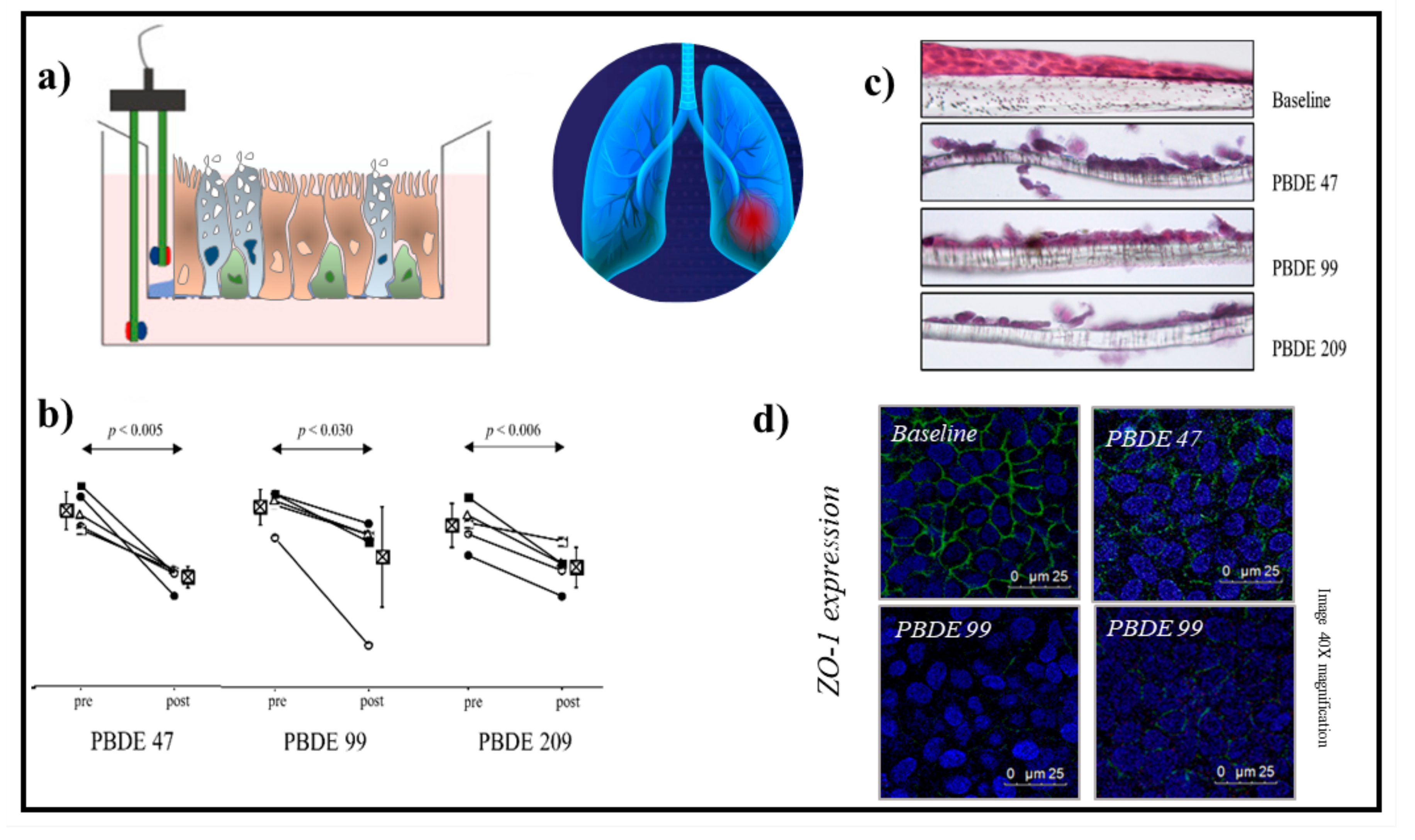
  40. Montalbano, A.M.; Albano, G.D.; Anzalone, G.; Moscato, M.; Gagliardo, R.; Di Sano, C.; Bonanno, A.; Ruggieri, S.; Cibella, F.; Profita, M. Cytotoxic and genotoxic effects of the flame retardants (PBDE-47, PBDE-99 and PBDE-209) in human bronchial epithelial cells. Chemosphere 2020, 245, 125600.
  41. Pozo, K.; Palmeri, M.; Palmeri, V.; Estellano, V.H.; Mulder, M.D.; Efstathiou, C.I.; Sará, G.L.; Romeo, T.; Lammel, G.; Focardi, S. Assessing persistent organic pollutants (POPs) in the Sicily island atmosphere, mediterranean, using PUF disk passive air samplers. Environ. Sci. Pollut. Res. Int. 2016, 23, 20796–20804.
  42. Santoro, A.; Ferrante, M.C.; Di Guida, F.; Pirozzi, C.; Lama, A.; Simeoli, R.; Clausi, M.T.; Monnolo, A.; Mollica, M.P.; Mattace Raso, G.; et al. Polychlorinated Biphenyls (PCB 101, 153, and 180) Impair Murine Macrophage Responsiveness to Lipopolysaccharide: Involvement of NF-κB Pathway. Toxicol Sci. 2015, 147, 255–269.
  43. Frederiksen, M.; Vorkamp, K.; Mathiesen, L.; Mose, T.; Knudsen, L.E. Placental transfer of the polybrominated di-phenyl ethers BDE-47, BDE-99 and BDE-209 in a human placenta perfusion system: An exper-imental study. Environ. Health 2010, 9, 32.
  44. La Guardia, M.J.; Hale, R.C.; Harvey, E. Detailed polybrominated diphenyl ether (PBDE) congener composition of the widely used penta-, octa-, and deca-PBDE technical flame-retardant mix-tures. Environ. Sci. Technol. 2006, 40, 6247–6254.
  45. ECD. Official Journal of the European Union Commission Decision 2005/717/EC—Exemption of DecaBDE from the Prohibition on Use, C 116 (9 May 2008). 2009. Available online: https://eur-lex.europa.eu/legal-content/EN/TXT/PDF/?uri=OJ:C:2008:116:FULL&from=PL (accessed on 24 January 2022).
  46. Crystal, R.G.; Randell, S.H.; Engelhardt, J.F.; Voynow, J.; Sunday, M.E. Airway epithelial cells: Current concepts and challenges. Proc. Am Thorac. Soc. 2008, 5, 772–777.
  47. Schechtman, L.M. Implementation of the 3Rs (refinement, reduction, and replacement): Vali-dation and regulatory acceptance considerations for alternative toxicological test methods. ILAR J. 2002, 43, S85–S94.
  48. Belliveau, M.E. The drive for a safer chemicals policy in the United States. New Solut. 2011, 21, 359–386.
  49. Impinen, A.; Nygaard, U.C.; Carlsen, K.L.; Mowinckel, P.; Carlsen, K.H.; Haug, L.S.; Granum, B. Prenatal exposure to perfluoralkyl sub-stances (PFASs) associated with respiratory tract infections but not allergy- and asthma-related health outcomes in childhood. Environ. Res. 2018, 160, 518–523.
  50. Tannenbaum, J.; Bennett, B.T. Russell and Burch’s 3Rs then and now: The need for clarity in definition and purpose. J. Am. Assoc. Lab. Anim Sci. 2015, 54, 120–132.
  51. Eagle, H. Nutrition needs of mammalian cells in tissue culture. Science 1955, 122, 501–514.
  52. Eagle, H. The specific amino acid requirements of a human carcinoma cell (Stain HeLa) in tis-sue culture. J. Exp. Med. 1955, 102, 3748.
  53. Ritter, D.; Knebel, J.; Niehof, M.; Loinaz, I.; Marradi, M.; Gracia, R.; Te Welscher, Y.; van Nostrum, C.F.; Falciani, C.; Pini, A.; et al. In vitro inhalation cytotoxicity testing of therapeutic nanosystems for pulmonary infection. Toxicol. In Vitro 2020, 63, 104714.
  54. BeruBe, K.; Aufderheide, M.; Breheny, D.; Clothier, R.; Combes, R.; Duffin, R.; Forbes, B.; Gaca, M.; Gray, A.; Hall, I.; et al. In vitro models of inhalation toxicity and disease. The report of a FRAME workshop. Altern. Lab. Anim. 2009, 37, 89–141.
  55. Hiemstra, P.S.; Grootaers, G.; van der Does, A.M.; Krul, C.A.M.; Kooter, I.M. Human lung epithelial cell cultures for analysis of inhaled toxicants: Lessons learned and future directions. Toxicol. In Vitro 2018, 47, 137–146.
  56. Lewinski, N.A.; Liu, N.J.; Asimakopoulou, A.; Papaioannou, E.; Konstandopoulos, A.; Riediker, M. Air-liquid interface cell exposures to nanoparticle aerosols. Methods Mol. Biol. 2017, 1570, 301–313.
  57. Polk, W.W.; Sharma, M.; Sayes, C.M.; Hotchkiss, J.A.; Clippinger, A.J. Aerosol generation and charac-terization of multi-walled carbon nanotubes exposed to cells cultured at the airliquid interface. Part. Fibre Toxicol. 2016, 13, 20.
  58. Kim, J.S.; Peters, T.M.; O’Shaughnessy, P.T.; Adamcakova-Dodd, A.; Thorne, P.S. Validation of an in vitro exposure system for toxicity assessment of air-delivered nanomaterials. Toxicol. In Vitro 2013, 27, 164–173.
  59. Ji, J.; Hedelin, A.; Malmlöf, M.; Kessler, V.; Seisenbaeva, G.; Gerde, P.; Palmberg, L. Development of combining of human bronchial mucosa models with XposeALI(R) for exposure of air pollution nanoparticles. PLoS ONE 2017, 12, e0170428.
  60. Dvorak, A.; Tilley, A.E.; Shaykhiev, R.; Wang, R.; Crystal, R.G. Do airway epithelium air-liquid cultures represent the in vivo airway epithelium transcriptome? Am. J. Respir. Cell Mol. Biol. 2011, 44, 465–473.
  61. Rock, J.R.; Onaitis, M.W.; Rawlins, E.L.; Lu, Y.; Clark, C.P.; Xue, Y.; Randell, S.H.; Hogan, B.L. Basal cells as stem cells of the mouse trachea and human airway epithelium. Proc. Natl. Acad. Sci. USA 2009, 106, 12771–12775.
  62. Tadokoro, T.; Wang, Y.; Barak, L.S.; Bai, Y.; Randell, S.H.; Hogan, B.L. IL-6/STAT3 promotes regenera-tion of airway ciliated cells from basal stem cells. Proc. Natl. Acad. Sci. USA 2014, 111, E3641–E3649.
  63. Polosukhin, V.V.; Cates, J.M.; Lawson, W.E.; Milstone, A.P.; Matafonov, A.G.; Massion, P.P.; Lee, J.W.; Randell, S.H.; Blackwell, T.S. Hypoxiainducible factor-1 signalling promotes goblet cell hyperplasia in airway epithelium. J. Pathol. 2011, 224, 203–211.
  64. Gao, X.; Bali, A.S.; Randell, S.H.; Hogan, B.L. GRHL2 coordinates regeneration of a polarized mucocil-iary epithelium from basal stem cells. J. Cell Biol. 2015, 211, 669–682.
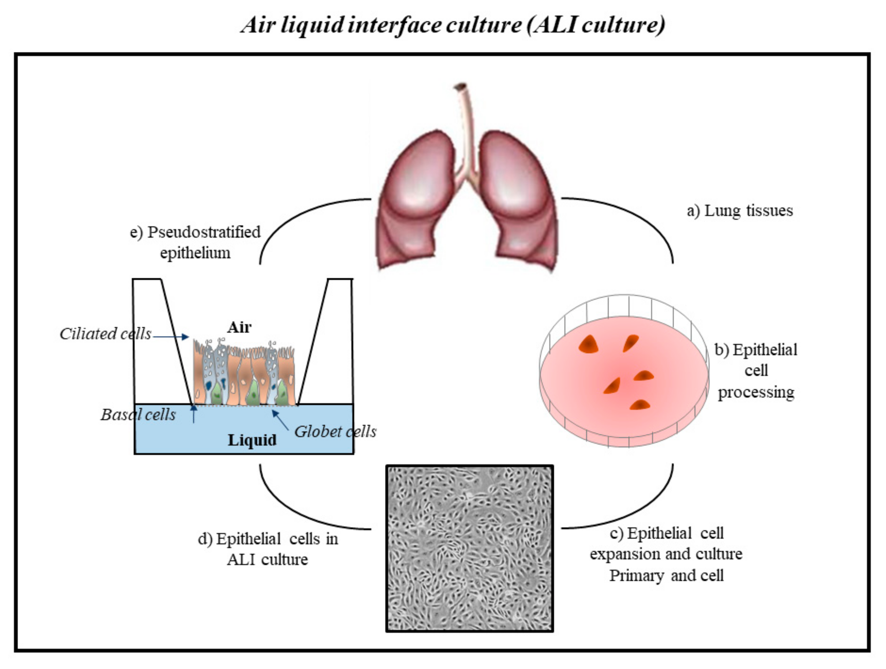
  65. Upadhyay, S.; Palmberg, L. Air-Liquid Interface: Relevant in vitro models for investigating air pollutant-induced pulmonary toxicity. Toxicol. Sci. 2018, 164, 21–30.
  66. King, P.T. Inflammation in chronic obstructive pulmonary disease and its role in cardiovascular disease and lung cancer. Clin. Transl. Med. 2015, 4, 68.
  67. Faber, S.C.; McCullough, S.D. Through the looking glass: In vitro models for inhalation toxicology and interindividual variability in the airway. Appl. In Vitro Toxicol. 2018, 4, 115–128.
  68. Gordon, S.; Daneshian, M.; Bouwstra, J.; Caloni, F.; Constant, S.; Davies, D.E.; Dandekar, G.; Guzman, C.A.; Fabian, E.; Haltner, E.; et al. Non-animal models of epithelial barriers (skin, in-testine and lung) in research, industrial applications and regulatory toxicology. Altex 2015, 32, 327–378.
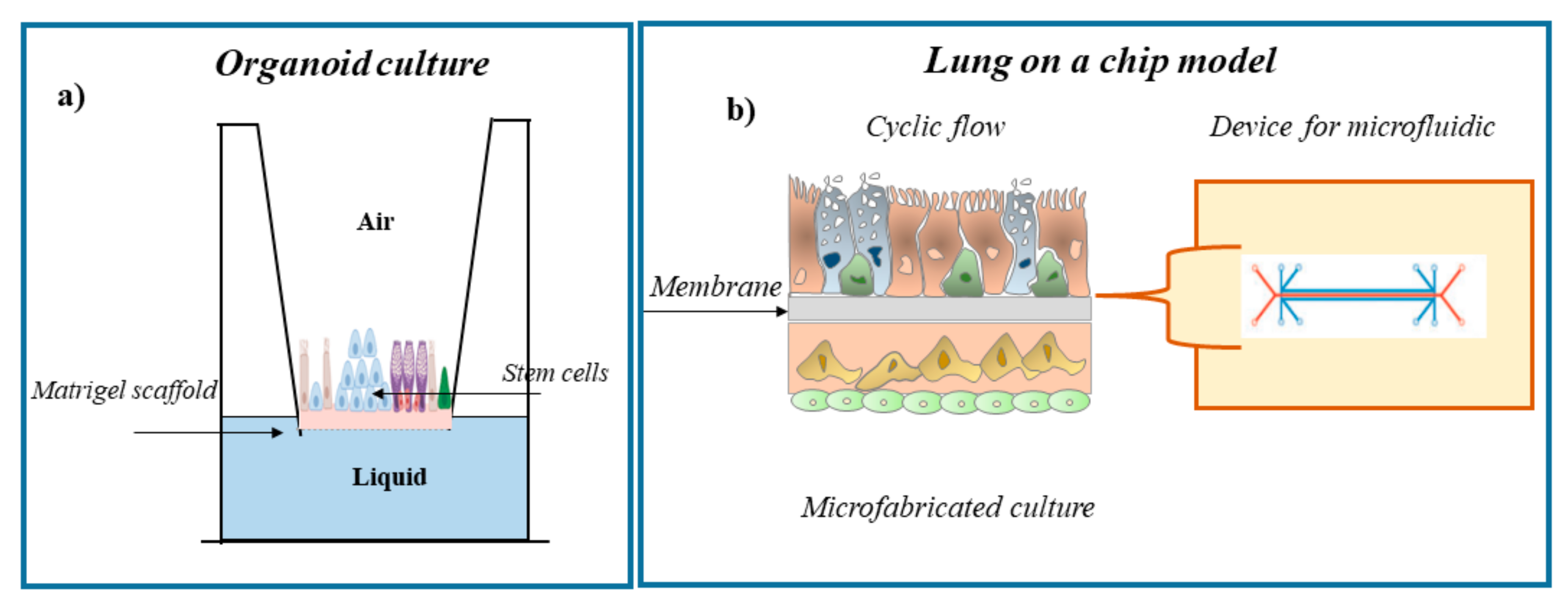
  69. Lancaster, M.A.; Knoblich, J.A. Organogenesis in a dish: Modeling development and disease using organoid technologies. Scence 2014, 345, 1247125.
  70. Fatehullah, A.; Tan, S.H.; Barker, N. Organoids as an in vitro model of human development and disease. Nat. Cell Biol. 2016, 18, 246–254.
  71. Pohl, C.; Hofmann, H.; Moisch, M.; Papritz, M.; Hermanns, M.I.; Dei-Anang, J.; Mayer, E.; Kehe, K.; Kirkpatrick, C.J. Acute cytotoxicity and apoptotic effects after l-Pam ex-posure in different cocultures of the proximal and distal respiratory system. J. Biotechnol. 2010, 148, 31–37.
  72. Papritz, M.; Pohl, C.; Wübbeke, C.; Moisch, M.; Hofmann, H.; Hermanns, M.I.; Thiermann, H.; Kirkpatrick, C.J.; Kehe, K. Side-specific effects by cadmium exposure: Apical and basolateral treatment in a coculture model of the blood-air barrier. Toxicol. Appl. Pharmacol. 2010, 245, 361–369.
  73. Emmler, J.; Hermanns, M.I.; Steinritz, D.; Kreppel, H.; Kirkpatrick, C.J.; Bloch, W.; Szinicz, L.; Kehe, K. Assessment of alterations in barrier functionality and induction of proinflammatory and cytotoxic effects after sulfur mustard exposure of an in vitro coculture model of the human alveolo-capillary barrier. Inhal. Toxicol. 2007, 19, 657–665.
  74. Miller, A.J.; Hill, D.R.; Nagy, M.S.; Aoki, Y.; Dye, B.R.; Chin, A.M.; Huang, S.; Zhu, F.; White, E.S.; Lama, V.; et al. In vitro induction and in vivo engraftment of lung bud tip progenitor cells derived from human pluripotent stem cells. Stem Cell Rep. 2018, 10, 101–119.
  75. Huh, D.; Matthews, B.D.; Mammoto, A.; Montoya-Zavala, M.; Hsin, H.Y.; Ingber, D.E. Reconstituting organ-level lung functions on a chip. Science 2010, 328, 1662–1668.
  76. Clevers, H. Modeling development and disease with organoids. Cell 2016, 165, 1586–1597.
  77. Paget, V.; Dekali, S.; Kortulewski, T.; Grall, R.; Gamez, C.; Blazy, K.; Aguerre-Chariol, O.; Chevillard, S.; Braun, A.; Rat, P.; et al. Specific uptake and genotoxicity induced by polystyrene nanobeads with distinct surface chemistry on human lung epithelial cells and macrophages. PLoS ONE 2015, 10, e0123297.
  78. Nalayanda, D.D.; Wang, Q.; Fulton, W.B.; Wang, T.H.; Abdullah, F. Engineering an artificial alveolar-capillary membrane: A novel continuously perfused model within microchannels. J. Pediatr. Surg. 2010, 45, 45–51.
  79. Stucki, A.O.; Stucki, J.D.; Hall, S.R.; Felder, M.; Mermoud, Y.; Schmid, R.A.; Geiser, T.; Guenat, O.T. A lung-on-a chip array with an integrated bio-inspired respiration mechanism. Lab. Chip 2015, 15, 1302–1310.
  80. Huh, D.D. A human breathing lung-on-a-chip. Ann. Am. Thorac. Soc. 2015, 12 (Suppl. 1), S42–S44.
  81. Fishler, R.; Sznitman, J. A microfluidic model of biomimetically breathing pulmonary acinar airways. J. Vis. Exp. 2016, 9, e53588.
  82. Cromwell, O.; Hamid, Q.; Corrigan, C.J.; Barkans, J.; Meng, Q.; Collins, P.D.; Kay, A.B. Expression and generation of interleukin-8, IL-6 and granulocyte-macrophage colony-stimulating factor by bronchial epithelial cells and enhance-ment by IL-1 beta and tumour necrosis factor-alpha. Immunology 1992, 77, 330–337.
  83. Kotas, M.E.; Medzhitov, R. Homeostasis, inflammation, and disease susceptibility. Cell 2015, 160, 816–827.
  84. Schleimer, R.P.; Kato, A.; Kern, R.; Kuperman, D.; Avila, P.C. Epithelium: At the interface of innate and adaptive immune responses. J. Allergy Clin. Immunol. 2007, 120, 1279–1284.
  85. Grilli, A.; Bengalli, R.; Longhin, E.; Capasso, L.; Proverbio, M.C.; Forcato, M.; Bicciato, S.; Gualtieri, M.; Battaglia, C.; Camatini, M. Transcriptional profiling of human bronchial epithelial cell BEAS-2B exposed to diesel and biomass ultrafine particles. BMC Genomics 2018, 19, 302.
  86. McCullough, S.D.; On, D.M.; Bowers, E.C. Using chromatin immunoprecipitation in toxicology: A Step-by-step guide to increasing efficiency, reducing variability, and expanding applications. Curr. Protoc. Toxicol. 2017, 72, 3.14.1–3.14.28.
  87. Lenz, A.G.; Karg, E.; Brendel, E.; Hinze-Heyn, H.; Maier, K.L.; Eickelberg, O.; Stoeger, T.; Schmid, O. Inflammatory and oxidative stress responses of an alveolar epithelial cell line to airborne zinc oxide nanoparticles at the air-liquid interface: A comparison with conventional, submerged cell-culture conditions. Biomed. Res. Int. 2013, 2013, 652632.
  88. Salthammer, T.; Bahadir, M. Occurrence, dynamics and reactions of organic pol-lutants in the indoor environment. Clean 2009, 37, 417–435.
  89. Petry, T.; Vitale, D.; Joachim, F.J.; Smith, B.; Cruse, L.; Mascarenhas, R.; Schneider, S.; Singal, M. Human health risk evaluation of selected VOC, SVOC and particulate emissions from scented candles. Regul. Toxicol. Pharmacol. 2014, 69, 55–70.
  90. Tsoutsoulopoulos, A.; Möhle, N.; Aufderheide, M.; Schmidt, A.; Thiermann, H.; Steinritz, D. Optimization of the CULTEX(®) radial flow system for in vitro investigation of lung damaging agents. Toxicol. Lett. 2016, 244, 28–34.
  91. Gminski, R.; Tang, T.; Mersch-Sundermann, V. Cytotoxicity and genotoxicity in human lung epithelial A549 cells caused by airborne volatile organic compounds emitted from pine wood and oriented strand boards. Toxicol. Lett. 2010, 196, 33–41.
  92. Dwivedi, A.M.; Upadhyay, S.; Johanson, G.; Ernstgård, L.; Palmberg, L. Inflammatory effects of acrolein, crotonaldehyde and hexanal vapors on human primary bronchial epithelial cells cultured at air-liquid interface. Toxicol. In Vitro 2018, 46, 219–228.
  93. Gostner, J.M.; Zeisler, J.; Alam, M.T.; Gruber, P.; Fuchs, D.; Becker, K.; Neubert, K.; Kleinhappl, M.; Martini, S.; Überall, F. Cellular reactions to long-term volatile organic compound (VOC) exposures. Sci. Rep. 2016, 6, 37842.
  94. Siroux, V.; Agier, L.; Slama, R. The exposome concept: A challenge and a potential driver for environmental health research. Eur. Respir. Rev. 2016, 25, 124–129.
  95. Rim, K. In vitro Models for Chemical Toxicity: Review of their applications and prospects. Toxicol. Environ. Health Sci. 2019, 11, 94–103.
More
Information
Contributor MDPI registered users' name will be linked to their SciProfiles pages. To register with us, please refer to https://encyclopedia.pub/register :
View Times: 790
Revisions: 2 times (View History)
Update Date: 15 Mar 2022
1000/1000