Submitted Successfully!
To reward your contribution, here is a gift for you: A free trial for our video production service.
Thank you for your contribution! You can also upload a video entry or images related to this topic.
Version Summary Created by Modification Content Size Created at Operation
1 + 11898 word(s) 11898 2022-02-16 07:57:50 |
2 format corrected. -6114 word(s) 5784 2022-02-23 02:44:37 | |
3 reference added. + 784 word(s) 6568 2022-02-23 03:40:04 | |
4 format corrected. + 784 word(s) 6568 2022-02-23 03:45:48 | |
5 format corrected. Meta information modification 6568 2022-02-24 04:27:55 |

Video Upload Options

Do you have a full video?

Confirm

Are you sure to Delete?
Cite
If you have any further questions, please contact Encyclopedia Editorial Office.
Thapa, R. Application of Sol–Gels for Treatment of Gynaecological Conditions. Encyclopedia. Available online: https://encyclopedia.pub/entry/19757 (accessed on 26 April 2024).
Thapa R. Application of Sol–Gels for Treatment of Gynaecological Conditions. Encyclopedia. Available at: https://encyclopedia.pub/entry/19757. Accessed April 26, 2024.
Thapa, Ritu. "Application of Sol–Gels for Treatment of Gynaecological Conditions" Encyclopedia, https://encyclopedia.pub/entry/19757 (accessed April 26, 2024).
Thapa, R. (2022, February 22). Application of Sol–Gels for Treatment of Gynaecological Conditions. In Encyclopedia. https://encyclopedia.pub/entry/19757
Thapa, Ritu. "Application of Sol–Gels for Treatment of Gynaecological Conditions." Encyclopedia. Web. 22 February, 2022.
Application of Sol–Gels for Treatment of Gynaecological Conditions
Edit

Approaches for effective and sustained drug delivery to the female reproductive tract (FRT) for treating a range of gynaecological conditions remain limited. The development of versatile delivery platforms, such as soluble gels (sol–gels) coupled with applicators/devices, holds considerable therapeutic potential for gynaecological conditions. Sol–gel systems, which undergo solution-to-gel transition, triggered by physiological conditions such as changes in temperature, pH, or ion composition, offer advantages of both solution- and gel-based drug formulations. Furthermore, they have potential to be used as a suitable drug delivery vehicle for other novel drug formulations, including micro- and nano-particulate systems, enabling the delivery of drug molecules of diverse physicochemical character. Hence, such systems are are of profound significance in delivering the drugs to various parts of FRT for optimal treatment of various gynecological conditions which was not achievable using conventional drug delivery technologies.

vaginal drug delivery sol–gel formulations stimuli-responsive polymers mucoadhesion vaginal applicators/devices

1. Introduction

Recent advances in pharmaceutical research and development have drawn considerable attention towards developing more effective, patient-friendly, and clinician-endorse treatment options for conditions of the FRT. Compared to the oral/parenteral routes, local and direct vaginal drug delivery (VDD) is preferred for both small and large molecules, owing to the mucosal surface area of the upper and lower FRT, with its rich blood supply, and avoidance of the gastrointestinal tract and hepatic first-pass metabolism. From the patients’ perspective, VDD minimizes off-target effects and enables self-administration in the case of lower FRT conditions, obviating the need for specially trained medical personnel [1][2][3].
The vagina offers a direct channel for delivering therapeutic agents for the treatment of various gynecological conditions that have, to date, been exclusively delivered via the oral/systemic route. However, effective VDD is challenged by the highly variable anatomical, physiological, and microbiological features of the FRT, which transition over a patient’s lifetime, consequently posing a great challenge to formulation scientists where catering to the varied needs of patients can be effectively met.
In this regard, various conventional formulations (e.g., creams and pessaries), although administered vaginally, result in limited delivery success, due to the physiological clearing mechanism of the vagina, which results in premature leakage and sub-therapeutic outcomes. These shortfalls stem from a short drug residence time on mucosa, with lowered drug absorption and unfavourable clinical outcomes [1][4][5][6]. Thus, an alternative vehicle that can tolerate a changing microenvironment and has good mucoadhesive/retention properties, while having appropriate flow properties to adequately disperse, reach, and coat target mucosa/structures in the FRT, is sought.
In this context, phase-transforming sol–gels offer a safe, practical, economical, and effective solution to the long-standing issues hampering drug delivery to the upper and lower FRT [1]. Sol–gels are initially in solution form, enabling ease of dispersion onto mucosa with an appropriate device, undergoing rapid gelation in response to physiological stimuli such as body temperature, changes in pH, and/or the presence of counter ions. Once gelled, the mucoadhesive properties of an appropriately engineered sol-gel pave the way for the sustained delivery of infused therapeutics (e.g., small molecules, peptides, proteins, genes) to underlying mucosal tissue [7][8]. Additionally, the hybridization of sol-gels with drug encapsulation strategies such as nanoparticles, microspheres, liposomes, and PEGylation can be used to further enhance/fine-tune the drug release characteristics [9]. Several studies confirm the successful delivery of drugs using sol-gel formulations in a number of FRT-related conditions, including infection treatment, the delivery of contraceptives, labor induction, supplementation of the microbiome, HRT, and prophylaxis of sexually transmitted diseases [10][11][12][13][14][15][16]. Given its immense potential, the highly versatile sol-gel platform technology has drawn great attention and interest from the scientific community in reimagining drug delivery to the FRT.

2. Sol–Gel Platform Technology in Vaginal Drug Delivery System

The vagina is an appropriate site for local and systemic drug delivery to treat/manage a broad range of gynecological conditions [17]. A range of intravaginal medications is available on the market, with most requiring frequent application/administration due to the associated key limitations of short residence time and inadequate drug distribution on/through the vaginal mucosa (Figure 1) [3][4]. In this context, there is a case to be made for sol-gels, which can be engineered to prolong the vaginal residence time, resulting in predictable drug disposition to the vaginal mucosa (Figure 1) [4][18]. Sol–gels, also referred to as in situ hydrogels, which involve phase transition simply in water, have shown promising results in terms of protein and peptide drugs, tissue engineering, and overcoming barriers to drug absorption [19]. With their combined attributes of both solutions and gels, they support polymer-induced drug solubilization, and uniform drug distribution, when applied to the vaginal mucosa with an appropriate applicator/device [11]. Once in the gelled state, increased viscosity and incorporation of excipients that promote mucoadhesion reduce vaginal outflow/leakage and mitigate the loss of the drug from the vaginal cavity, providing greater opportunity for drug absorption in/through the vaginal mucosa and favourable drug release kinetics [20][21]. Thus, overall, an appropriately engineered mucoadhesive in situ sol–gel system can bring a multitude of benefits that support the attainment of desired clinical outcomes in the FRT.
/media/item_content/202202/62159d44a5a08gels-08-00099-g002.png
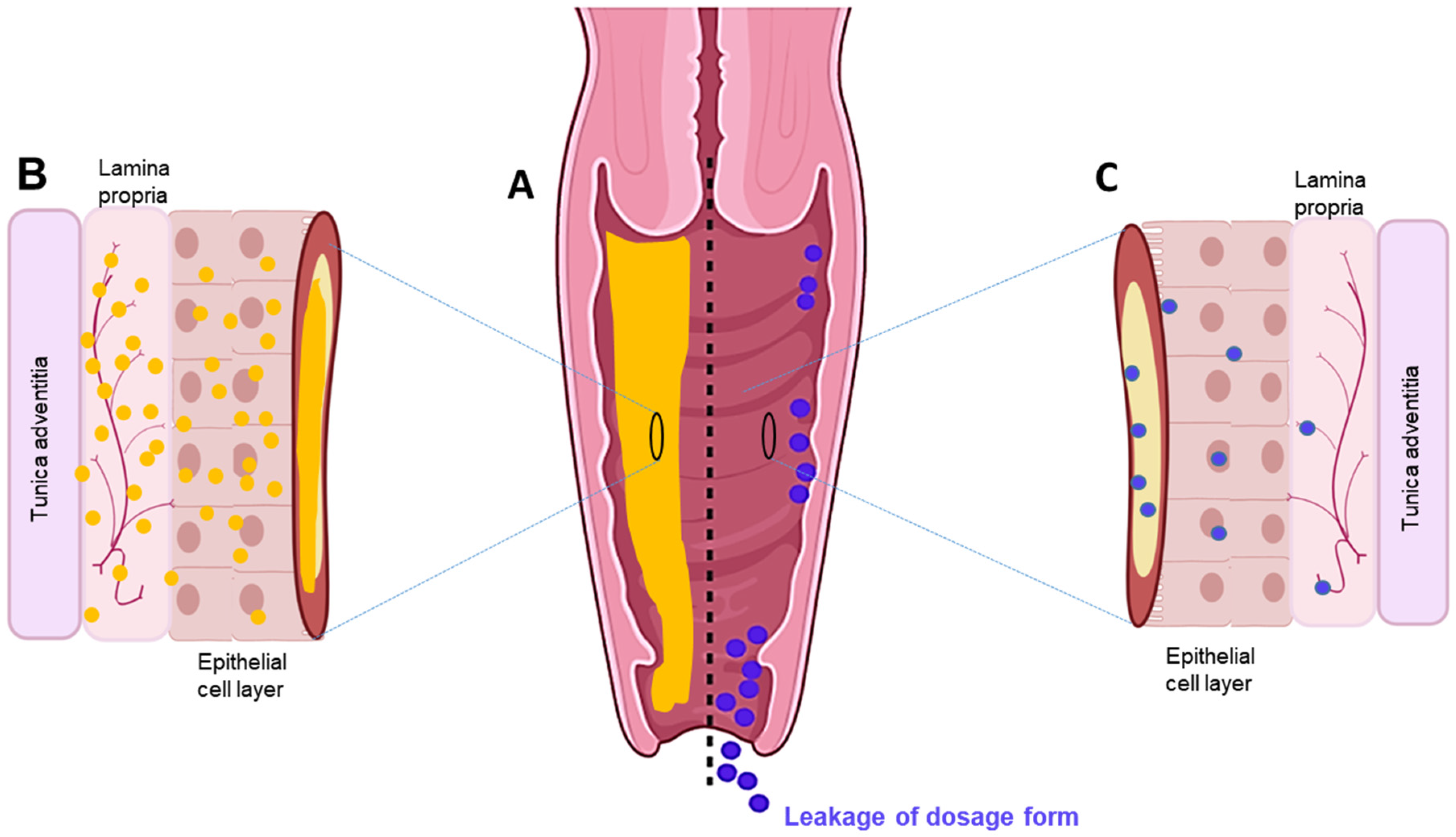
Figure 1. (A) Cross-section of the vaginal tract. (B) Uniform distribution and diffusion of drug throughout mucosal–epithelial layer with sustained delivery using an in situ sol–gel system. (C) Poor and sparse drug distribution through mucosal-epithelial layer and leakage via conventional dosage forms.

2.1. Features and Use of Vaginal Sol–Gel Formulations

The development of a drug delivery system using stimuli-reactive smart polymers, as is the case with sol–gels, allows fine-tuning of the rheomechanical properties aligned to the use, be it localised (deposition) or systemic (permeation) drug delivery that is sought [21][22]. These smart polymers tend to transform their characteristics in response to physiological changes, with the extent/degree of transformation determined by the nature of the monomer(s), charge density, pendant chains, and the degree of polymer crosslinking [23]. Stimuli-responsive in situ sol–gel systems are a unique dosage form, being a clear, low-viscosity polymeric liquid/solution, which, through a given trigger (pH, temperature, ions), converts to a viscous gel upon administration into a body cavity [21]. From a thermodynamic viewpoint, the balance between the hydrophobic and hydrophilic groups on the polymer chain and the free energy of mixing ( Δ G = Δ H T Δ S ) result in marked alteration of the aqueous solubility of the polymer and cause sol-to-gel phase transition [1][6]. Since the enthalpy (ΔH) is smaller than the entropy (ΔS), an increase in temperature (T) results in negative free energy of association (−ΔG), which increases the preference of polymer-polymer and water-water interactions to polymer–water interactions and causes the dehydration of solvated polymers [1][24]. In the case of amphiphilic polymers, an increase in polymer concentration above CMC results in the packaging of micelles in an ordered manner, forming a hydrophobic core and hydrophilic shell, and ultimately forms a gel [1][24][25]. Various mechanisms of stimuli-responsive gelation are shown in Table 1.
Table 1. Marketed VDD formulations, their indication, and the manufacturer.
Active Drug Brand Name® Dosage Form Indication Manufacturer Reference
Oestradiol Vagifem Tablet Atrophic vaginitis Novo Nordisk Health Care AG [26]
Dinoprostone Prostin E2, Tablet Cervical ripening and labour induction Pfizer [27]
Dinoprostone Cervidil Insert Cervical ripening and labour induction Forest
Laboratories
[28]
Misoprostol Misodel Insert Labour induction Ferring
Pharmaceuticals
[29]
Progesterone Endometrin Insert Assists embryo
transplantation
Ferring
Pharmaceuticals
[30]
Oestradiol Imvexxy Inserts Atrophic vagina Therapeutics MD [31]
Clotrimazole Gino-Canesten Cream Vulovaginal candidiasis Bayer [32]
Sertaconazole Sertopic Cream Vulovaginal candidiasis CPH [32]
Clindamycin Dalacin V Cream Antibacterial Pfizer [32]
Z. multiflora Leucorex Cream Trichomoniasis Barijessence [33]
Oestriol Ovestin Cream Oestrogen hormone
supplement
Aspen [32]
Etonogestrel/
Ethinyloestradiol
Nuvaring Ring Endometriosis, cervical cancer Organon [3][34][35][36]
Progesterone Progering Ring Release progesterone Laboratorios
Andrómaco
[36]
Oestradiol Estring Ring Oestrogen replacement therapy, cervical cancer Pfizer [3][35]
Nonoxyl-9 Today Sponge Spermicide Almatica
Pharma, Inc.
[3]
Progesterone Crinone Gel Assisted reproductive procedures Merck [37]
Nonoxynol-9 Vaginal Contraceptive Film Film Spermicide Apothecus [3]
Lactobacilli gasser and Lactobacilli rhamnosus EcoVag Capsule Bacterial vaginosis HÄLSA Pharma GmbH [38]
Progesterone Utrogestran Capsule Luteal phase support Laboratories Besins International [39]
The stimuli-responsive polymers exhibit pseudo-plastic behaviour, which is highly desirable to formulate sol–gels as they offer better distribution on the mucosal tissue [25]. For the drug formulation exhibiting such behaviour, viscosity is reduced during application on the mucosa due to the change in alignment of tangled polymeric chains in the shear direction. However, the original rheological profile is regained immediately after application on the vaginal mucosa in response to the physiological stimuli [6][25]. This leads to the formation of a viscous gel in the vaginal cavity, which is essential for prolonged drug residence time in the vaginal cavity and ultimately results in sustained and controlled drug release [21]. The sustained and controlled drug release profiles exhibited in the examples of sol–gel systems included in Table 1 are attributed to the increased vaginal residence time of the drug formulations. Further, drug distribution in the mucosal tissue is also assisted by the ability of the gel to permeate the mucosa [6]. For instance, a significant increase in drug permeation was observed with a vaginal in situ gel formulation of fluconazole, which could be attributed to both the improved drug release profile of the drug in the formulation as well as the interaction between chitosan used in the formulation and mucin present in the vaginal mucosal layer [40]. Studies on VDDS report an increase in drug permeability by the use of polyethylene glycol [41].
Mucoadhesive in situ gelling systems are those (containing natural or synthetic mucoadhesive polymers) that interact with and adhere to the mucosal epithelial surface components, particularly mucin, via hydrogen bonding, electrostatic interaction, and van der Waals forces, and, once administered in soluble form, they rapidly undergo in situ gelation (Figure 2) [8][42][17]. Published results suggest that at least 6 h of vaginal drug residence time is desired to represent the mucosal clearance turnover rate for the drug to cross the mucosal barrier (30–100 µm thick), epithelium layer (200–300 µm), and other adjacent layers before reaching the blood vessels in the vagina. This target can be met by a gel using mucoadhesive polymers containing suitable surface characteristics, charge, and functional groups, such as hydroxyl, sulphate, carboxyl, and amine groups—for example, polymers such as polyacrylic acid, cellulose, chitosan, hyaluronic acid, carrageenan, alginate, gums, and sulphated polysaccharides [6][7][20][17]. Use of these polymers results in optimum mucoadhesive strength, sustained drug release, and increased drug uptake by the vaginal mucosa [7]. Mucoadhesive systems not only improve bioavailability through localised action but also alter tissue permeability and enhance the absorption of protein and peptide-based drugs [43]. This makes mucoadhesion an essential parameter to consider and optimise as per the requirements to obtain formulations for controlled and sustained deliveries [44][45]. For instance, in a study comprising a thermosensitive formulation, mucoadhesive formulation, and thermosensitive–mucoadhesive system, the in situ mucoadhesive gel was considered the optimum formulation, with a longer vaginal residence time compared to the two other systems (>8 h). The study suggests that both gelation and mucoadhesiveness together result in a robust vaginal formulation [41].
/media/item_content/202202/62159d667c180gels-08-00099-g003.png
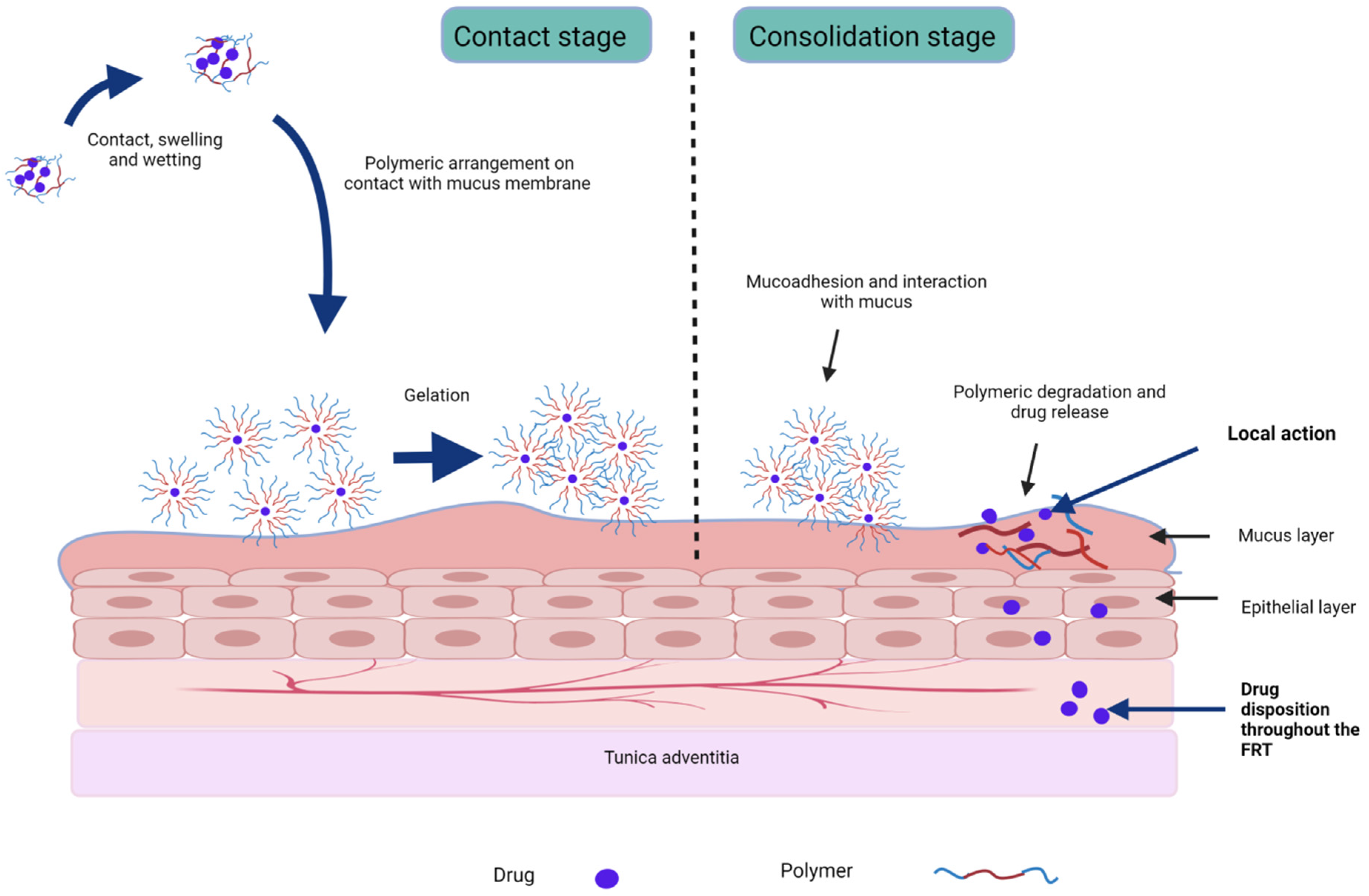
Figure 2. Stages of mucoadhesion and drug release from stimuli-responsive sol–gel formulations.
Mucoadhesion occurs in two stages: (i) contact stage involving hydration, wetting, and spreading; (ii) consolidation stage involving strengthening polymer and mucin interactions through hydrogen bonds, hydrophobic interactions, van der Waals forces, electrostatic interactions driven by negatively charged mucin, and/or mucoadhesive or polymer chain interpenetration into the cervicovaginal mucus gel (Figure 2) [6][7][45]. Mucoadhesive intravaginal formulations should be engineered considering the nature and physico-chemical characteristics of drugs and their transportation route to overcome the associated challenges and improve their therapeutic efficacy [46]. In addition, for vaginal application, these polymers should be non-toxic, non-irritating, flexible, comfortable, and ideally remain unabsorbed in the vaginal epithelium [47]. The various polymers used in VDDS are represented. Natural polymers used in these systems typically respond to single or multiple stimuli, while synthetic polymers respond to specific stimuli. However, a major issue with the use of synthetic polymers is that they can result in irritation and toxicity to the underlying tissue [21]. Hence, diligent polymer and excipient selection is necessary before formulation into sol–gel systems, for either localised or systemic action.
In this context, the World Health Organization (WHO) has issued guidance for vaginal preparations, such as lubricants, recommending that they be mildly acidic (pH 4.5), with an upper limit of osmolality not exceeding 1200 mOsm/kg, to minimise any risk of mucosal/epithelial damage [44][48]. In the context of tonicity, while hypotonic vaginal products enhance muco-penetration, hyperosmolar vaginal products raise safety concerns with respect to vaginal tissue health and sperm viability and mobility [32]. This was corroborated in a phase 1 clinical trial for a vaginal microbicide developed against HIV, which was discontinued, with the sponsor citing unacceptable side effects resulting from the high osmolality of the gel, reinforcing the need to factor in osmolality when developing vaginal semi-solid formulations [49]. In addition, the size of the carriers/particles infused in semi-solid formulations has a direct impact on cervicovaginal mucosal and epithelium penetration, with a 200–500 nm particle size range recommended for VDD mucus [44]. Surprisingly, lowering the particle size range to 100–150 nm results in them becoming trapped and immobilised in the numerous tiny pores/pockets of the cervicovaginal mucosa, rendering them appropriate for localised, deep mucosal drug delivery. In contrast, larger-sized carriers >1000 nm are unable to diffuse into such pores/pockets and so remain on the outer mucosal surface, where they are susceptible to more rapid clearance by ciliated mucosa [44][50].
Considering the biological and physicochemical challenges, nanotechnology-based delivery systems have proven a promising means of improving drug distribution, retention, and therapeutic efficacy in VDD [51]. Nanoparticulate systems can enhance the solubility, bioavailability, and targeting of drugs, while increasing the rates of dissolution and surface area that can be reached. This can be achieved through the design of particulates including, but not limited to, micelles, carbon nanotubes, polymeric lipid nanoparticles, nanocapsules, nanogels, nanofibers, dendrimers, quantum dots, and nanocomposites, which are extensively reviewed elsewhere [19][50][52]. The literature suggests that these nanocarriers have demonstrable drug solubility enhancement properties, while also protecting against rapid drug degradation and enhancing drug concentrations in target tissues, further masking them from the harsh conditions of the FRT and addressing the many shortfalls of conventional VDDS [53][54][55]. For instance, the aqueous solubility, stability, and mucosal permeability of antifungal drugs have been addressed using inclusion complexes and gel flakes [56][57]. Antifungal drugs prepared using hydroxypropyl β-cyclodextrin (HPβ-CD) have been readily incorporated into sol–gel formulations, and exhibited sustained drug release without any detrimental effect on the vaginal tissue [56]. Similarly, enhanced drug permeation/bioavailability was achieved through the vaginal epithelium, alongside improved epithelial drug viability, when in situ nanoparticles of acyclovir, a highly water-insoluble drug, were formed, in comparison to the pure drug [58]. Likewise, atorvastatin, a BCS class II drug, when formulated into nanoparticles exhibited significant improvements in solubility and efficacy, compared to the native powdered atorvastatin [59]. Hence, the use of vaginal sol–gel formulations as vehicles for such tailored and innovative micro- and nano-encapsulated drug forms is a highly promising proposition to address the shortfalls of conventional dosage forms for a range of conditions, as elaborated in Table 1 [1][9][60][61].
The range of gynaecological conditions receiving significant attention through innovative formulation development include vaginal infection and atrophy, neoplasia, labour induction, prophylaxis of HIV, and contraception [32][62][63][64]. Vaginal infections in particular are of growing concern, and, in this context, several studies have focused on the development of sol–gels to treat a range of STIs of bacterial and fungal origin [3][61]. A few studies on vaginal sol–gel systems are represented in Table 1. These studies suggest that the sol–gel formulations, when used in vaginal infections, not only have better efficacy compared to their conventional formulations but also are associated with reduced toxicity towards the underlying tissues of the FRT. Hence, they present strong potential to solve the problems of high reinfection rate and incomplete treatment with the current treatment regimen of vaginal infections. For instance, in a pilot, randomised, controlled trial with confirmed bacterial vaginosis, which has a very high rate of reoccurrence, the initial treatment rate for an in situ gel and gel was 85% and 71.24%, respectively. However, the difference was more prominent with treatment for 4 weeks, with values of 80% with the in situ gel and 47.4% with gel application, indicating the higher long-term efficacy of in situ gels, which can be ascribed to the increased mucoadhesiveness, increased vaginal residence time, and sustained release nature exhibited by the use of poloxamers in in situ gel formulation [61].
Current strategies for HIV prophylaxis utilise vaginal microbicides that act specifically on the critical steps of HIV transmission, and several such formulations are in different stages of development [4][65]. In this regard, vaginal sol–gels provide an opportunity for early intervention to the sexual transmission of HIV in females, with pH-induced gelation triggered by exposure to semen, effectively shielding underlying epithelial cells, and restricting the entry of virions into the systemic circulation [64]. For instance, a pH-responsive polymeric network comprising phenylboronic acid, salicylhydroxamic acid, and 2-hydroxypropyl methacrylamide impeded the migration of HIV at pH ≥ 4.8 [66]. This mechanism gives rise to the concept of “molecular condoms”, where the temperature and pH responsiveness of formulations can be applied to the vaginal mucosa setting, with the gel form effectively covering the mucosal tissue and releasing microbicides [67]. Moreover, this concept of shielding the mucosal tissue helps drug concentration and retention at the vaginal mucosa surface to facilitate mucopenetration and enhance pharmacokinetics at the target tissue [14]. In cases where the microbiome and/or vaginal mucosal tissue integrity is compromised, susceptibility to HIV infection is elevated. Therefore, co-administration of anti-HIV drugs alongside the localised delivery of mucosal barrier formulations is a combination approach to the prophylaxis of HIV infection that warrants widespread use [14]. For instance, thermosensitive nanoparticles of the combination of hydrophilic drug Raltegravir, a HIV integrase inhibitor, and lipophilic drug efaviren (non-nucleoside reverse transcriptase inhibitor), prepared using a poloxamer (Table 2), resulted in thermogelation at 32.5 °C and exhibited anti-HIV activity at a concentration lower than that exhibited by the solution of the combination of these drugs. Moreover, nanoparticles were taken up rapidly by HeLa cells (within 30 min) and exhibited sustained drug release without exhibiting cytotoxicity for a period of 14 days, which indicates that the formulation is a suitable candidate for the prevention of prolonged vaginal pre-exposure of HIV. Furthermore, the incorporation of (RAL + EFV) nanoparticles did not result in the aggregation of nanoparticles, suggesting that the thermosensitive gel is an effective drug delivery vehicle for these anti-HIV drug-loaded nanoparticles [13].
Similarly, intravaginal dendrimer-based sol–gels have also earned considerable attention in recent years for the treatment of the highly challenging HPV infection, particularly in pregnant women, where systemic drug exposure is not desirable for either the mother or the growing foetus [53]. An in situ hydrogel infused with amoxicillin using a generation 4 poly(amidoamine) dendrimer with polyethylene glycol provided in vitro drug release for 240 h and a sustained antibiotic effect through a dual mechanism, i.e., the antibiotic effect of the dendrimer itself and the sustained release of the drug. Moreover, the dendrimer complex targeted the inflammatory cells and reduced cytotoxicity and hence no change in vaginal pH or tissue necrosis was observed while the formulation was retained in the vaginal mucosa (72 h), after which the hydrogel started to become degraded [68].
With these established benefits of VDDS, and the many limitations/unwanted effects associated with current modes of hormonal contraceptive administration, safer and more patient-friendly intravaginal hormonal contraceptives—specifically, stimuli-responsive in situ hydrogels—are gaining attention and interest [62][69]. It has been demonstrated that formulations containing multiple drugs are more efficient contraceptives compared to single-drug formulations [70][69]. In this context, an in situ pH-responsive hydrogel containing indomethacin, gestodene, and ethinyl estradiol prevented pregnancy completely compared to a control group, which presented 100% pregnancy. However, surprisingly, another group receiving hydrogels without any drugs had a 60% pregnancy rate, indicating that the hydrogel components also play a role in contraception [69]. Likewise, in situ hydrogels of non-hormonal agents have also been explored for vaginal contraceptives [10][63]. For instance, a chitosan-based in situ hydrogel of iron (II) gluconate dihydrate, used prior to sexual intercourse, releases iron rapidly in the vagina and exhibits spermicidal properties [10]. In another study, poloxamer-based temperature-responsive in situ hydrogels of nonoxynol-9 resulted in up to 10 h of vaginal residence time [71]. With the rising interest in using multi-drug treatment regimens for more comprehensive therapeutic coverage, drugs with complementary modes of action have been proposed (e.g., anti-HIV + anti- HPV + spermicide), and so the development of sol–gels in this context would be an important milestone in advancing VDD and women’s health and well-being more broadly [14][72]. For instance, vaginal administration of a nanoparticle formulation containing an antimicrobial and spermicidal agent, curcumin, and the anti-HIV agent efavirenz exhibited better encapsulation efficiency compared to single-drug nanoparticles and exhibited better efficacy compared to their solution form, without affecting lactobacilli viability or vaginal tissue, hence indicating the formulation as an efficient example of multiple prevention technology (MPT)-based VDDS [73]. In this context, the development of an efficient delivery vehicle such as sol–gels would enhance the therapeutic benefits [74]. Additionally, the intravaginal administration of hormones infused in sol–gels is also being investigated for hormonal replacement therapy and fertility treatment, while its application in cervical cancer has shown promise (Table 2 and Table 3) [75][76].
Table 2. Sol–gel formulations designed for gynaecological indications.
Indication API Drug Form Stimuli-Sensitive and Mucoadhesive Polymers (w/v) Gelation Trigger Gelation
Mechanism
Comments References
Bacterial vaginosis Metronidazole Free drug 20% poloxamer 407 and 10% poloxamer 188 Temperature Swelling due to polymeric crosslinking Increased prolonged curative rate with sol–gel (80%) compared to conventional gel (47.4%) [61]
Clotrimazole Free drug 15% poloxamer 407, 15% and/or 20% poloxamer 188, and 0.2% w/v polycarbophil Temperature Micelle formation Antifungal effect for 10 days; reduced toxicity to epithelium cells of
human cervix
[18][21]
Secnidazole Aerosol foam 0.45% carbopol 940 with 0.35% HPMC K4 M and 0.35% carbopol 940 with 0.35% HPC pH Hydrogen bonding Less than 50% of drug released by 8 h, indicating controlled drug release [77]
Secnidazole Free drug 20% poloxamer 407, 1% poloxamer 188, and 1 or 2.5% chitosan Temperature Micelle formation Approximately 1–2-fold increase in mucoadhesiveness with chitosan [11]
  Clindamycin Free drug 1% gellan gum and 1% HPMC Ion Polymeric crosslinking Good gelling capacity; good mucoadhesion and adequate inhibition of microbial growth [17][78]
  Voriconazole Drug-
hydroxypropyl
β-cyclodextrin inclusion complex
Poloxamer 407, poloxamer 188 HPMC, HEC, polycarbophil, and carrageenan Temperature Formation of closely packed micelles in aqueous medium Increased vaginal tissue uptake by the use of
cyclodextrin and
sustained drug
release for 8 h using in situ gel in female Wistar rats compared to conventional formulation
[60]
  Amphotericin B Drug-
Hydroxypropyl ϒ-cyclodextrin complex
25% poloxamer-based multiblock
copolymers
pH and
temperature
Hydrogen bonding Toxicity reduced by complexation; dissolution controlled drug release rate; prolonged drug release observed at pH 7.4 and pH 9.0 [56]
Herpes simplex virus (HSV) infection Acyclovir Nanoparticle 18% poloxamer 407 pH and temperature Polymeric crosslinking Drug’s therapeutic level achieved with 10 times smaller amount of drug;
relative bioavailability increased twice compared to suspension dosage form of pure drug
[58][79]
Infertility Fetilty-Promoting intrauterine infusion liquid (FPL) Icariin extracted from Epimedium, safflower, and motherwort 19% poloxamer 407, 2.5% poloxamer 188, and 0.3% HPMC Temperature Hydrogen bonding Uterus and ovarian
indices significantly increased in the rats receiving the sol–gel formulation compared to control group;
oestradiol levels increased after day 7 to day 22
[80]
Sildenafil citrate Free drug 15% poloxamer 407 and 1% HEC Temperature Entanglement and condensed
micelle packing at increased polymer concentration
Sol–gel transition temperature reduced by addition of HEC; increased endometrial thickness as well as uterine flow with reduced dosing length compared to vaginal suppositories [81]
Pre-
exposure prophylaxis of HIV
Raltegravir + efaviren (RAL + EFV) Nanoparticles 20% poloxamer 407 and 1% poloxamer 188 Temperature Hydrogen bonding Inhibitory concentration of RAL + EFV–NPs less than the solution form; sol–gel proved an efficient delivery vehicle of NPs [13][82]
Tenofovir Microsphere α,β-glycerophosphate (GP), chitosan, sodium alginate Temperature Electrostatic interaction between polymers Viscosity of chitosan–GP complex strengthened by sodium alginate; initial burst release (30%) in the first 30 min followed by cumulative release (87.82%) after 24 hrs [83]
Contraceptive Nonoxynol-9 Free drug 18% poloxamer 407 and 1% or 6% poloxamer Temperature Micelle
formation
Increased vaginal residence time compared to solution form; rapid hydrogel erosion and drug release [11][71]
Intrauterine device
insertion for contraception
Lidocaine Free drug 18% poloxamer 407, 5% poloxamer 188, and 0.3% gellan gum Temperature and ionic strength Hydrogen bonding between the polymers Better acceptance and pain management by sol–gel formulation compared to conventional gel [82]
Hormone replacement therapy, preterm birth Progesterone Free drug 5% glycol chitin Temperature Hydrophobic interaction No significant effect on
gel property by viscosity reduction after dilution by vaginal fluid but not
recommended in presence of semen; prolonged vaginal residence time and controlled drug release
[76][84]
Cervical cancer Doxorubicin Free drug 7% glycol chitin Temperature Hydrophobic interaction Initial 20% burst release followed by sustained release for 13 days [75][84]
HPMC—hydroxypropyl methyl cellulose, HPC—hydroxypropyl cellulose, HEC—hydroxyethyl cellulose.
Table 3. Types of polymers used in VDD.
Source Polymers Role/Feature References
Plant Cellulose derivatives e.g., HPMC, HPC, HEC, MC, EC Thermo responsive gelation;
Mucoadhesive; non-biodegradable
 
Pectin Mucoadhesive  
Alginate Biocompatible; biodegradable; anionic; ion-responsive gelation  
Carrageenan Mucoadhesive; antimicrobial and antiviral activity [17][85]
Animal Chitosan Polycationic copolymer;
Mucoadhesive; biocompatible; biodegradable; antibacterial activity
[47][17]
Gelatin Biocompatible; biodegradable; [17]
Hyaluronic acid Negatively charged [47]
Microbial Gellan gum Ion-responsive gelation [17]
Xanthan gum Form physical gel [17]
Synthetic Poloxamers Non-ionic triblock copolymer; amphiphilic; multi-stimuli responsive gelation [47][17][86]
Polyacrylates Viscosity affected by formulation pH [47]
Polyethylene glycol Water soluble [17]
Polyvinylpyrrolidone Linear; water soluble [17]
HPMC—hydroxypropyl methylcellulose, HPC—hydroxypropyl cellulose, HEC—hydroxyethyl cellulose, MC—methyl cellulose, EC—ethyl cellulose.

2.2. In Situ Sol-to-Gel Phase Transition Stimuli

2.2.1. Thermoresponsive Gelation

Thermosensitive sol–gel systems comprise polymers that undergo gelation at/approaching body temperature. Gelation occurs physically by the entanglement of polymer chains, micelle packing due to self-assembly of the polymeric micelles at elevated temperatures, physical crosslinking due to the dehydration of the polymeric block above a lower critical solution temperature (LCST), hydrophobic interaction, and the transition of a coil into helix form [6][8]. The aqueous solutions of thermogelling polymers have a perfect balance of hydrophilic and hydrophobic groups, which is disturbed with the slightest change in temperature, and they undergo phase separation at the critical solution temperature (CST) [8][21]. For the polymers exhibiting LCST, phase separation occurs above CST, whie the opposite is true for polymers exhibiting an upper critical solution temperature (UCST) [87]. The hydrophilic polymers become hydrophobic and insoluble above their LCST, resulting in gel formation (Figure 3A). LCST determines the thermo-reversibility of thermoresponsive systems and depends upon the polymer concentration [8]. There is an inverse relationship between polymer concentration and gelation temperature, driven by the hydrophobic force [8][88]. At a higher polymer concentration, hydrophobic interaction increases due to molecular crowding, resulting in gelation at a lower temperature [79]. Polymers are typically used in concentrations that trigger gelation in the 25–37 °C range, in the context of VDD. Using an appropriate applicator, an appropriately engineered sol–gel can provide ease of application, while its rapid transformation to a viscous gel can reduce leakage, enhancing retention on the vaginal mucosa [6]. Here, a gelling temperature close to the physiological temperature is ideal for the stabilisation, solubilisation, and controlled release of hydrophobic drugs, as the polymeric monomers aggregate to form micelles within their hydrophobic core, wherein the solubilised hydrophobic drug resides [21][25]. The concentration of thermogelling polymers, co-solutes, and dilution by fluid in the vagina affect the gelation temperature and the viscosity of the gel formed. Hence, it is essential to characterise thermosensitive systems in simulated conditions to help predict their in vivo performance [8][18]. Temperature-stimulated sol–gel transition is a commonly employed phenomenon in several studies of VDDS, even though dual stimuli are also employed for sol–gel transition (see examples in Table 2).
/media/item_content/202202/62159d951baabgels-08-00099-g004.png
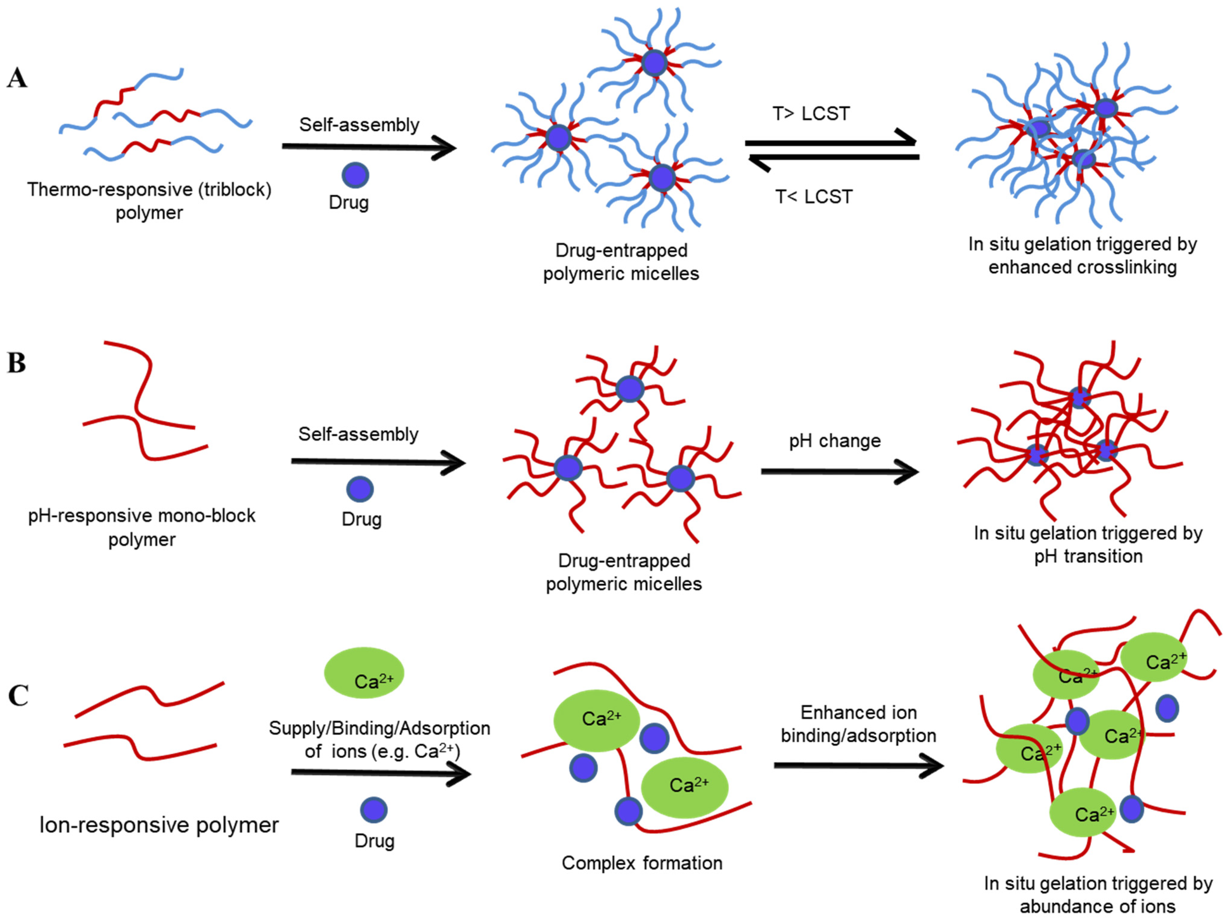
Figure 3. Sol–gel transition of various stimuli-sensitive polymeric systems: temperature-sensitive (A), pH-sensitive (B), and ion-sensitive (C) systems. T—transition temperature, LCST—lower critical solution temperature.
  • Poloxamers
Poloxamers (Pluronic®) are triblock copolymers of poly(ethylene oxide)-poly(propylene oxide)-poly(ethyleneoxide) (PEO-PPO-PEO) units. They are amphiphilic in nature, with two outer hydrophilic PEO segments and an inner hydrophobic PPO segment that can partly solubilise hydrophobic drugs [79]. However, such characteristics result in inconsistent drug release profiles since the drug loaded in the PEO portion is released prior to gel dissolution, in contrast to the drug loaded in the PPO portion, which is released after gel dissolution; hence, modification of the formulation is required for better drug release characteristics [86]. An increase in temperature causes a change in the orientation of the methyl group of the side chain and dehydration of the PPO segment, as well as water extrusion from the micellar core of poloxamers, resulting in gelation [21]. Above the critical micellar concentration of polymers (CMC), the hydrophobic cores of the micelles absorb water and can also accommodate and solubilise hydrophobic drugs [21]. This encapsulation process can also protect drugs from cellular interactions and degradation [89]. Although poloxamers are water-soluble at room temperature and have excellent gelling properties at body temperature, their lack of inherent mucoadhesiveness warrants the use of mucoadhesive polymers (e.g., chitosan, carbopol, HPMC, which are discussed below), although their addition can disrupt the gels’ rheomechanical properties; thus, further fine-tuning of the poloxamer composition is usually warranted [11][25][90]. For instance, supplementation of poloxamer 407 with poloxamer 188 increases the mechanical strength of the gels and hence slows polymer erosion and modulates drug release. On the other hand, the higher hydrophilicity of poloxamer 188 can result in increased polymer erosion and rapid drug release, thus balancing the concentrations, and tailoring them to the infused drug is needed to ensure optimal mucoadhesion, polymer erosion, and drug release [71].
  • Cellulose derivatives
MC and HPMC exhibit thermoresponsive behaviour at 40–50 °C and 75–90 °C, respectively [91]. Gelation occurs by polymer–polymer hydrophobic interactions at higher temperatures due to the loss of incomplete but sufficient water for the hydration of the polymers, leading to the association of polymer units and gel formation. When the temperature of these polymers is increased, the viscosity of the polymers is reduced, which, on further heating, increases again, driving gel formation [20][92]. However, the gelation temperature can be reduced by the use of physical and chemical methods—for example, the addition of NaCl to MC solution reduces its transition temperature to 32–34 °C [24]. In the context of VDD, ethyl(hydroxyethyl) cellulose, whose viscosity is reduced on increasing temperature, has a reverse character after incorporating an ionic surfactant such as sodium dodecyl sulfate, cetyl triammonium bromide, etc., and undergoes gelation at a temperature of 30–40 °C, making it a suitable polymer for VDDS [21][24].
  • Gelatin
Gelatin forms a gel when the temperature is lowered, due to the conversion of coils into helices through hydrogen bonding as well as van der Waals forces, and hence is grafted with other polymers to ensure the desired sol–gel transition in the human body [93]. For instance, gelatin combined with poly-N-isopropylacrylamide produces a thermoresponsive matrix, which undergoes rapid gelation at 37 °C [8].

2.2.2. pH Sensitive Sol–Gel Systems

Here, polymers contain weakly acidic or basic groups capable of donating or accepting H+ ions depending upon the environmental pH, leading to the ionisation, association, and binding of ions to the polymer chains, resulting in changes in polymer conformation and solubility, both of which are drivers of gelation (Figure 3B) [22][94]. Such changes occur at a specific pH known as the transition/critical pH and it depends upon the pKa of the polymer [1][22]. pH-responsive delivery is a promising approach for the delivery of poorly water-soluble drugs such as paclitaxel, for the treatment of ovarian and cervical cancer. Here, the elevated pH of tumour cells triggers the release of chemotherapeutic agents from the drug formulation containing the pH-responsive polymer mPEG2000-Isopropylideneglycerol [95]. pH-responsive gelation has also been employed in the prophylaxis of STIs and HIV, wherein drug activity is delayed by the vaginal pH and only triggered in the presence of a higher pH once semen is detected [22]. Human semen, with a pH of 6.5 to 7.0, has a high buffering capacity and hence acts a trigger for gelation and drug release, resulting in the inactivation of HIV or other pathogens. The resulting gel acts as a protective microbicide, coating virus particles at the vaginal epithelium, although a short mucosal residence time usually warrants co-formulation of the gel with mucoadhesive polymers [14][22].
  • Chitosan
Chitosan, a naturally derived glucosamine and N-acetylglucosamine polymer, is widely used in the pharmaceutical sector owing to its cationic-based mucoadhesiveness and antimicrobial activity [9][45]. The positively charged groups in chitosan interact with the negatively charged mucin layer, developing a strong attractive force resulting from the hydrogen bonding, coulombic force, and hydrophobic interactions between chitosan and mucin. Prolonged adhesion of chitosan gels in the vaginal mucosa results in sustained and comprehensive drug release, wherein it disrupts intracellular junctions on the vaginal mucosa, providing mucopeneterating characteristics [45]. It promotes gelation in the pH 6–7 range due to deprotonation of the amine groups, which is an advantage for VDD. However, chitosan also becomes insoluble in the basic pH range, which presents practical challenges to its use in sol–gel systems [96]. Interestingly, the pH sensitivity of chitosan systems can be transformed into a thermosensitive nature by supplementation with polyol salts [24][45]. For instance, a combination of chitosan and alginate at the ratio of 1:2 w/w provides an improved antibiotic effect and better control of drug release compared to the use of chitosan alone [97]. Due to such features, chitosan has been widely employed in formulations for treating vaginal infections. Furthermore, it has been found that microparticles prepared using chitosan effectively encapsulate both hydrophilic and hydrophobic drugs for VDD, paving the way for multi-drug delivery [98][45].
  • Polyacrylates (PA)
PAs are esters of acrylic and methacrylic acids and are commercially available as Eudragit®, Kollicoat®, and Eudispert® [17]. Carbopol and polycarbophil are the most commonly used PAs for VDDS and are found to be effective for both local and systemic effects [47]. A drawback, however, includes the limited drug loading capacity for poorly aqueous soluble drugs [99]. Carbopol is highly versatile, serving as a mucoadhesive agent, viscosity modifier, and hydrophilising agent in various liquid and semi-solid formulations for VDDS [17]. Phase transition of carbopol occurs when the pH increases beyond its pKa value of 5.5. In the acidic environment of the vagina, the carboxylic group of carbopol dissociates, resulting in increased intra-polymeric ionic repulsion, which causes swelling of the uncoiled polymeric chain, eventually forming a completely packed gel structure [21][100]. The mucoadhesive nature of carbopol is ascribable to its ability of forming hydrogen bonds with mucin of the vaginal mucosa [101]. Polycarbophil is found to possess a normalising effect on the vaginal pH during menopause and vaginitis, and is often employed as the mucoadhesive polymer of choice [7].

2.2.3. Ion-Sensitive Sol–Gel Systems

Anionic polysaccharides, which undergo gelation by crosslinking in the presence of ions, are employed to create ion-sensitive systems [1]. Here, the solution forms of drug–polysaccharide complexes undergo gelation in the presence of ions existing in vaginal fluid, most typically sodium (Na+), calcium (Ca2+), potassium (K+), and chloride (Cl¯) [2]. Although limited studies have been published that use ion-responsive systems, they provide another avenue for investigation to circumvent the shortfalls related to conventional formulations for VDD.
  • Gellan gum
Being an anionic polymer, gellan gum undergoes gelation via hydrogen bonding between the ions and water through the formation and subsequent aggregation of double helical structures in the presence of monovalent, divalent, and trivalent ions [2][102]. The role of cations is crucial during this process and divalent cations are found to have greater gelling capacity than monovalent cations [2]. Vaginal sol–gel formulations of clindamycin have been prepared using gellan gum and supplemented with HPMC, the latter of which aligned the gelation temperature of gellan gum close to body temperature, providing a well-tolerated formulation and a viable alternative to conventional VDDSs [78].
  • Alginate
Alginate is an acidic polysaccharide that contains residues of (1,4)-linked β-D-mannuronate (M) and α-L-guluronate (G), undergoing gelation on binding with divalent (e.g., Ca2+) and trivalent (e.g., Al3+ and Fe3+) ions [96]. Ions drive the dimerisation of two G chains oriented in opposite directions, forming a hydrophilic cavity, serving as the binding site for ions, while each ion is capable of binding four G chains. The resulting orientation resembles an “egg-box”, and an interconnecting gel network forms, resembling a “zip” (Figure 3C) [103]. Sodium alginate, when used in a thermosensitive polymeric microsphere of tenofovir, did not impact gelation time; however, it strengthened the gel, supporting adherence to the vaginal mucosa and resulting in extended drug absorption kinetics [83].
  • Pectin
Pectin is another polysaccharide, consisting of methoxylated galacturonic acid units, with gelation related to the degree of methoxylation; low methoxylation content is desirable for appropriate responses to ionic changes [21]. A pseudo-“egg-box” model has been proposed as a gelation mechanism of pectin wherein Ca2+ ions bind to the antiparallel pectin chains, forming egg-box dimers [104]. Studies using pectin for localised VDD of fungistatic/fungicidal agents have shown promise and warrant further clinical investigation [47][105].

3. Applicators for Intravaginal Administration of Dosage Forms

The effectiveness of VDDSs is largely influenced by the patients’ acceptance and adherence to treatment regimens, which is ultimately determined by the overall user experience. Acceptable user experience can be achieved by ensuring ease of use and patient comfort when administering any vaginal product [106]. Applicators make the vaginal drug administration convenient and drug delivery more reliable. They are classified as class I medical devices and hence possess low risk to the user and are subjected to minimal regulatory control [107]. Although vaginal products can be administered without an applicator, studies suggest the preference of an applicator, despite the associated elevated costs to patients/consumers. Moreover, the physical attributes of the applicator, including the length, width, colour, comfort, ease of grip and use, overall appearance, and environmental friendliness, have been found to influence the choice of applicator [106][107].
Generally, applicators are an optional tool for administering solid dosage forms such as tablets and capsules. However, their use becomes critical when administering liquids, semi-solids, and foams, which typically require deep insertion of the formulation, and applicators offer the advantage of more uniform drug distribution and localised targeted delivery while mitigating leakage and systemic effect [3][108]. Semi-solid formulations, such as creams and sol–gels, need to be sufficiently free-flowing to be used in syringe applicator-based devices, so that the formulation can be ejected via a plunger with ease [28]. Historically, vaginal applicators were developed to deliver contraceptives to the cervix and hence drug exposure to the entire vaginal mucosal tissue was not considered critical [109]. However, increasingly so, the focus has shifted more towards the development of vaginal microbicides, wherein the applicator’s role has become more critical in ensuring delivery to a larger proportion of the lower FRT [109]. As a result, device manufacturers have designed applicators with pores along their length, which ensures that the formulation spreads across a larger surface area of the vaginal mucosa when actuated; this is in contrast to the delivery profile of conventional applicators that aim to deliver the drug into the cervix and upper FRT [6][109]. Recently, a non-hormonal contraceptive with a pre-filled applicator and multiple-pore design was approved by the U.S. FDA, providing on-demand contraception when used 1 h before or immediately after sexual intercourse [110]. Similarly, dinoprostone is used to induce labour and is administered deep in the endocervical canal using an applicator inserted intravaginally by trained personnel [108].
The lack of a suitably designed applicator can seriously hamper the effectiveness of even the best therapeutics, and so patient experience/acceptability must go hand in hand with dosage form and applicator design if expected clinical outcomes are to be met [100]. Selection of a suitable applicator design for VDD has historically been somewhat of an afterthought, although the tide is turning with new vaginal applicators on the horizon, some of which are highlighted in Table 4.
Table 4. Summary of vaginal applicators used in clinical practice.
Applicator Type Dimensions (mm) Features Advantages Disadvantages Product Examples Reference
Single use 114 × 12.7 with a tapered, rounded tip Comprises plunger, barrel, and cap fabricated from PP and a piston inside the barrel made of non-latex rubber;
pre-filled or manual filling
Reduced cost due to bulk production Higher plastic waste KY-gel;
Canesten® cream
[109][111][112]
Multiple use 114.5 × 11.3 Comprises barrel and plunger fabricated from PE Can be refilled and reusable, reducing packaging, storage, and transportation costs Sanitary concerns Ovestin® intravaginal cream [106][111][112]
Single-use squeeze tube 105 × 29 tube, plus 5-mm-wide applicator tip Single-piece device fabricated from PE Pre-filled, cost-effective Cannot be filled
manually
Norden-Pac
applicator
[111]
Multiple pores - Presence of PE-fabricated membrane around the reservoir, infused with drug product and with perforations Covers entire vaginal mucosa immediately after application; uniform drug delivery; pre-filled; biodegradable High manufacturing cost Universal vaginal applicator [109]
PP—polypropylene, PE—polyethylene.

References

  1. Vigani, B.; Rossi, S.; Sandri, G.; Bonferoni, M.C.; Caramella, C.M.; Ferrari, F. Recent Advances in the Development of In Situ Gelling Drug Delivery Systems for Non-Parenteral Administration Routes. Pharmaceutics 2020, 12, 859.
  2. Pandey, M.; Choudhury, H.; Abdul-Aziz, A.; Bhattamisra, S.K.; Gorain, B.; Carine, T.; Wee Toong, T.; Yi, N.J.; Win Yi, L. Promising Drug Delivery Approaches to Treat Microbial Infections in the Vagina: A Recent Update. Polymers 2021, 13, 26.
  3. Neves, J.D.; de Oliveira, R.P.; de Oliveira, A.P.; Rodrigues, F.; Sarmento, B. Vaginal mucosa and drug delivery. In Mucoadhesive Materials and Drug Delivery Systems, 1st ed.; Wiley: Chichester, UK, 2014; pp. 99–132.
  4. Wong, T.W.; Dhanawat, M.; Rathbone, M.J. Vaginal drug delivery: Strategies and concerns in polymeric nanoparticle development. Expert Opin. Drug Deliv. 2014, 11, 1419–1434.
  5. Mirza, M.A.; Panda, A.K.; Asif, S.; Verma, D.; Talegaonkar, S.; Manzoor, N.; Khan, A.; Ahmed, F.J.; Dudeja, M.; Iqbal, Z. A vaginal drug delivery model. Drug Deliv. 2016, 23, 3123–3134.
  6. Cook, M.T.; Brown, M.B. Polymeric gels for intravaginal drug delivery. J. Control. Release 2018, 270, 145–157.
  7. Caramella, C.M.; Rossi, S.; Ferrari, F.; Bonferoni, M.C.; Sandri, G. Mucoadhesive and thermogelling systems for vaginal drug delivery. Adv. Drug Deliv. Rev. 2015, 92, 39–52.
  8. Matanović, M.R.; Kristl, J.; Grabnar, P.A. Thermoresponsive polymers: Insights into decisive hydrogel characteristics, mechanisms of gelation, and promising biomedical applications. Int. J. Pharm. 2014, 472, 262–275.
  9. Alexander, A.A.; Khan, J.; Giri, T.K.; Tripathi, D.K.; Saraf, S.; Saraf, S. Advancement in stimuli triggered in situ gelling delivery for local and systemic route. Expert Opin. Drug Deliver. 2012, 9, 1573–1592.
  10. Jalalvandi, E.; Shavandi, A. In situ-forming and pH-responsive hydrogel based on chitosan for vaginal delivery of therapeutic agents. J. Mater. Sci. Mater. Med. 2018, 29, 158.
  11. Argenta, D.F.; Bernardo, B.D.C.; Chamorro, A.F.; Matos, P.R.; Caon, T. Thermosensitive hydrogels for vaginal delivery of secnidazole as an approach to overcome the systemic side-effects of oral preparations. Eur. J. Pharm. Sci. 2021, 159, 105722.
  12. Mennini, N.; Casella, G.; Cirri, M.; Maestrelli, F.; Mura, P. Development of cyclodextrin hydrogels for vaginal delivery of dehydroepiandrosterone. J. Pharm. Pharmacol. 2016, 68, 762–771.
  13. Date, A.A.; Shibata, A.; Goede, M.; Sanford, B.; la Bruzzo, K.; Belshan, M.; Destache, C.J. Development and evaluation of a thermosensitive vaginal gel containing raltegravir+ efavirenz loaded nanoparticles for HIV prophylaxis. Antiviral Res. 2012, 96, 430–436.
  14. Antimisiaris, S.G.; Mourtas, S. Recent advances on anti-HIV vaginal delivery systems development. Adv. Drug Deliv. Rev. 2015, 92, 123–145.
  15. Dorr, M.L.; Pierson, R.C.; Daggy, J.; Quinney, S.K.; Haas, D.M. Buccal versus vaginal misoprostol for term induction of labor: A retrospective cohort study. Am. J. Perinatol. 2019, 36, 765.
  16. Tosti, C.; Biscione, A.; Morgante, G.; Bifulco, G.; Luisi, S.; Petraglia, F. Hormonal therapy for endometriosis: From molecular research to bedside. Eur. J. Obstet. Gynecol. Reprod. Biol. 2017, 209, 61–66.
  17. Osmałek, T.; Froelich, A.; Jadach, B.; Tatarek, A.; Gadziński, P.; Falana, A.; Gralińska, K.; Ekert, M.; Puri, V.; Wrotyńska-Barczyńska, J. Recent Advances in Polymer-Based Vaginal Drug Delivery Systems. Pharmaceutics 2021, 13, 884.
  18. Palmeira-de-Oliveira, R.; Palmeira-de-Oliveira, A.; Martinez-de-Oliveira, J. New strategies for local treatment of vaginal infections. Adv. Drug Deliv. Rev. 2015, 92, 105–122.
  19. Jeong, B.; Kim, S.W.; Bae, Y.H. Thermosensitive sol–gel reversible hydrogels. Adv. Drug Deliv. Rev. 2012, 64, 154–162.
  20. Taurin, S.; Almomen, A.A.; Pollak, T.; Kim, S.J.; Maxwell, J.; Peterson, C.M.; Owen, S.C.; Janát-Amsbury, M.M. Thermosensitive hydrogels a versatile concept adapted to vaginal drug delivery. J. Drug Target. 2018, 26, 533–550.
  21. Agrawal, M.; Saraf, S.; Saraf, S.; Dubey, S.K.; Puri, A.; Gupta, U.; Kesharwani, P.; Ravichandiran, V.; Kumar, P.; Naidu, V.G.M.; et al. Stimuli-responsive In situ gelling system for nose-to-brain drug delivery. J. Control. Release 2020, 327, 235–265.
  22. Bazban-Shotorbani, S.; Hasani-Sadrabadi, M.M.; Karkhaneh, A.; Serpooshan, V.; Jacob, K.I.; Moshaverinia, A.; Mahmoudi, M. Revisiting structure-property relationship of pH-responsive polymers for drug delivery applications. J. Control. Release 2017, 253, 46–63.
  23. Bahram, M.; Mohseni, N.; Moghtader, M. An introduction to hydrogels and some recent applications. In Emerging Concepts in Analysis and Applications of Hydrogels; IntechOpen: London, UK, 2016.
  24. Ruel-Gariepy, E.; Leroux, J.-C. In situ-forming hydrogels—review of temperature-sensitive systems. Eur. J. Pharm. Biopharm. 2004, 58, 409–426.
  25. da Silva, J.B.; Cook, M.T.; Bruschi, M.L. Thermoresponsive systems composed of poloxamer 407 and HPMC or NaCMC: Mechanical, rheological and sol-gel transition analysis. Carbohydr. Polym. 2020, 240, 116268.
  26. Pickar, J.; Amadio, J.; Bernick, B.; Mirkin, S. Pharmacokinetic studies of solubilized estradiol given vaginally in a novel softgel capsule. Climacteric 2016, 19, 181–187.
  27. Abdelaziz, A.; Mahmoud, A.A.; Ellaithy, M.I.; Abees, S.H. Pre-induction cervical ripening using two different dinoprostone vaginal preparations: A randomized clinical trial of tablets and slow release retrievable insert. Taiwan. J. Obstet. Gynecol. 2018, 57, 560–566.
  28. Bernkop-Schnürch, A.; Hornof, M. Intravaginal Drug Delivery Systems: Design, Challenges, and Solutions. Am. J. Adv. Drug Deliv. 2003, 1, 241–254.
  29. Bolla, D.; Weissleder, S.V.; Radan, A.P.; Gasparri, M.L.; Raio, L.; Müller, M.; Surbek, D. Misoprostol vaginal insert versus misoprostol vaginal tablets for the induction of labour: A cohort study. BMC Pregnancy Childbirth 2018, 18, 149.
  30. Peet, M.M.; Agrahari, V.; Anderson, S.M.; Hanif, H.; Singh, O.N.; Thurman, A.R.; Doncel, G.F.; Clark, M.R. Topical Inserts: A Versatile Delivery Form for HIV Prevention. Pharmaceutics 2019, 11, 374.
  31. Liu, J.H.; Bernick, B.; Mirkin, S. Estradiol softgel inserts for the treatment of VVA symptoms: An expert opinion. Expert Opin. Drug Deliv. 2020, 17, 1573–1581.
  32. Machado, R.M.; Palmeira-de-Oliveira, A.; Martinez-de-Oliveira, J.; Palmeira-de-Oliveira, R. Vaginal semisolid products: Technological performance considering physiologic parameters. Eur. J. Pharm. Sci. 2017, 109, 556–568.
  33. Küng, E.; Fürnkranz, U.; Walochnik, J. Chemotherapeutic options for the treatment of human trichomoniasis. Int. J. Antimicrob. Agents 2019, 53, 116–127.
  34. Friend, D.R. Drug delivery for the treatment of endometriosis and uterine fibroids. Drug Deliv. Transl. Res. 2017, 7, 829–839.
  35. Wang, X.; Liu, S.; Guan, Y.; Ding, J.; Ma, C.; Xie, Z. Vaginal drug delivery approaches for localized management of cervical cancer. Adv. Drug Deliv. Rev. 2021, 174, 114–126.
  36. Brache, V.; Faundes, A. Contraceptive vaginal rings: A review. Contraception 2010, 82, 418–427.
  37. Velázquez, N.S.; Turino, L.N.; Luna, J.A.; Mengatto, L.N. Progesterone loaded thermosensitive hydrogel for vaginal application: Formulation and in vitro comparison with commercial product. Saudi Pharm. J. 2019, 27, 1096–1106.
  38. Marcotte, H.; Krogh Andersen, K.; Lin, Y.; Zuo, F.; Zeng, Z.; Larsson, P.G.; Brandsborg, E.; Brønstad, G.; Hammarström, L. Characterization and complete genome sequences of L. rhamnosus DSM 14870 and L. gasseri DSM 14869 contained in the EcoVag® probiotic vaginal capsules. Microbiol. Res. 2017, 205, 88–98.
  39. Child, T.; Leonard, S.A.; Evans, J.S.; Lass, A. Systematic review of the clinical efficacy of vaginal progesterone for luteal phase support in assisted reproductive technology cycles. Reprod. Biomed. Online 2018, 36, 630–645.
  40. Sarwal, A.; Singh, G.; Singh, S.; Singh, K.; Sinha, V. Novel and effectual delivery of an antifungal agent for the treatment of persistent vulvovaginal candidiasis. J. Pharm. Investig. 2019, 49, 135–147.
  41. Enggi, C.K.; Isa, H.T.; Sulistiawati, S.; Ardika, K.A.R.; Wijaya, S.; Asri, R.M.; Mardikasari, S.A.; Donnelly, R.F.; Permana, A.D. Development of thermosensitive and mucoadhesive gels of cabotegravir for enhanced permeation and retention profiles in vaginal tissue: A proof of concept study. Int. J. Pharm. 2021, 609, 121182.
  42. Vigani, B.; Faccendini, A.; Rossi, S.; Sandri, G.; Bonferoni, M.C.; Grisoli, P.; Ferrari, F. Development of a mucoadhesive in situ gelling formulation for the delivery of Lactobacillus gasseri into vaginal cavity. Pharmaceutics 2019, 11, 511.
  43. Choi, S.G.; Lee, S.-E.; Kang, B.-S.; Ng, C.L.; Davaa, E.; Park, J.-S. Thermosensitive and mucoadhesive sol-gel composites of paclitaxel/dimethyl-β-cyclodextrin for buccal delivery. PLoS ONE 2014, 9, e109090.
  44. Smoleński, M.; Karolewicz, B.; Gołkowska, A.M.; Nartowski, K.P.; Małolepsza-Jarmołowska, K. Emulsion-Based Multicompartment Vaginal Drug Carriers: From Nanoemulsions to Nanoemulgels. Int. J. Mol. Sci. 2021, 22, 6455.
  45. Araujo, V.H.S.; de Souza, M.P.C.; Carvalho, G.C.; Duarte, J.L.; Chorilli, M. Chitosan-based systems aimed at local application for vaginal infections. Carbohydr. Polym. 2021, 261, 117919.
  46. Zierden, H.C.; Josyula, A.; Shapiro, R.L.; Hsueh, H.T.; Hanes, J.; Ensign, L.M. Avoiding a Sticky Situation: Bypassing the Mucus Barrier for Improved Local Drug Delivery. Trends Mol. Med. 2021, 27, 436–450.
  47. Lalan, M.S.; Patel, V.N.; Misra, A. Polymers in vaginal drug delivery: Recent advancements. In Applications of Polymers in Drug Delivery, 2nd ed.; Elsevier: Amsterdam, The Netherlands, 2021; pp. 281–303.
  48. Devadasu, V.R.; Deb, P.K.; Maheshwari, R.; Sharma, P.; Tekade, R.K. Physicochemical, pharmaceutical, and biological considerations in GIT absorption of drugs. In Dosage Form Design Considerations; Academic Press: Cambridge, MA, USA, 2018; pp. 149–178.
  49. Lacey, C.; Woodhall, S.; Qi, Z.; Sawant, S.; Cowen, M.; McCormack, S.; Jiang, S. Unacceptable side-effects associated with a hyperosmolar vaginal microbicide in a phase 1 trial. Int. J. STD AIDS 2010, 21, 714–717.
  50. Liu, C.; Jiang, X.; Gan, Y.; Yu, M. Engineering nanoparticles to overcome the mucus barrier for drug delivery: Design, evaluation and state-of-the-art. Med. Drug Discov. 2021, 12, 100110.
  51. Tuğcu-Demiröz, F.; Saar, S.; Kara, A.A.; Yıldız, A.; Tunçel, E.; Acartürk, F. Development and characterization of chitosan nanoparticles loaded nanofiber hybrid system for vaginal controlled release of benzydamine. Eur. J. Pharm. Sci. 2021, 161, 105801.
  52. Halder, J.; Pradhan, D.; Kar, B.; Ghosh, G.; Rath, G. Nanotherapeutics approaches to overcome P-glycoprotein-mediated multi-drug resistance in cancer. Nanomed. Nanotechnol. Biol. Med. 2021, 40, 102494.
  53. Vanić, Ž.; Škalko-Basnet, N. Nanopharmaceuticals for improved topical vaginal therapy: Can they deliver? Eur. J. Pharm. Sci. 2013, 50, 29–41.
  54. El-Hammadi, M.M.; Arias, J.L. Nanomedicine for vaginal drug delivery. In Theory and Applications of Nonparenteral Nanomedicines; Elsevier: Amsterdam, The Netherlands, 2021; pp. 235–257.
  55. El-Hammadi, M.M.; Arias, J.L. Nanotechnology for vaginal drug delivery and targeting. In Nanoengineered Biomaterials for Advanced Drug Delivery; Elsevier: Amsterdam, The Netherlands, 2020; pp. 647–682.
  56. Kim, Y.-T.; Shin, B.-K.; Garripelli, V.K.; Kim, J.-K.; Davaa, E.; Jo, S.; Park, J.-S. A thermosensitive vaginal gel formulation with HPγCD for the pH-dependent release and solubilization of amphotericin B. Eur. J. Pharm. Sci. 2010, 41, 399–406.
  57. Chindamo, G.; Sapino, S.; Peira, E.; Chirio, D.; Gallarate, M. Recent Advances in Nanosystems and Strategies for Vaginal Delivery of Antimicrobials. Nanomaterials 2021, 11, 311.
  58. Ramyadevi, D.; Rajan, K.S.; Vedhahari, B.N.; Ruckmani, K.; Subramanian, N. Heterogeneous polymer composite nanoparticles loaded in situ gel for controlled release intra-vaginal therapy of genital herpes. Colloids Surf. B 2016, 146, 260–270.
  59. Iqbal, R.; Qureshi, O.S.; Yousaf, A.M.; Raza, S.A.; Sarwar, H.S.; Shahnaz, G.; Saleem, U.; Sohail, M.F. Enhanced solubility and biopharmaceutical performance of atorvastatin and metformin via electrospun polyvinylpyrrolidone-hyaluronic acid composite nanoparticles. Eur. J. Pharm. Sci. 2021, 161, 105817.
  60. Deshkar, S.S.; Palve, V.K. Formulation and development of thermosensitive cyclodextrin-based in situ gel of voriconazole for vaginal delivery. J. Drug Deliv. Sci. Technol. 2019, 49, 277–285.
  61. Shaaban, O.M.; Fetih, G.N.; Abdellah, N.H.; Ismail, S.; Ibrahim, M.A.; Ibrahim, E.S.A. Pilot randomized trial for treatment of bacterial vaginosis using in situ forming metronidazole vaginal gel. J. Obstet. Gynaecol. Res. 2011, 37, 874–881.
  62. Jalalvandi, E.; Jafari, H.; Amorim, C.A.; Petri, D.F.S.; Nie, L.; Shavandi, A. Vaginal Administration of Contraceptives. Sci. Pharm. 2021, 89, 3.
  63. Major, I.; McConville, C. Vaginal drug delivery for the localised treatment of cervical cancer. Drug Deliv. Transl. Res. 2017, 7, 817–828.
  64. Traore, Y.L.; Chen, Y.; Ho, E.A. Current state of microbicide development. Clin. Pharmacol. Ther. 2018, 104, 1074–1081.
  65. Shi, S.; Nguyen, P.K.; Cabral, H.J.; Diez-Barroso, R.; Derry, P.J.; Kanahara, S.M.; Kumar, V.A. Development of peptide inhibitors of HIV transmission. Bioact. Mater. 2016, 1, 109–121.
  66. Mahalingam, A.; Jay, J.I.; Langheinrich, K.; Shukair, S.; McRaven, M.D.; Rohan, L.C.; Herold, B.C.; Hope, T.J.; Kiser, P.F. Inhibition of the transport of HIV in vitro using a pH-responsive synthetic mucin-like polymer system. Biomaterials 2011, 32, 8343–8355.
  67. Shaikh, R.P.; Pillay, V.; Choonara, Y.E.; du Toit, L.C.; Ndesendo, V.M.; Bawa, P.; Cooppan, S. A review of multi-responsive membranous systems for rate-modulated drug delivery. AAPS PharmSciTech 2010, 11, 441–459.
  68. Navath, R.S.; Menjoge, A.R.; Dai, H.; Romero, R.; Kannan, S.; Kannan, R.M. Injectable PAMAM dendrimer–PEG hydrogels for the treatment of genital infections: Formulation and in vitro and in vivo evaluation. Mol. Pharm. 2011, 8, 1209–1223.
  69. Nie, L.; Zou, P.; Dong, J.; Sun, M.; Ding, P.; Han, Y.; Ji, C.; Zhou, Q.; Yuan, H.; Suo, J. Injectable vaginal hydrogels as a multi-drug carrier for contraception. Appl. Sci. 2019, 9, 1638.
  70. Bahamondes, L.; Bahamondes, M.V. New and emerging contraceptives: A state-of-the-art review. Int. J. Womens Health 2014, 6, 221.
  71. Liu, Y.; Yang, F.; Feng, L.; Yang, L.; Chen, L.; Wei, G.; Lu, W. In vivo retention of poloxamer-based in situ hydrogels for vaginal application in mouse and rat models. Acta Pharm. Sin. B 2017, 7, 502–509.
  72. das Neves, J.; Nunes, R.; Rodrigues, F.; Sarmento, B. Nanomedicine in the development of anti-HIV microbicides. Adv. Drug Deliv. Rev. 2016, 103, 57–75.
  73. Lakshmi, Y.S.; Kumar, P.; Kishore, G.; Bhaskar, C.; Kondapi, A.K. Triple combination MPT vaginal microbicide using curcumin and efavirenz loaded lactoferrin nanoparticles. Sci. Rep. 2016, 6, 25479.
  74. Mesquita, L.; Galante, J.; Nunes, R.; Sarmento, B.; das Neves, J. Pharmaceutical vehicles for vaginal and rectal administration of anti-HIV microbicide nanosystems. Pharmaceutics 2019, 11, 145.
  75. Li, Z.; Cho, S.; Kwon, I.C.; Janát-Amsbury, M.M.; Huh, K.M. Preparation and characterization of glycol chitin as a new thermogelling polymer for biomedical applications. Carbohydr. Polym. 2013, 92, 2267–2275.
  76. Almomen, A.; Cho, S.; Yang, C.-H.; Li, Z.; Jarboe, E.A.; Peterson, C.M.; Huh, K.M.; Janát-Amsbury, M.M. Thermosensitive progesterone hydrogel: A safe and effective new formulation for vaginal application. Pharm. Res. 2015, 32, 2266–2279.
  77. Arun Karthick, R.; Ramya Devi, D.; Vedha Hari, B.N. Investigation of sustained release mucoadhesive in-situ gel system of Secnidazole for the persistent treatment of vaginal infections. J. Drug Deliv. Sci. Technol. 2018, 43, 362–368.
  78. Patel, P.; Patel, P. Formulation and evaluation of clindamycin HCL in situ gel for vaginal application. Int. J. Pharm. Investig. 2015, 5, 50.
  79. Vandenhaute, M.; Schelfhout, J.; Van Vlierberghe, S.; Mendes, E.; Dubruel, P. Cross-linkable, thermo-responsive Pluronic® building blocks for biomedical applications: Synthesis and physico-chemical evaluation. Eur. Polym. J. 2014, 53, 126–138.
  80. Lu, C.; Liu, M.; Fu, H.; Zhang, W.; Peng, G.; Zhang, Y.; Cao, H.; Luo, L. Novel thermosensitive in situ gel based on poloxamer for uterus delivery. Eur. J. Pharm. Sci. 2015, 77, 24–28.
  81. Soliman, G.M.; Fetih, G.; Abbas, A.M. Thermosensitive bioadhesive gels for the vaginal delivery of sildenafil citrate: In vitro characterization and clinical evaluation in women using clomiphene citrate for induction of ovulation. Drug Dev. Ind. Pharm. 2017, 43, 399–408.
  82. Abd Ellah, N.H.; Abouelmagd, S.A.; Abbas, A.M.; Shaaban, O.M.; Hassanein, K.M.A. Dual-responsive lidocaine in situ gel reduces pain of intrauterine device insertion. Int. J. Pharm. 2018, 538, 279–286.
  83. Yang, T.-T.; Cheng, Y.-Z.; Qin, M.; Wang, Y.-H.; Yu, H.-L.; Wang, A.-L.; Zhang, W.-F. Thermosensitive chitosan hydrogels containing polymeric microspheres for vaginal drug delivery. Biomed. Res. Int. 2017, 2017, 3564060.
  84. Hu, X.; Tang, Y.; Wang, Q.; Li, Y.; Yang, J.; Du, Y.; Kennedy, J.F. Rheological behaviour of chitin in NaOH/urea aqueous solution. Carbohydr. Polym. 2011, 83, 1128–1133.
  85. Dos Santos, A.M.; Carvalho, S.G.; Araujo, V.H.S.; Carvalho, G.C.; Gremião, M.P.D.; Chorilli, M. Recent advances in hydrogels as strategy for drug delivery intended to vaginal infections. Int. J. Pharm. 2020, 590, 119867.
  86. Akash, M.S.H.; Rehman, K. Recent progress in biomedical applications of Pluronic (PF127): Pharmaceutical perspectives. J. Control. Release 2015, 209, 120–138.
  87. Echeverria, C.; Fernandes, S.N.; Godinho, M.H.; Borges, J.P.; Soares, P.I. Functional stimuli-responsive gels: Hydrogels and microgels. Gels 2018, 4, 54.
  88. Pandey, P.; Cabot, P.J.; Wallwork, B.; Panizza, B.J.; Parekh, H.S. Formulation, functional evaluation and ex vivo performance of thermoresponsive soluble gels-A platform for therapeutic delivery to mucosal sinus tissue. Eur. J. Pharm. Sci. 2017, 96, 499–507.
  89. Russo, E.; Villa, C. Poloxamer hydrogels for biomedical applications. Pharmaceutics 2019, 11, 671.
  90. Ci, L.; Huang, Z.; Liu, Y.; Liu, Z.; Wei, G.; Lu, W. Amino-functionalized poloxamer 407 with both mucoadhesive and thermosensitive properties: Preparation, characterization and application in a vaginal drug delivery system. Acta Pharm. Sin. B 2017, 7, 593–602.
  91. Barros, S.C.; da Silva, A.A.; Costa, D.B.; Cesarino, I.; Costa, C.M.; Lanceros-Méndez, S.; Pawlicka, A.; Silva, M.M. Thermo-sensitive chitosan–cellulose derivative hydrogels: Swelling behaviour and morphologic studies. Cellulose 2014, 21, 4531–4544.
  92. Klouda, L. Thermoresponsive hydrogels in biomedical applications: A seven-year update. Eur. J. Pharm. Biopharm. 2015, 97, 338–349.
  93. Chatterjee, S.; Hui, P.C.-L. Review of Applications and Future Prospects of Stimuli-Responsive Hydrogel Based on Thermo-Responsive Biopolymers in Drug Delivery Systems. Polymers 2021, 13, 2086.
  94. Chai, Q.; Jiao, Y.; Yu, X. Hydrogels for biomedical applications: Their characteristics and the mechanisms behind them. Gels 2017, 3, 6.
  95. Chen, D.; Yu, H.; Sun, K.; Liu, W.; Wang, H. Dual thermoresponsive and pH-responsive self-assembled micellar nanogel for anticancer drug delivery. Drug Deliv. 2014, 21, 258–264.
  96. Qin, C.; Zhou, J.; Zhang, Z.; Chen, W.; Hu, Q.; Wang, Y. Convenient one-step approach based on stimuli-responsive sol-gel transition properties to directly build chitosan-alginate core-shell beads. Food Hydrocoll. 2019, 87, 253–259.
  97. Cirri, M.; Maestrelli, F.; Scuota, S.; Bazzucchi, V.; Mura, P. Development and microbiological evaluation of chitosan and chitosan-alginate microspheres for vaginal administration of metronidazole. Int. J. Pharm. 2021, 598, 120375.
  98. Moreno, M.A.; Gómez-Mascaraque, L.G.; Arias, M.; Zampini, I.C.; Sayago, J.E.; Ramos, L.L.P.; Schmeda-Hirschmann, G.; López-Rubio, A.; Isla, M.I. Electrosprayed chitosan microcapsules as delivery vehicles for vaginal phytoformulations. Carbohydr. Polym. 2018, 201, 425–437.
  99. Rodriguez-Tenreiro, C.; Diez-Bueno, L.; Concheiro, A.; Torres-Labandeira, J.J.; Alvarez-Lorenzo, C. Cyclodextrin/carbopol micro-scale interpenetrating networks (ms-IPNs) for drug delivery. J. Control. Release 2007, 123, 56–66.
  100. Migliozzi, S.; Angeli, P.; Mazzei, L. Gelation kinetics of non-aqueous Carbopol dispersions. Colloids Surf. B 2019, 577, 84–95.
  101. Singh, V.K.; Anis, A.; Banerjee, I.; Pramanik, K.; Bhattacharya, M.K.; Pal, K. Preparation and characterization of novel carbopol based bigels for topical delivery of metronidazole for the treatment of bacterial vaginosis. Mater. Sci. Eng. C 2014, 44, 151–158.
  102. Sassi, A.B.; McCullough, K.D.; Cost, M.R.; Hillier, S.L.; Rohan, L.C. Permeability of tritiated water through human cervical and vaginal tissue. J. Pharm. Sci. 2004, 93, 2009–2016.
  103. Ching, S.H.; Bansal, N.; Bhandari, B. Alginate gel particles–A review of production techniques and physical properties. Crit. Rev. Food Sci. Nutr. 2017, 57, 1133–1152.
  104. Cao, L.; Lu, W.; Mata, A.; Nishinari, K.; Fang, Y. Egg-box model-based gelation of alginate and pectin: A review. Carbohydr. Polym. 2020, 242, 116389.
  105. Mishra, R.; Soni, K.; Mehta, T. Mucoadhesive vaginal film of fluconazole using cross-linked chitosan and pectin. J. Therm. Anal. Calorim. 2017, 130, 1683–1695.
  106. Bakke, A.J.; Zaveri, T.; Higgins, M.J.; Ziegler, G.R.; Hayes, J.E. Design aspects of vaginal applicators that influence acceptance among target users. Sci. Rep. 2021, 11, 9802.
  107. Montesino, M.; Labrie, F.; Archer, D.F.; Zerhouni, J.; Côté, I.; Lavoie, L.; Beauregard, A.; Martel, C.; Vaillancourt, M.; Moyneur, E. Evaluation of the acceptability of intravaginal prasterone ovule administration using an applicator. Gynecol. Endocrinol. 2016, 32, 240–245.
  108. Das Neves, J.; Notario-Pérez, F.; Sarmento, B. Women-specific routes of administration for drugs: A critical overview. Adv. Drug Deliv. Rev. 2021, 176, 113865.
  109. Omar, R.F.; Trottier, S.; Brousseau, G.; Ouellet, C.; Danylo, A.; Ong, T.; Bergeron, M.G. Universal vaginal applicator for the uniform distribution of vaginal gel and cream formulations: A magnetic resonance imaging study. J. Obstet. Gynaecol. Can. 2014, 36, 42–50.
  110. Coppola, J.S. A New Vaginal pH Regulator for Hormone-Free, On-Demand Contraception. Nurs. Womens Health 2021, 25, 152–155.
  111. Brache, V.; Cohen, J.A.; Cochon, L.; Alvarez, F. Evaluating the clinical safety of three vaginal applicators: A pilot study conducted in the Dominican Republic. Contraception 2006, 73, 72–77.
  112. Brunner, H.; Theodor, R.A. Multiple use applicator for vaginal tablets/vaginal inserts: Compliance verification and suitability studies. BMC Womens Health 2020, 20, 235.
More
Information
Contributor MDPI registered users' name will be linked to their SciProfiles pages. To register with us, please refer to https://encyclopedia.pub/register :
View Times: 872
Entry Collection: Biopharmaceuticals Technology
Revisions: 5 times (View History)
Update Date: 24 Feb 2022
1000/1000