Submitted Successfully!
To reward your contribution, here is a gift for you: A free trial for our video production service.
Thank you for your contribution! You can also upload a video entry or images related to this topic.
Version Summary Created by Modification Content Size Created at Operation
1 + 1362 word(s) 1362 2022-01-14 06:35:34 |
2 The format is correct Meta information modification 1362 2022-02-23 02:27:33 | |
3 The format is correct + 51 word(s) 1413 2022-02-23 02:30:12 | |
4 The format is correct + 51 word(s) 1413 2022-02-23 02:31:08 |

Video Upload Options

Do you have a full video?

Confirm

Are you sure to Delete?
Cite
If you have any further questions, please contact Encyclopedia Editorial Office.
Della Valle, G. Canine Seventh Lumbar Vertebra Fracture. Encyclopedia. Available online: https://encyclopedia.pub/entry/19744 (accessed on 28 April 2024).
Della Valle G. Canine Seventh Lumbar Vertebra Fracture. Encyclopedia. Available at: https://encyclopedia.pub/entry/19744. Accessed April 28, 2024.
Della Valle, Giovanni. "Canine Seventh Lumbar Vertebra Fracture" Encyclopedia, https://encyclopedia.pub/entry/19744 (accessed April 28, 2024).
Della Valle, G. (2022, February 22). Canine Seventh Lumbar Vertebra Fracture. In Encyclopedia. https://encyclopedia.pub/entry/19744
Della Valle, Giovanni. "Canine Seventh Lumbar Vertebra Fracture." Encyclopedia. Web. 22 February, 2022.
Canine Seventh Lumbar Vertebra Fracture
Edit

Lumbosacral traumatic injuries are reported as 39% of canine vertebral lesions. This area is prone to fracture and luxation. Non-ambulatory paraparesis/plegia, sciatic nerve involvement, faecal/urinary incontinence, and severe back lumbar pain were the most reported signs. Survey radiographs were the most reported technique to confirm the diagnoses. The seventh lumbar vertebra fracture, despite the different surgical techniques performed, had a favourable prognosis for long-term outcome and neurological recovery. 

spinal fracture L7 fracture traumatic lumbosacral joint dislocation spine stabilization dog

1. Introduction

Spinal fracture/luxation in small animals is relatively common and usually associated with a road car injury and falls from heights [1]. Other causes are represented by animal attacks, gunshot wounds, and nontraumatic causes, such as neoplasia, infection, and metabolic disease [2]. The lumbar tract represents the second most common region of the spine affected by fractures and luxation after the thoracolumbar region [3].
The lumbosacral area is considered a high mobile functional unit, in which the stress concentration at the junction of relatively mobile and not the mobile spinal tract, makes this area prone to joint displacement accounting the 39% of all vertebral lesions [4]. Lumbosacral traumatic injuries essentially are represented by the fracture of the seventh lumbar (L7) vertebra with a short oblique vertebral body fracture [5] and a traumatic lumbosacral joint dislocation (TLSJD) with a cranioventral displacement of the sacrum without the typical fracture of L7 and articular facets [6].
The first published report of fractures of L7 in the dog was in 1975 [7], and, in the last 46 years, different techniques have been described to stabilize these fractures [5][6][7][8][9][10][11][12][13][14].

2. Canine Seventh Lumbar Vertebra Fracture

2.1. Anatomical and Biomechanical Considerations

The LS joint is the junction between the last lumbar vertebra L7 and the sacrum (S1-2-3). Unlike other spine tracts, the LS area is significantly wider and stiffer and able to guarantee all the necessary support for the propulsive forces. However, the junction between the mobile lumbar spine and the rigid and immobile sacrum is a stress concentrator, and consequently one of the most critical sites in case of traumatic events [15]. According to the classification of the spinal fracture principle, the so-called “three-column spine”, the LS can be divided into dorsal, middle, and ventral compartments.
The dorsal compartment includes the spinous process of L7, the vertebral lamina, the articular process stabilized by a robust joint capsule, the mamillary process, and the supra and interspinous ligament that act as tension band between the junctions. The middle compartment is represented by the dorsal longitudinal ligament, the floor of the vertebral canal, and the dorsal part of the annulus fibrosus. The ventral compartment includes the lateral and ventral aspect of the annulus fibrosus, the nucleus pulposus, and the ventral longitudinal ligament [16].
The spine stability is provided by a complex network of supporting tissues. The intervertebral disc (IVD), articular process, and vertebral body are principally involved in spine stability [15]. The IVD failure, usually due to a flexion loading force, significantly increase the lateral bending and the rotational instability.
Flexion and rotation, acting as simultaneous loading forces, are associated with the vertebral body and articular facet fractures, as in L7 fractures resulting from a combination of compression, lateral translation, and rotational forces [3][7]. The terminal part of the spinal cord lodged within the vertebral canal is anatomically represented by the conus medullaris with its nerve roots that caudally transverse the LS and are defined as Cauda Equina.
Generally, the spinal cord ends at the level of the L5 or L6. Although, in some small dogs and cats, the cord can end at the level of the L7 or even S1. At this level, nerve roots of the cauda equina are more resilient to trauma than the cord itself [12][17]. The neurological deficits of nerve roots inside the LS vertebral canal are basically due to traction, compression, or avulsion of the nervous fibre resulting in functional deficits of the pudendal nerve and sacral plexus [15].

2.2. Clinical Presentation

The lumbosacral joint fracture causes severe back lumbar pain due to mechanical instability and secondary neurological deficits. The clinical presentation may consist of an ambulatory paraparesis, non-ambulatory paraparesis, or plegia from sciatic nerve involvement and faecal/urinary incontinence. Moreover, concomitant injuries (i.e., hindlimb fractures, concomitant pelvic fractures and coxofemoral luxation, and severe soft tissues damage) are often reported and need to be addressed before the LS surgical approach.
Ambulatory paraparesis was detected in 31.5% (12/38) of the cases, non/ambulatory paraparesis or plegia in 60.5% (23/38), sciatic nerve involvement in 21.0% (8/38), and faecal/urinary incontinence in 42.1% (16/38). Moreover, at the rump inspection, different authors reported a typical dorsal displacement of the spinous process of L7 as compared to the level of the ilial wing in L7 fracture [5][6][8][11][12][13][18][19][20]. Decreased or absent pelvic limb withdrawal reflex secondary to sciatic nerve dysfunction as well as faecal and urinary incontinence due to damage of the pudendal nerve and sacral plexus were detected and reported by several authors. As reported in the literature, all dogs presented with L7 fracture had neurological signs consistent with an L6-S2 myelopathy [5][6].

2.3. Imaging

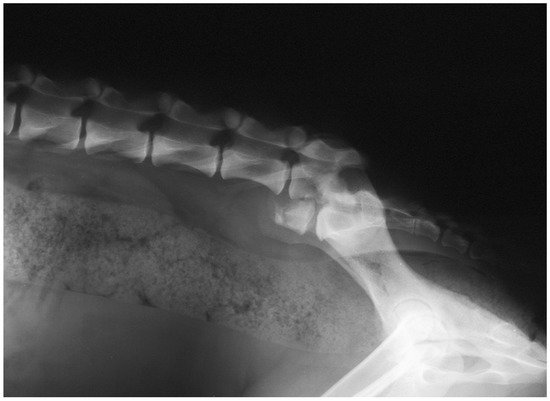
All patients with L7 fractures underwent survey radiographs in latero-lateral (LL) recumbency to confirm the diagnoses and assess the fracture or luxation [5][6][8][11][12][13][18][19][20] (Figure 1). The ventro-dorsal (VD) view was not performed for L7 fracture. In 4/38 patients, Computer Tomography (CT)-myelography was used [18].

Figure 1. Post-trauma lateral radiographic projection of the seventh lumbar vertebra fracture showing cranio-ventral displacement of the sacrum.

2.4. Treatment

None of the patients with L7 fractures underwent to conservative treatment. Several surgical techniques were performed to reduce and stabilize the L7 fracture. Dulisch and colleagues, in 1981, used a double transilial pins and plastic plates to treat the L7 fracture in one patient [19]. McAnulty and colleagues in 1986 described the use of the Steinmann pins placed transversely through both ilial wings at the level of the sacral dorsal lamina, bent at a right angle, and then placed alongside the laminae and attached to the articular facets and spinous process by a stainless steel wire [8].

In 1993, Ullman and Boudrieau reported the use of two crossed transilial pins secured with double Kirschner clamps placed adjacent to the ilial wings under the muscular plane [10]. Beaver and colleagues in 1996 reported the use of screws bilaterally placed into the ilial wing and into the bodies of sixth and fifth lumbar vertebra incorporated into Polymethyl-methacrylate (PMMA) [12]. Harrington and Bagley, in 1998 in two dogs with L7 fractures, used Steinmann pins inserted across the L7-S1 articular facets and a different number of pins placed as anchors and embedded in PMMA [11].
In 2007, Weh and colleagues reported the use of four positive-profile threaded pins into a L7-L6 pedicle body embedded in PMMA [13]. Wheeler and colleagues in 2007 described the use of trans-sacral and transilial pins [20]. Di Dona and colleagues in 2016 reports the use of double transilial pins externally fixed with two Kirschner clamps in 17 patients [5]. Segal and colleagues in 2018 addressed the L7 fracture in six dogs by using bilateral string-of-pearls (SOP) fixed to the lateral aspect of the vertebral body cranial to L7 and ilio-sacral joints [18].

2.5. Complications and Prognosis

All patients surgically treated regain the ability to deambulate [5][6][8][11][12][13][18][19][20]. The failure of fixation is the primary complication detected, and incorrect choice of fixation technique, incorrect execution of fixation technique, and incorrect post-operative management are the factors leading to instability. Bone lysis around pins and pin tract infection due to inflammation/infection and breakage of pins/screw were reported as the main causes of implant failure [8][13][20].
However, the long-term prognosis can be considered favourable for outcome and neurological recovery. Caudal lumbar lesions had a better neurological status as compared to more cranially located vertebral injuries [12]. Moreover, the bone healing via callus of L7 body vertebra quickly stabilizes the fractured site ensuring a rapid solution of the orthopaedic injuries and a good neurological recovery [6].

3. Conclusions

Despite the diversity of surgical techniques applied, the prognosis is good for this type of injury. The bone healing that is achieved, via callus, results in a rapid resolution of orthopaedic injuries and a few neurological complications. Therefore, it can assume that the L7 fracture can be considered a problem of orthopaedic instability with limited long term neurological consequences. The neurological improvement and outcome are not related to the shortening of the involved vertebral body or residual vertebral canal compromise but is rather related to the initial neurological damage.
It is likely that the use of internal osteosynthesis systems, such as contourable plates, which allow reducing the width of the surgical access and a better adaptation to the irregular surface of the vertebrae, could optimize post-operative management and accelerate the healing processes of the fracture; however, further studies are needed.

References

  1. Turner, W.D. Fractures and fracture-luxations of the lumbar spine: A retrospective study in the dog. J. Am. Anim. Hosp. Assoc. 1987, 23, 459–464.
  2. Selcer, R.R.; Bubb, W.J.; Walker, T.L. Management of vertebral column fractures in dogs and cats: 211 cases (1977–1985). J. Am. Vet. Med. Assoc. 1991, 198, 1965–1968.
  3. Jeffery, N.D. Vertebral fracture and luxation in small animals. Vet. Clin. N. Am. Small Anim. Pract. 2010, 40, 809–828.
  4. Bali, M.S.; Lang, J.; Jaggy, A.; Spreng, D.; Doherr, M.G.; Forterre, F. Comparative study of vertebral fractures and luxations in dogs and cats. Vet. Comp. Orthop. Traumatol. 2009, 22, 47–53.
  5. Di Dona, F.; Della Valle, G.; Lamagna, B.; Balestriere, C.; Murino, C.; Santangelo, B.; Lamagna, F.; Fatone, G. Percutaneous transilial pinning for treatment of seventh lumbar vertebral body fracture: A retrospective analysis of 17 cases. Vet. Comp. Orthop. Traumatol. 2016, 29, 164–169.
  6. Della Valle, G.; Di Dona, F.; Mennonna, G.; Lamagna, B.; Pasolini, M.P.; Caterino, C.; Lamagna, F.; Fatone, G. Traumatic lumbosacral joint dislocation in 3 dogs: Clinical presentation, diagnosis, treatment and short-term follow-up. Pak. Vet. J. 2021, 41, 97–101.
  7. Slocum, B.; Rudy, R.L. Fractures of the seventh lumbar vertebra in the dog. J. Am. Anim. Hosp. Assoc. 1975, 11, 167–174.
  8. McAnulty, J.F.; Lenehan, T.M.; Maletz, L.M. Modified Segmental Spinal Instrumentation in Repair of Spinal Fractures and Luxations in Dogs. Vet. Surg. 1986, 15, 143–149.
  9. Shores, A.; Nichols, C.; Rochat, M.; Fox, S.M.; Burt, G.J.; Fox, W.R. Combined Kirschner-Ehmer device and dorsal spinal plate fixation technique for caudal lumbar vertebral fractures in dogs. J. Am. Vet. Med. Assoc. 1989, 195, 335–339.
  10. Ullman, S.L.; Boudrieau, R.J. Internal Skeletal Fixation Using a Kirschner Apparatus for Stabilization of Fracture/Luxations of the Lumbosacral Joint in Six Dogs: A Modification of the Transilial Pin Technique. Vet. Surg. 1993, 22, 11–17.
  11. Harrington, M.L.; Bagley, R.S. Realignment of a Seventh Lumbar Vertebral Fracture/Luxation Using a Senn Retractor in Two Puppies. J. Am. Anim. Hosp. Assoc. 1998, 34, 377–380.
  12. Beaver, D.P.; MacPhersont, G.C.; Muir, P.; Johnson, K.A. Methyl-methacrylate and bone screw repair of seventh lumbar vertebral fracture-luxations in dogs. J. Small Anim. Pract. 1996, 37, 381–386.
  13. Weh, J.M.; Kraus, K.H. Use of a four pin and methylmethacrylate fixation in L7 and the iliac body to stabilize lumbosacral fracture-luxations: A clinical and anatomic study. Vet. Surg. 2007, 36, 775–782.
  14. Krauss, M.W.; Theyse, L.F.H.; Tryfonidou, M.A.; Hazewinkel, H.A.W.; Meij, B.P. Treatment of spinal fractures using Lubra plates: A retrospective clinical and radiological evaluation of 15 cases. Vet. Comp. Orthop. Traumatol. 2012, 25, 326–331.
  15. Hermanson, J.W.; de Lahunta, A. Spinal Cord and Meninges. In Miller’s Anatomy of the Dog, 5th ed.; Elsevier Inc.: Amsterdam, The Netherlands, 2018; pp. 589–610.
  16. Denis, F. Spinal instability as defined by the three-column spine concept in acute spinal trauma. Clin. Orthop. Relat. Res. 1984, 189, 65–76.
  17. Barquet, A.; Menendez, J.; Dubra, A.; Masliah, R.; Pereyra, D. Anterolateral dislocation of the lumbosacral junction. Can. Assoc. Radiol. J. 1993, 44, 129–132.
  18. Segal, U.; Bar, H.; Shani, J. Repair of lumbosacral fracture–luxation with bilateral twisted string-of-pearls locking plates. J. Small Anim. Pract. 2018, 59, 501–507.
  19. Dulisch, M.L.; Nichols, J.B. A Surgical Technique for Management of Lower Lumbar Fractures: Case Report. Vet. Surg. 1981, 10, 90–93.
  20. Wheeler, J.L.; Lewis, D.D.; Cross, A.R.; Sereda, C.W. Closed fluoroscopic-assisted spinal arch external skeletal fixation for the stabilization of vertebral column injuries in five dogs. Vet. Surg. 2007, 36, 442–448.
More
Information
Contributor MDPI registered users' name will be linked to their SciProfiles pages. To register with us, please refer to https://encyclopedia.pub/register :
View Times: 1.2K
Revisions: 4 times (View History)
Update Date: 23 Feb 2022
1000/1000