Submitted Successfully!
To reward your contribution, here is a gift for you: A free trial for our video production service.
Thank you for your contribution! You can also upload a video entry or images related to this topic.
Version Summary Created by Modification Content Size Created at Operation
1 + 2393 word(s) 2393 2022-01-04 02:25:22 |
2 format correction Meta information modification 2393 2022-02-17 02:32:06 |

Video Upload Options

Do you have a full video?

Confirm

Are you sure to Delete?
Cite
If you have any further questions, please contact Encyclopedia Editorial Office.
Wei, J. Ribosome Interactions with SARS-CoV-2 and COVID-19 mRNA Vaccine. Encyclopedia. Available online: https://encyclopedia.pub/entry/19546 (accessed on 04 July 2024).
Wei J. Ribosome Interactions with SARS-CoV-2 and COVID-19 mRNA Vaccine. Encyclopedia. Available at: https://encyclopedia.pub/entry/19546. Accessed July 04, 2024.
Wei, Jiao. "Ribosome Interactions with SARS-CoV-2 and COVID-19 mRNA Vaccine" Encyclopedia, https://encyclopedia.pub/entry/19546 (accessed July 04, 2024).
Wei, J. (2022, February 16). Ribosome Interactions with SARS-CoV-2 and COVID-19 mRNA Vaccine. In Encyclopedia. https://encyclopedia.pub/entry/19546
Wei, Jiao. "Ribosome Interactions with SARS-CoV-2 and COVID-19 mRNA Vaccine." Encyclopedia. Web. 16 February, 2022.
Ribosome Interactions with SARS-CoV-2 and COVID-19 mRNA Vaccine
Edit

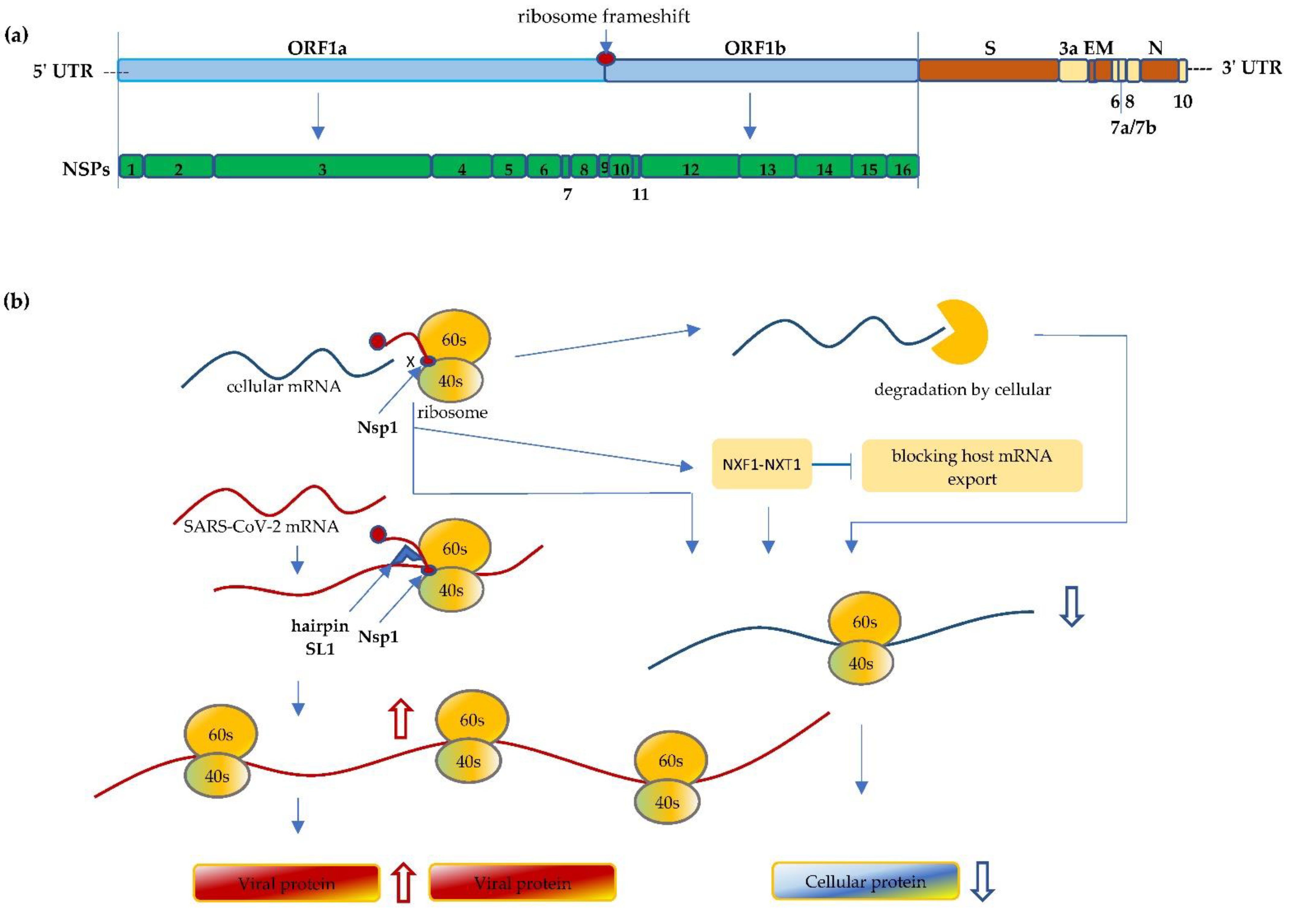
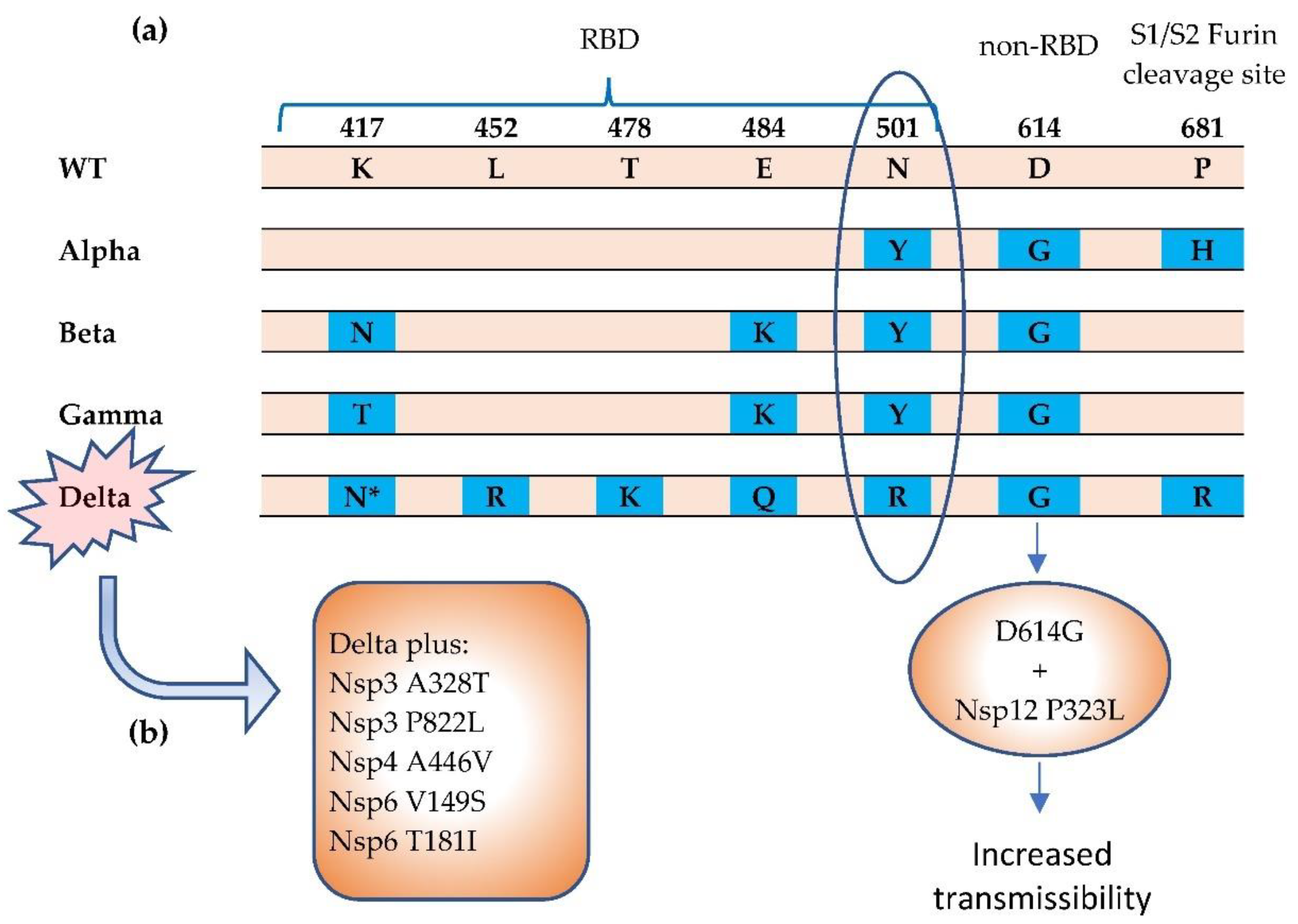
Severe Acute Respiratory Syndrome Coronavirus 2 (SARS-CoV-2) is the causing pathogen of the unprecedented global Coronavirus Disease 19 (COVID-19) pandemic. Upon infection, the virus manipulates host cellular machinery and ribosomes to synthesize its own proteins for successful replication and to facilitate further infection. SARS-CoV-2 executes a multi-faceted hijacking of the host mRNA translation and cellular protein synthesis. Viral nonstructural proteins (NSPs) interact with a range of different ribosomal states and interfere with mRNA translation. Concurrent mutations on NSPs and spike proteins contribute to the epidemiological success of variants of concern (VOCs). The interactions between ribosomes and SARS-CoV-2 represent attractive targets for the development of antiviral therapeutics and vaccines.

ribosome SARS-CoV-2 COVID-19 mRNA vaccines

1. Introduction

SARS-CoV-2 is the causing pathogen of the COVID-19 pandemic that has resulted in more than 250 million cases and 5 million deaths [1][2][3]. Viruses employ the host cellular translation machinery to synthesize their own proteins. Consequently, they have developed specialized mechanisms to commandeer the host machinery. SARS-CoV-2 uses a multipronged procedure to manipulate host cellular machinery, to reduce global protein translation and engage cellular resources in order to regulate their own protein production. As the factory for protein synthesis in human cells, ribosomes play a critical role in infection and human antiviral responses.
Each human ribosome consists of 2 unequal sized subunits; one is a 40S small subunit and the other one is a large subunit (60S) [4]. The 40S small subunit is the decoding site that consists of the 18S ribosomal RNA (rRNA) and 33 proteins. At the decoding site, sequence information of the messenger RNA (mRNA) is translated into a protein sequence.

2. SARS-CoV-2 Interfere with Ribosome mRNA Translation

The SARS-CoV-2 is a novel beta-coronavirus, a category that also includes two already known virulent coronaviruses, namely SARS-CoV-1 and MERS-CoV, that have resulted in serious outbreaks in 2002 and 2012, respectively [5]. SARS-CoV-2 is an enveloped, positive-sense and single-stranded RNA virus; its genome is approximately 30 kb in length. The SARS-CoV-2 genome comprises a 5′-cap, 5′ untranslated region (5′ UTR), followed by -replicase (ORF1a/ORF1b)-Spike (S)-Envelope (E)-Membrane (M)-Nucleocapsid (N)-3′ UTR-poly(A) tail [6] (Figure 1). Though it shares more than 80% homology with SARS-CoV-1 and ~50% with MERS-CoV, the mortality rates of these infections are slightly different, ranging from 15% for SARS-CoV-1 and 34.4–37% for MERS-CoV, to around 2–13% for SARS-CoV-2 [1][7][8][9][10].
Figure 1. (a) Schematic illustration of the SARS-CoV-2 genome. The size of the coronavirus genome is approximately 30 kb in length and comprises a 5′-cap, 5′ untranslated region (5′ UTR), ORF1a/ORF1b, spike (S), envelope (E), membrane (M), nucleocapsid (N), and 3′ UTR-poly(A) tail. The first ORF comprises of an approximated 2/3 of the genome that encodes the nonstructural proteins (Nsp1 to Nsp16). (b) Nsp1 of SARS-CoV-2 binds to the 40S mRNA and block the mRNA entrance channel. Following viral infection, SARS-CoV-2 performs a multifaceted hijack on host machinery, including blocking the mRNA entry channel, accelerating host mRNA degradation, and inhibiting host mRNA nucleus export. Furthermore, Nsp1 interacts with 5′ UTR of SARS-CoV-2 and facilitates the translation of its own protein, resulting in viral replication and protein accumulation, and inhibiting anti-viral immune responses.
At the 5′ end of SARS-CoV-2 genome, it starts with two large overlapping ORFs (ORF1a and ORF1b) representing two-thirds of its genome, with the other one-third encoding the structural proteins and accessory proteins [11][12]. Upon entering host cells, the ORF1a and 1b of the viral genomic RNA are translated and continually produce polyprotein, which is then cleaved into functional NSPs. These NSPs play pivotal roles, like evading the host immune system. Multiple NSPs interact with each other or form complex structures to modify cellular conditions for efficient mRNA translation and viral replication [13]. The 440–500 kDa Polyprotein 1a (pp1a) is first translated from ORF1a and then cleaved from Nsp1 to Nsp11. Between ORF1a and 1b, the programmed ribosomal -1 frameshift (PRF) of the reading frame immediately takes place preceding the stop codon of ORF1a, which enables the downstream translation of pp1ab from ORF1b. The pp1ab is subsequently processed into functional Nsp1 through Nsp16 [14].
The PRF, a well-preserved process among coronaviruses, is essential for functioning translation of NSPs. An efficient PRF engages a conserved slippery sequence (U_UUA_AAC) that changes the reading frame to UUU_AAA_C after the frameshifting [15]. When a ribosome approaches the slippery site during translation, a stimulatory RNA folds into a stable pseudoknot structure that slows down translation and promotes the PRF [15]. Importantly, Nsp12 and downstream NSPs that are involved in RNA capping, modification, and proofreading, rely on the PRF process as they are translated after the frameshifting [15]. It has been reported that multiple factors, including, for example, the position of the ORF1a stop codon, and interactions between ribosomal tunnels and RNA elements, including pseudoknot and nascent chain, modulate the optimum efficiency of frameshifting [15]. In addition to RNA regulation, the zinc-finger antiviral protein (ZAP-s) has been observed to interact with viral RNA and interfere with PRF [16]. PRF is one of the crucial steps in ribosome translation of virus genomes and viral replication, and thus presents a viable potential target for antiviral intervention therapeutics [15][17][18].
Among the 16 NSPs, Nsp1 is one of the first functional coronaviral nonstructural proteins translated in infected cells. Nsp1 is a protein comprised of 180 amino acids that targets cellular processes to inhibit translation, triggers host mRNA cleavage and decay, and down-regulates type I interferon (IFN) response [19][20][21][22]. NSP1 is a major virulence factor that is essential for viral replication, and is thus emphasized here [13][17][20][23].
Type I interferon induction and innate interferon response represent one of the major innate antiviral host defenses against viral infections [24]. SARS-CoV-2 efficiently suppresses the IFN-I signaling, likely mediated by the inhibition of STAT1 and STAT2, resulting in lack of efficient IFN-dependent antiviral innate immune responses. In addition to the shutting-off of host mRNA translation, the inhibition of INF-I antiviral responses leads to higher viral replication, viral protein accumulation, and pathogenesis [24]. Collectively, a fully functional Nsp1 is necessary for virulence; thus, the targeting of Nsp1 proteins and the Nsp1–ribosome interactions presents an attractive therapeutic opportunity for future studies [11][20][25].

3. Mutations Impact Replication and Virulence

Although SARS-CoV-2 has proof-reading processes, mutations arise naturally during viral replication, which causes new variants to form. Since late 2020, several novel variants have been named SARS-CoV-2 variants of concern/interest (VOC/I) due to their greater risk of enhanced transmissibility, pathogenicity and/or ability to evade host response [26]. The Alpha (B.1.1.7) variant, first detected in England in September 2020, appears to have a higher reproduction number and transmits more efficiently from person to person [27]. The Beta (B.1.351) variants, first reported in South America, and Gamma (P.1) variants share some of the same genetic changes that are associated with the increased transmission, and higher viral load. It is reported that these mutations lead to immune escape from neutralizing antibodies [28][29]. The Delta (B.1.617.2) variant is believed to be highly transmissible with more than twice as the original strain of SARS-CoV-2 [30]. Since first appearing in India in late 2020, it has spread worldwide and became the dominant variant of SARS-CoV-2 virus in the U.S. in late 2021 [31]. The recent emergence of Omicron has raised significant concern due to its extensive mutations, including more than 30 mutations on the Spike protein [32].
The virus is able to mutate in a dangerous and clever way to become structurally more infectious or cause more severe disease, in which multiple mechanisms may be operating. The major VOCs have shared mutations in the spike protein of SARS-CoV-2 genome, mostly on the S1 subunit, which is the unit that possesses the receptor-binding domain (RBD) that binds to cellular receptor ACE2 through six key amino acid residues [33] (Figure 2). N501 on the RBD domain is one of these amino acids that has a specific interaction with ACE2 receptor. Observed in three variants (Alpha, Beta, and Gamma), the N501Y mutation raises considerable concern due to its greater ACE2 binding affinity and enhanced transmissibility [34][35]. The Beta variant carries K417N and E484K mutations while Gamma shares E484K, along with the mutation of K417 to K417T [35]. These S1 mutations increase the binding affinity to the ACE2 receptor, thereby possibly enhancing transmissibility, which can affect disease severity and clinical outcomes [33].
Figure 2. The Alpha, Beta, Gamma, and Delta variants have been termed SARS-CoV-2 COVs. (a) Notable mutations in spike protein [33][34][35]. The N501Y mutation results in greater affinity for ACE2 receptor, which can increase transmissibility. (b) The Delta variant carries a more diverse repertoire of mutations [36][37]. The Delta Plus variant carries increased mutations in NSPs. * = K417N. The K417N mutation is significantly more prevalent in the Delta Plus (AY.1 or B.1.617.2.1) variant than in the Delta (B.1.617.2) variant. RBD = receptor binding domain.

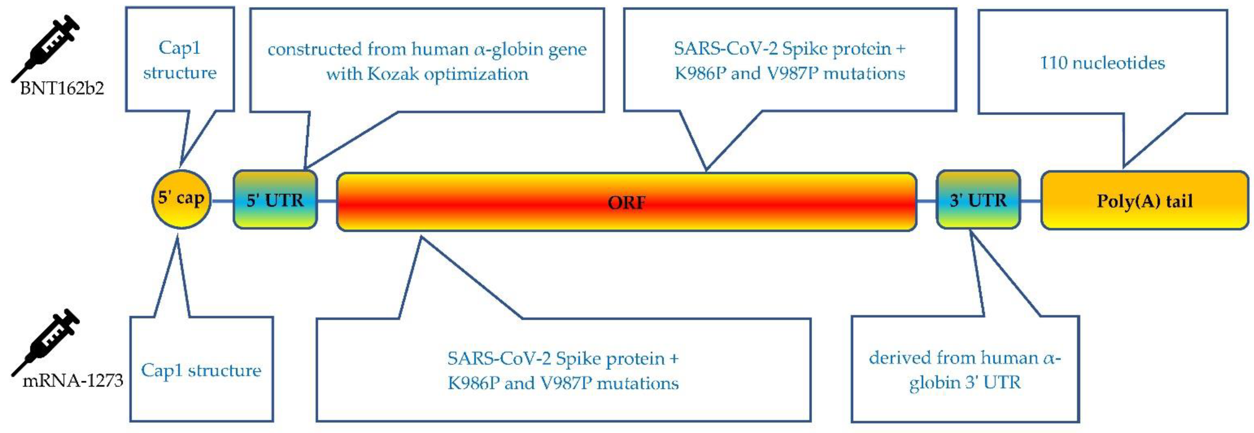
4. Molecule Design Impacts Ribosome Vaccine mRNA Translation

During the COVID-19 pandemic, scientists spent the past year developing vaccines and treatments to lessen the disease’s damage. The vaccine is a critical tool to help stop the pandemic. To date, two COVID-19 mRNA vaccines (BNT162b2 and mRNA-1273) are authorized and distributed worldwide [38][39]. In principle, an mRNA vaccine comprises synthetic mRNA molecules coded with the sequence of immunogen, that direct the cell machine, ribosomes, to produce vaccine protein antigens and generate an immune response. Once the vaccine is delivered into the cells, the ribosomes translate the mRNA vaccine sequence and produce the antigen, which is the spike protein of SARS-CoV-2 for the COVID-19 vaccine. The produced spike protein then triggers the immune response, including the production of antibodies and the cellular immune response [40].
Both BNT162b2 and mRNA-1273 utilize modified mRNA technology to code an optimized version of the spike protein sequence. The nucleoside-modified mRNA is encapsulated in a lipid nanoparticle with different lipids and formulations. Through intramuscular injection, BNT162b2 is administered in 21 days apart with a 30 µg dose; while mRNA-1273 is given in two 100 µg doses with 28 days in between each dose. Both BNT162b2 and mRNA-1273 have been shown to be safe and highly effective with 95% and 94.1% efficacy, respectively, in large Phase 3 double-blind clinical trials [41][42][43][44].
The mRNA vaccines contain 5 essential components, including 5′ cap, 5′ UTR, an open reading frame (ORF) that encodes the antigen, 3′ UTRs and a 3′ poly(A) tail [45] (Figure 3). Although both mRNA vaccines share the same antigen sequence of SARS-CoV-2 spike protein, each vaccine involves many different types of optimizations. In addition to the ongoing efforts for optimizing the delivery technology, optimizing the technical basis of the design features is critical in regulating the interaction of the vaccine and ribosomes can reduce dosage to be injected, lead to more efficient immunization, and improve safety [40].
Figure 3. Schematic presentation of the BNT162b2 and mRNA-1273 COVID-19 mRNA vaccine.
Each functional component in the mRNA vaccine can be independently optimized. It is well known that the 5′ cap structure of the mRNA molecule is the essence for the efficient translation of mRNA on the ribosome. The 5′ cap protects mRNA from degradation and interacts with the eukaryotic initiation factor (eIF) 4E to recruit 40S ribosome subunits and promotes the translation initiation complex of ribosomes [46][47][48]. In addition, it plays a prominent role in antigen production and prevents unintended immune responses by preventing recognition by cytosolic sensors of viral RNA [49][50]. In eukaryotes, a cap structure, either Cap 0 [m7G(5′)pppN1pN2p] or Cap 1 [m7G(5′)pppN1mpNp], is associated with the 5′ end of the mRNA [47][48]. The addition of the 5′ cap in mRNA vaccines can be achieved either co-transcriptionally, in which the cap is attached when the rest of the mRNA is assembled, or post-transcriptionally, in an additional process step after transcription [51]. Capping of BNT162b2 uses a trinucleotide Cap 1 analogue that is co-transcriptionally produced, while for the mRNA-1273 vaccine, the capping process is performed using a vaccinia capping enzyme post transcription [45][52].
The 5′ and 3′ UTR regions are engaged in the half-life, localization, translation efficacy, and recruiting of mRNAs to the ribosomes [47][53][54]. Therefore, the molecular design and optimization of the UTRs are critical for mRNA vaccine to ensure efficient antigen production and immune responses [55]. The highly expressed and naturally existing UTRs from human genes are often advantaged and employed for mRNA vaccines [56]. The 5′ UTR of BNT162b2 is constructed from the human α-globin gene using Kozak sequence optimization [57]. Similar approaches were naturally followed when designing the 3′ UTR, by incorporating regulatory elements for stability from human α-globin and β-globin [57][58]. The mRNA-1273 has a 110-nt 3′ UTR of the human α--globin gene (HBA1) inserted into its 3′ UTR region. [57].
There is a growing body of knowledge on alternative UTR sequences, such alteration in the UTRs, which can affect the functionality of the ribosomes [59]. For example, the length and structures of the 5′ and 3′ UTR, and regulatory components in the UTR sequences alter mRNA translation characteristics and protein synthesis [60]. Length is critical because the 5′ UTR serves as the ribosome landing zone and physically provides “lead-in”. In addition, the nucleotide composition of A, C, G, and U are considered important factors that would impact structure and mRNA stability. High GC content is likely to induce secondary structural formation and increased mRNA stability, while an AU rich region is associated with translation regulation [56][61]. In addition, microRNA binding sites should be avoided as it can bind to target sites within 5′ UTR and interfere with the ribosomal scanning [62]. The length of the 3′ UTR can be optimized, for example, as mRNAs with longer 3′ UTRs is commonly associated with shorter half-life, whereas shorter 3′ UTRs are associated with decreased translation efficiency [63]. The secondary structures can prevent ribosomes from skipping the stop codon and benefit protein production [64][65].
The mRNA sequence encodes trimerized SARS-CoV-2 S protein with K986P and V987P mutation optimizations for prefusion stabilization of the translated spike protein [66][67]. As mammalian host cells attack unmodified exogenous RNA as it activates innate immune response [68]. The modifications of N1-methyl-pseudouridine (N1m) lead to increased stability and low immunogenicity. Incorporation of N1m nucleotides facilitates ribosome loading and increases its density on mRNA, resulting in the alteration of translation dynamics. [46]. In COVID-19 mRNA vaccines, uridine bases are replaced with N1m to improve safety and translation efficacy [52][69].
The very end of mRNA is polyadenylated. The proper length and properties of the poly(A) tail are important for mRNA translation. In general, a 100–150 bp poly(A) tail is considered a sufficiently long enough tail to interact with poly(A) binding proteins, which is necessary for translation initiation [70][71]. In addition, the poly(A) tail is crucial for protection of the cap from degradation by de-capping enzymes [72]. The poly(A) tail of the BNT162b2 vaccine includes 30 A’s and 70A’s with a “10 nucleotide linker” (GCAUAUGACU) in between [73].

References

  1. Zhou, P.; Yang, X.-L.; Wang, X.-G.; Hu, B.; Zhang, L.; Zhang, W.; Si, H.-R.; Zhu, Y.; Li, B.; Huang, C.-L.; et al. A pneumonia outbreak associated with a new coronavirus of probable bat origin. Nature 2020, 579, 270–273.
  2. Zhu, N.; Zhang, D.; Wang, W.; Li, X.; Yang, B.; Song, J.; Zhao, X.; Huang, B.; Shi, W.; Lu, R.; et al. A Novel Coronavirus from Patients with Pneumonia in China. N. Engl. J. Med. 2020, 382, 727–733.
  3. WHO. Coronavirus Disease (COVID-1) Dashboard; WHO: Geneva, Switzerland, 2021.
  4. Nieto, B.; Gaspar, S.G.; Moriggi, G.; Pestov, D.G.; Bustelo, X.R.; Dosil, M. Identification of distinct maturation steps involved in human 40S ribosomal subunit biosynthesis. Nat. Commun. 2020, 11, 156.
  5. Coronaviridae Study Group of the International Committee on Taxonomy of Viruses. The species severe acute respiratory syndrome-related coronavirus: Classifying 2019-nCoV and naming it SARS-CoV-2. Nat. Microbiol. 2020, 5, 536–544.
  6. Chan, J.F.-W.; Kok, K.-H.; Zhu, Z.; Chu, H.; To, K.K.-W.; Yuan, S.; Yuen, K.-Y. Genomic characterization of the 2019 novel human-pathogenic coronavirus isolated from a patient with atypical pneumonia after visiting Wuhan. Emerg. Microbes Infect. 2020, 9, 221–236.
  7. Kim, D.; Kim, S.; Park, J.; Chang, H.R.; Chang, J.; Ahn, J.; Park, H.; Park, J.; Son, N.; Kang, G.; et al. A high-resolution temporal atlas of the SARS-CoV-2 translatome and transcriptome. Nat. Commun. 2021, 12, 5120.
  8. WHO. Tracking SARS-CoV-2 Variants; WHO: Geneva, Switzerland, 2021.
  9. Abdelghany, T.; Ganash, M.; Bakri, M.M.; Qanash, H.; Al-Rajhi, A.M.; Elhussieny, N.I. SARS-CoV-2, the other face to SARS-CoV and MERS-CoV: Future predictions. Biomed. J. 2021, 44, 86–93.
  10. Abdelrahman, Z.; Li, M.; Wang, X. Comparative Review of SARS-CoV-2, SARS-CoV, MERS-CoV, and Influenza A Respiratory Viruses. Front. Immunol. 2020, 11, 552909.
  11. Mariano, G.; Farthing, R.J.; Lale-Farjat, S.L.M.; Bergeron, J.R.C. Structural Characterization of SARS-CoV-2: Where We Are, and Where We Need to Be. Front. Mol. Biosci. 2020, 7, 605236.
  12. Finkel, Y.; Gluck, A.; Nachshon, A.; Winkler, R.; Fisher, T.; Rozman, B.; Mizrahi, O.; Lubelsky, Y.; Zuckerman, B.; Slobodin, B.; et al. SARS-CoV-2 uses a multipronged strategy to impede host protein synthesis. Nature 2021, 594, 240–245.
  13. Schubert, K.; Karousis, E.D.; Jomaa, A.; Scaiola, A.; Echeverria, B.; Gurzeler, L.-A.; Leibundgut, M.; Thiel, V.; Mühlemann, O.; Ban, N. SARS-CoV-2 Nsp1 binds the ribosomal mRNA channel to inhibit translation. Nat. Struct. Mol. Biol. 2020, 27, 959–966.
  14. Krichel, B.; Falke, S.; Hilgenfeld, R.; Redecke, L.; Uetrecht, C. Processing of the SARS-CoV pp1a/ab nsp7–10 region. Biochem. J. 2020, 477, 1009–1019.
  15. Bhatt, P.R.; Scaiola, A.; Loughran, G.; Leibundgut, M.; Kratzel, A.; Meurs, R.; Dreos, R.; O’Connor, K.M.; McMillan, A.; Bode, J.W.; et al. Structural basis of ribosomal frameshifting during translation of the SARS-CoV-2 RNA genome. Science 2021, 372, 1306–1313.
  16. Zimmer, M.M.; Kibe, A.; Rand, U.; Pekarek, L.; Ye, L.; Buck, S.; Smyth, R.P.; Cicin-Sain, L.; Caliskan, N. The short isoform of the host antiviral protein ZAP acts as an inhibitor of SARS-CoV-2 programmed ribosomal frameshifting. Nat. Commun. 2021, 12, 7193.
  17. Simeoni, M.; Cavinato, T.; Rodriguez, D.; Gatfield, D. I(nsp1)ecting SARS-CoV-2–ribosome interactions. Commun. Biol. 2021, 4, 715.
  18. Sun, Y.; Abriola, L.; Niederer, R.O.; Pedersen, S.F.; Alfajaro, M.M.; Monteiro, V.S.; Wilen, C.B.; Ho, Y.-C.; Gilbert, W.V.; Surovtseva, Y.V.; et al. Restriction of SARS-CoV-2 replication by targeting programmed −1 ribosomal frameshifting. Proc. Natl. Acad. Sci. USA 2021, 118, e2023051118.
  19. Lokugamage, K.G.; Narayanan, K.; Huang, C.; Makino, S. Severe Acute Respiratory Syndrome Coronavirus Protein nsp1 Is a Novel Eukaryotic Translation Inhibitor That Represses Multiple Steps of Translation Initiation. J. Virol. 2012, 86, 13598–13608.
  20. Thoms, M.; Buschauer, R.; Ameismeier, M.; Koepke, L.; Denk, T.; Hirschenberger, M.; Kratzat, H.; Hayn, M.; Mackens-Kiani, T.; Cheng, J.; et al. Structural basis for translational shutdown and immune evasion by the Nsp1 protein of SARS-CoV-2. Science 2020, 369, 1249–1255.
  21. Baggen, J.; Vanstreels, E.; Jansen, S.; Daelemans, D. Cellular host factors for SARS-CoV-2 infection. Nat. Microbiol. 2021, 6, 1219–1232.
  22. Zhang, K.; Miorin, L.; Makio, T.; Dehghan, I.; Gao, S.; Xie, Y.; Zhong, H.; Esparza, M.; Kehrer, T.; Kumar, A.; et al. Nsp1 protein of SARS-CoV-2 disrupts the mRNA export machinery to inhibit host gene expression. Sci. Adv. 2021, 7, eabe7386.
  23. Min, Y.-Q.; Mo, Q.; Wang, J.; Deng, F.; Wang, H.; Ning, Y.-J. SARS-CoV-2 nsp1: Bioinformatics, Potential Structural and Functional Features, and Implications for Drug/Vaccine Designs. Front. Microbiol. 2020, 11, 587317.
  24. Xia, H.; Cao, Z.; Xie, X.; Zhang, X.; Chen, J.Y.-C.; Wang, H.; Menachery, V.D.; Rajsbaum, R.; Shi, P.-Y. Evasion of Type I Interferon by SARS-CoV-2. Cell Rep. 2020, 33, 108234.
  25. Tidu, A.; Janvier, A.; Schaeffer, L.; Sosnowski, P.; Kuhn, L.; Hammann, P.; Westhof, E.; Eriani, G.; Martin, F. The viral protein NSP1 acts as a ribosome gatekeeper for shutting down host translation and fostering SARS-CoV-2 translation. RNA 2021, 27, 253–264.
  26. Boehm, E.; Kronig, I.; Neher, R.A.; Eckerle, I.; Vetter, P.; Kaiser, L.; Geneva Centre for Emerging Viral Diseases. Novel SARS-CoV-2 variants: The pandemics within the pandemic. Clin. Microbiol. Infect. 2021, 27, 1109–1117.
  27. Davies, N.G.; Abbott, S.; Barnard, R.C.; Jarvis, C.I.; Kucharski, A.J.; Munday, J.D.; Pearson, C.A.B.; Russell, T.W.; Tully, D.C.; Washburne, A.D.; et al. Estimated transmissibility and impact of SARS-CoV-2 lineage B.1.1.7 in England. Science 2021, 372, eabg3055.
  28. Tegally, H.; Wilkinson, E.; Giovanetti, M.; Iranzadeh, A.; Fonseca, V.; Giandhari, J.; Doolabh, D.; Pillay, S.; San, E.J.; Msomi, N.; et al. Detection of a SARS-CoV-2 variant of concern in South Africa. Nature 2021, 592, 438–443.
  29. Hoffmann, M.; Arora, P.; Groß, R.; Seidel, A.; Hörnich, B.F.; Hahn, A.S.; Krüger, N.; Graichen, L.; Hofmann-Winkler, H.; Kempf, A.; et al. SARS-CoV-2 variants B.1.351 and P.1 escape from neutralizing antibodies. Cell 2021, 184, 2384–2393.e12.
  30. Reardon, S. How the Delta variant achieves its ultrafast spread. Nat. Cell Biol. 2021, 21, 134.
  31. Scudellari, M. How the coronavirus infects cells- and why Delta is so dangerous. Nat. Cell Biol. 2021, 595, 640–644.
  32. CDC. SARS-CoV-2 Variant Classifications and Definitions. Available online: https://www.cdc.gov/coronavirus/2019-ncov/variants/variant-classifications.html#anchor_1632154493691 (accessed on 31 December 2021).
  33. Khateeb, J.; Li, Y.; Zhang, H. Emerging SARS-CoV-2 variants of concern and potential intervention approaches. Crit. Care 2021, 25, 244.
  34. Singh, J.; Samal, J.; Kumar, V.; Sharma, J.; Agrawal, U.; Ehtesham, N.; Sundar, D.; Rahman, S.; Hira, S.; Hasnain, S. Structure-Function Analyses of New SARS-CoV-2 Variants B.1.1.7, B.1.351 and B.1.1.28.1: Clinical, Diagnostic, Therapeutic and Public Health Implications. Viruses 2021, 13, 439.
  35. Sanches, P.R.; Charlie-Silva, I.; Braz, H.L.; Bittar, C.; Calmon, M.F.; Rahal, P.; Cilli, E.M. Recent advances in SARS-CoV-2 Spike protein and RBD mutations comparison between new variants Alpha (B.1.1.7, United Kingdom), Beta (B.1.351, South Africa), Gamma (P.1, Brazil) and Delta (B.1.617.2, India). J. Virus Erad. 2021, 7, 100054.
  36. Kannan, S.R.; Spratt, A.N.; Cohen, A.R.; Naqvi, S.H.; Chand, H.S.; Quinn, T.P.; Lorson, C.L.; Byrareddy, S.N.; Singh, K. Evolutionary analysis of the Delta and Delta Plus variants of the SARS-CoV-2 viruses. J. Autoimmun. 2021, 124, 102715.
  37. Ilmjärv, S.; Abdul, F.; Acosta-Gutiérrez, S.; Estarellas, C.; Galdadas, I.; Casimir, M.; Alessandrini, M.; Gervasio, F.L.; Krause, K.-H. Concurrent mutations in RNA-dependent RNA polymerase and spike protein emerged as the epidemiologically most successful SARS-CoV-2 variant. Sci. Rep. 2021, 11, 13705.
  38. FDA. Emergency Use Authorization (EUA) for Emergency Use of mRNA-1273. Available online: https://www.fda.gov/media/144636/download (accessed on 31 December 2021).
  39. FDA. Comirnaty (COVID-19 Vaccine, mRNA) Letter of Authorization. Available online: https://www.fda.gov/media/150386/download (accessed on 31 December 2021).
  40. Chaudhary, N.; Weissman, D.; Whitehead, K.A. mRNA vaccines for infectious diseases: Principles, delivery and clinical translation. Nat. Rev. Drug Discov. 2021, 20, 817–838.
  41. Walsh, E.E.; Frenck, R.W., Jr.; Falsey, A.R.; Kitchin, N.; Absalon, J.; Gurtman, A.; Lockhart, S.; Neuzil, K.; Mulligan, M.J.; Bailey, R.; et al. Safety and Immunogenicity of Two RNA-Based COVID-19 Vaccine Candidates. N. Engl. J. Med. 2020, 383, 2439–2450.
  42. Chu, L.; McPhee, R.; Huang, W.; Bennett, H.; Pajon, R.; Nestorova, B.; Leav, B.; on behalf of the mRNA-1273 Study Group. A preliminary report of a randomized controlled phase 2 trial of the safety and immunogenicity of mRNA-1273 SARS-CoV-2 vaccine. Vaccine 2021, 39, 2791–2799.
  43. Polack, F.P.; Thomas, S.J.; Kitchin, N.; Absalon, J.; Gurtman, A.; Lockhart, S.; Perez, J.L.; Marc, G.P.; Moreira, E.D.; Zerbini, C.; et al. Safety and efficacy of the BNT162b2 mRNA COVID-19 vaccine. N. Engl. J. Med. 2020, 383, 2603–2615.
  44. Baden, L.R.; El Sahly, H.M.; Essink, B.; Kotloff, K.; Frey, S.; Novak, R.; Diemert, D.; Spector, S.A.; Rouphael, N.; Creech, C.B.; et al. Efficacy and Safety of the mRNA-1273 SARS-CoV-2 Vaccine. N. Engl. J. Med. 2021, 384, 403–416.
  45. Corbett, K.S.; Edwards, D.K.; Leist, S.R.; Abiona, O.M.; Boyoglu-Barnum, S.; Gillespie, R.A.; Himansu, S.; Schäfer, A.; Ziwawo, C.T.; DiPiazza, A.T.; et al. SARS-CoV-2 mRNA vaccine design enabled by prototype pathogen preparedness. Nature 2020, 586, 567–571.
  46. Svitkin, Y.V.; Cheng, Y.M.; Chakraborty, T.; Presnyak, V.; John, M.; Sonenberg, N. N1-methyl-pseudouridine in mRNA enhances translation through eIF2α-dependent and independent mechanisms by increasing ribosome density. Nucleic Acids Res. 2017, 45, 6023–6036.
  47. Wadhwa, A.; Aljabbari, A.; Lokras, A.; Foged, C.; Thakur, A. Opportunities and Challenges in the Delivery of mRNA-Based Vaccines. Pharmaceutics 2020, 12, 102.
  48. Ramanathan, A.; Robb, G.B.; Chan, S.-H. mRNA capping: Biological functions and applications. Nucleic Acids Res. 2016, 44, 7511–7526.
  49. Fechter, P.; Brownlee, G.G. Recognition of mRNA cap structures by viral and cellular proteins. J. Gen. Virol. 2005, 86, 1239–1249.
  50. Kozak, M. An analysis of vertebrate mRNA sequences: Intimations of translational control. J. Cell Biol. 1991, 115, 887–903.
  51. Pardi, N.; Hogan, M.J.; Weissman, D. Recent advances in mRNA vaccine technology. Curr. Opin. Immunol. 2020, 65, 14–20.
  52. Sahin, U.; Muik, A.; Vogler, I.; Derhovanessian, E.; Kranz, L.M.; Vormehr, M.; Quandt, J.; Bidmon, N.; Ulges, A.; Baum, A.; et al. BNT162b2 vaccine induces neutralizing antibodies and poly-specific T cells in humans. Nat. Cell Biol. 2021, 595, 572–577.
  53. Chatterjee, S.; Pal, J.K. Role of 5′- and 3′-untranslated regions of mRNAs in human diseases. Biol. Cell 2009, 101, 251–262.
  54. Berkovits, B.D.; Mayr, C. Alternative 3′ UTRs act as scaffolds to regulate membrane protein localization. Nat. Cell Biol. 2015, 522, 363–367.
  55. Jackson, N.A.C.; Kester, K.E.; Casimiro, D.; Gurunathan, S.; DeRosa, F. The promise of mRNA vaccines: A biotech and industrial perspective. NPJ Vaccines 2020, 5, 11.
  56. Weng, Y.; Li, C.; Yang, T.; Hu, B.; Zhang, M.; Guo, S.; Xiao, H.; Liang, X.-J.; Huang, Y. The challenge and prospect of mRNA therapeutics landscape. Biotechnol. Adv. 2020, 40, 107534.
  57. Xia, X. Detailed Dissection and Critical Evaluation of the Pfizer/BioNTech and Moderna mRNA Vaccines. Vaccines 2021, 9, 734.
  58. Weissman, D. mRNA transcript therapy. Expert Rev. Vaccines 2015, 14, 265–281.
  59. Trepotec, Z.; Aneja, M.K.; Geiger, J.; Hasenpusch, G.; Plank, C.; Rudolph, C. Maximizing the Translational Yield of mRNA Therapeutics by Minimizing 5′-UTRs. Tissue Eng. Part A 2019, 25, 69–79.
  60. Sample, P.J.; Wang, B.; Reid, D.W.; Presnyak, V.; McFadyen, I.J.; Morris, D.R.; Seelig, G. Human 5′ UTR design and variant effect prediction from a massively parallel translation assay. Nat. Biotechnol. 2019, 37, 803–809.
  61. Leppek, K.; Das, R.; Barna, M. Functional 5′ UTR mRNA structures in eukaryotic translation regulation and how to find them. Nat. Rev. Mol. Cell Biol. 2018, 19, 158–174.
  62. Ricci, E.; Limousin, T.; Rifo, R.S.; Rubilar, P.S.; Decimo, D.; Ohlmann, T. miRNA repression of translation in vitro takes place during 43S ribosomal scanning. Nucleic Acids Res. 2012, 41, 586–598.
  63. von Niessen, A.G.O.; Poleganov, M.A.; Rechner, C.; Plaschke, A.; Kranz, L.; Fesser, S.; Diken, M.; Löwer, M.; Vallazza, B.; Beissert, T.; et al. Improving mRNA-Based Therapeutic Gene Delivery by Expression-Augmenting 3′ UTRs Identified by Cellular Library Screening. Mol. Ther. 2019, 27, 824–836.
  64. Klauer, A.A.; Van Hoof, A. Degradation of mRNAs that lack a stop codon: A decade of nonstop progress. Wiley Interdiscip. Rev. RNA 2012, 3, 649–660.
  65. Jackson, R.J.; Hellen, C.U.T.; Pestova, T.V. The mechanism of eukaryotic translation initiation and principles of its regulation. Nat. Rev. Mol. Cell Biol. 2010, 11, 113–127.
  66. Hsieh, C.-L.; Goldsmith, J.A.; Schaub, J.M.; DiVenere, A.M.; Kuo, H.-C.; Javanmardi, K.; Le, K.C.; Wrapp, D.; Lee, A.G.; Liu, Y.; et al. Structure-based design of prefusion-stabilized SARS-CoV-2 spikes. Science 2020, 369, 1501–1505.
  67. Xia, X. Domains and Functions of Spike Protein in SARS-CoV-2 in the Context of Vaccine Design. Viruses 2021, 13, 109.
  68. Karikó, K.; Buckstein, M.; Ni, H.; Weissman, D. Suppression of RNA Recognition by Toll-like Receptors: The Impact of Nucleoside Modification and the Evolutionary Origin of RNA. Immun. 2005, 23, 165–175.
  69. Roncati, L.; Corsi, L. Nucleoside-modified messenger RNA COVID-19 vaccine platform. J. Med Virol. 2021, 93, 4054–4057.
  70. Linares-Fernández, S.; Lacroix, C.; Exposito, J.-Y.; Verrier, B. Tailoring mRNA Vaccine to Balance Innate/Adaptive Immune Response. Trends Mol. Med. 2020, 26, 311–323.
  71. Xiong, Q.; Lee, G.Y.; Ding, J.; Li, W.; Shi, J. Biomedical applications of mRNA nanomedicine. Nano Res. 2018, 11, 5281–5309.
  72. Mugridge, J.S.; Coller, J.; Gross, J.D. Structural and molecular mechanisms for the control of eukaryotic 5′-3′ mRNA decay. Nat. Struct. Mol. Biol. 2018, 25, 1077–1085.
  73. Granados-Riveron, J.T.; Aquino-Jarquin, G. Engineering of the current nucleoside-modified mRNA-LNP vaccines against SARS-CoV-2. Biomed. Pharmacother. 2021, 142, 111953.
More
Information
Contributor MDPI registered users' name will be linked to their SciProfiles pages. To register with us, please refer to https://encyclopedia.pub/register :
View Times: 634
Entry Collection: COVID-19
Revisions: 2 times (View History)
Update Date: 18 Feb 2022
1000/1000
Video Production Service