Submitted Successfully!
To reward your contribution, here is a gift for you: A free trial for our video production service.
Thank you for your contribution! You can also upload a video entry or images related to this topic.
Version Summary Created by Modification Content Size Created at Operation
1 + 1958 word(s) 1958 2022-02-10 04:52:38 |
2 corrected the format -21 word(s) 1937 2022-02-11 01:57:36 |

Video Upload Options

Do you have a full video?

Confirm

Are you sure to Delete?
Cite
If you have any further questions, please contact Encyclopedia Editorial Office.
Sergi, C. Biliary Atresia. Encyclopedia. Available online: https://encyclopedia.pub/entry/19332 (accessed on 02 July 2024).
Sergi C. Biliary Atresia. Encyclopedia. Available at: https://encyclopedia.pub/entry/19332. Accessed July 02, 2024.
Sergi, Consolato. "Biliary Atresia" Encyclopedia, https://encyclopedia.pub/entry/19332 (accessed July 02, 2024).
Sergi, C. (2022, February 10). Biliary Atresia. In Encyclopedia. https://encyclopedia.pub/entry/19332
Sergi, Consolato. "Biliary Atresia." Encyclopedia. Web. 10 February, 2022.
Biliary Atresia
Edit

The term biliary atresia has substituted the original term of “extrahepatic biliary atresia”, which has been in use for several generations. The concept was related to the often-identified absence of gallbladder with an obliterated cord at the site the extrahepatic biliary system. It is now known that biliary atresia is a necro-inflammatory and fibro-obliterative process of both intrahepatic and extrahepatic biliary tract.

biliary atresia cholestasis newborn genetics virus

1. Introduction

Despite the fact that the etiology is far from being known, pediatric surgeries conducted last century worldwide were incredibly supportive for the clarification of the nosology of this disease [1][2][3]. Mainly, Morio Kasai, a Japanese pediatric surgeon, may be considered the pioneer of a specific pediatric surgery that may be offered to parents of a child with biliary atresia. His name is indelibly bound to the palliative surgical procedure he ideated in Japan [4]. The porto-enterostomy is indeed the major palliative procedure, which may be implemented before a liver is available for transplantation (orthotopic transplant or transplant of a liver from a recently deceased donor, a living donor transplant, or a split type of liver transplant) can be put forward. The biliary atresia is a very complex disease, not just complicated, and several genes have been investigated thoroughly. Some of these genes may be highly relevant to this disease, although their role is mostly not known. 

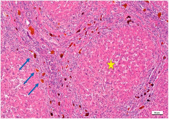
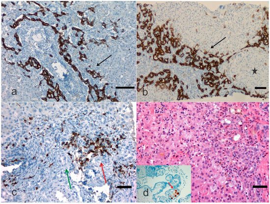
2. Pathological Anatomy of the Biliary Atresia

The histology of the liver with biliary atresia is variable and depends on the stage when the biopsy is done substantially. At the early stage, fibrosis and biliary ductular proliferation, two main essential criteria, are present but may be very focal and missed at first glance. A subsequent liver biopsy may harbor more conclusive findings. However, the liver biopsy is essential to indicate if the main inflammatory component is at the level of the portal tracts or at acinar level, which may indicate neonatal hepatitis [5][6]. Thus, in biliary atresia, there are prominent histologic features, including a limited involvement of the liver acinus with very few multinucleated giant hepatocytes, which should have six or more nuclei. The limitation of acinus involvement is critical. At the late stage, fibrosis becomes more prominent, and the chances to be successful with a hepatic portoenterostomy are limited [7][8]. At the late stage, liver cirrhosis is usually encountered with pseudolobules and lobules surrounded by expanded portal tracts with biliary ductular proliferation (Figure 1).
Figure 1. Biliary atresia in a child treated with the Kasai portoenterostomy, which failed. In this microphotograph, the formation of lobules and pseudo-lobules (yellow star) is apparent indicating stage IV fibrosis/cirrhosis. The portal tracts are fused highlighting the porto-portal and centro-portal bridges. In these bridges (blue arrows), the intrahepatic biliary tract shows biliary ductal proliferations and lumens filled with bile, which appears brown (hematoxylin and eosin staining, 50× as original magnification, bar: 50 μm).
One of the most detailed studies on biliary atresia was performed at the Division of Surgery, Children’s Research Hospital, Kyoto Prefectural University of Medicine, Kyoto, Japan. In this clinic-pathological investigation, 31 patients with uncorrectable obstructive cholangiopathy underwent hepatic portoenterostomy and steroid therapy between 1988 and 2005 [9]. An immunohistochemical investigation was performed using an antibody against keratin 19 of the intermediate filament family of the cytoskeleton. Ductal plate malformation (DPM) typing was performed according to generally accepted criteria [10]. Only type II DPM was considered relevant, and specimens were deemed as DPM-positive if a concentric cellular arrangement was detected. Shimadera et al. found that the presence of DPM in the liver of patients with biliary atresia predicts poor bile flow after hepatoportoenterostomy [9]. This research was retrospective and included comparisons of preoperative characteristics, the postoperative jaundice period, and cumulative steroid doses between patients with and without DPM. Despite numerous reports on the DPM of biliary atresia, the influence of biliary remnants remains controversially discussed in the literature. During the re-examination of the microphotographic material, researchers identified abnormalities, such as cytomegalovirus expression in the placenta (Figure 2). Visualization of cytokeratins (keratins) and neural cell adhesion molecules in the immature ductal cells by means of the immunohistochemical method can be a useful tool for the microscopic examination of the immature biliary structures in the liver. Russo et al. [11] assessed the relative value of histologic features in 227 liver needle biopsies in discriminating between biliary atresia and other cholestatic disorders in infants enrolled in a prospective Childhood Liver Disease Research and Education Network (ChiLDReN) cohort study. These authors correlated histology with clinical findings in infants with and without biliary atresia. Also, Russo et al. reviewed 316 liver biopsies from clinically proven biliary atresia patients and correlated histologic features with total serum bilirubin 6 months post-Kasai or hepatoportoenterostomy and transplant-free survival up to 6 years. Logistic regression analysis determined that bile plugs in portal bile ducts/ductules, moderate to marked ductular reaction and portal stromal edema had the largest odds ratio for predicting biliary atresia vs. non- biliary atresia. Remarkably, the diagnostic accuracy of the needle liver biopsy was assessed to be 90.1%, whereas sensitivity and specificity for a diagnosis of biliary atresia were 88.4% and 92.7%, respectively. Russo et al. found that higher stages of fibrosis, a ductal plate configuration, moderate to marked bile duct injury, an older age at hepatoportoenterostomy and an elevated INR (international normalized ratio), which is a type of calculation based on prothrombin test results, were independently associated with a higher risk of transplantation [11].
Figure 2. Biliary atresia with different clinical background. In this microphotograph panel, the bile ductular proliferations (arrows) are highlighted with a monoclonal antibody against cytokeratin or keratin 19 (a) and against cytokeratin or keratin 7 (b) by immunohistochemistry. Keratin-19 or cytokeratin-19 is a 40 kDa type I cytoskeletal protein that in humans is encoded by the KRT19 gene. In (b) the bile ductular proliferation is particularly pronounced (arrow) and a pseudonodule is noted (black star). In (c), an antibody against myeloperoxidase is carried out in an early case of biliary atresia highlighting the neutrophilic inflammation (red arrow) close to bile ducts (green arrow). Staining is either by immunohistochemistry (this case) or enzyme cytochemistry. Myeloperoxidase is the most sensitive and specific stain for myeloid leukemias and granulocytic sarcoma, but it is also very useful to stain neutrophils, particularly when their identification may be difficult to catch when other cells confuse the histology. In (d) a child with biliary atresia and positive cytomegalovirus (CMV) serology, but negative CMV immunohistochemistry, may disclose CMV positive cells in the placenta ((a), Bar 50 μm, 100× original magnification; (b), Bar 50 μm, 50× original magnification; (c), Bar 10 μm, 200× original magnification; (d), Bar 10 μm, 200× original magnification, and inset, 200× original magnification).
Apart from biliary atresia major causes of cholestatic liver disease in infants include choledochal cyst, Caroli’s syndrome, Alagille syndrome, and neonatal infection (Cytomegalovirus Herpesvirus Hepatotropic viruses, Human immunodeficiency virus, Parvovirus B19, Paramyxovirus, Enteric viruses, Rubella, Bacterial sepsis, Listeriosis, Toxoplasmosis, Syphilis). Also, disorders of carbohydrate metabolism, disorders of amino acid metabolism, disorders of glycolipid and lipid metabolism, disorders of glycoprotein metabolism, metal storage disorders, peroxisomal disorders, mitochondrial cytopathies, hereditary disorders of bilirubin metabolism, hereditary disorders of bile formation, disorders of bile acid biosynthesis, disorders of protein biosynthesis and targeting (e.g., α1-Antitrypsin deficiency) and miscellaneous disorders (e.g., Aagenaes syndrome, citrullinemia, type II X-linked adrenoleukodystrophy, shock/hypoperfusion, parenteral nutrition, fetal alcohol syndrome, drugs, Budd-Chiari syndrome, multiple hemangiomas) as well as neoplastic diseases (e.g., neonatal leukemia, neuroblastoma, hepatoblastoma, Langerhans cell histiocytosis, and erythrophagocytic lymphohistiocytosis). The broad differential diagnosis of jaundice in infants and children presupposes a good clinical and pathological correlation and periodic liver rounds. Since the evaluation of pediatric liver biopsies is quite distinct from that of adults, the liver pathologist needs to possess a good knowledge of biochemistry and metabolic disorders as well as dysmorphology understanding. The performance of all potentially necessary tests when a liver biopsy is carried out, prior arrangements should be in place between the clinician, the radiologist, and the laboratory to ensure adequate specimen processing and rapid workout. The laboratory should have liquid nitrogen on site or, better, at the bedside. Cores of tissue, up to 2 cm in length, should be snap frozen in liquid nitrogen refrigerated isopentane immediately to preserve tissue integrity and mRNA (messenger ribonucleic acid). The tissue fragments should then be placed within a specimen vial and a small portion of tissue may be placed in glutaraldehyde for electron microscopy processing, and a portion can be sent to the biochemistry laboratory for specific requests.

3. Treatment

Although initially considered an extrahepatic pathology of the biliary tract, this disease is now correctly considered a pathology of the intra- and extrahepatic biliary tract. Currently and at least in several children’s hospitals, biliary atresia is surgically classified as type I (atresia of the common bile duct, 10%), type II (atresia of the hepatic duct, 2%), and type III (atresia of the porta hepatis, 88%), but also as embryonic with DPM and fetal type without DPM. Nevertheless, classifications may have limited value in biliary atresia. The porto-enterostomy ideated by the Japanese Morio Kasai, who introduced an operative procedure, called portoenterostomy, aimed to ‘‘correct the uncorrectable’’ [4]. It represents a breakthrough for the outcome of patients affected with biliary atresia. Although the risk of cholangitis may jeopardize the outcome, several factors have also been suggested. Among others, the presence of ductal plate malformation with the inadequacy of the intrahepatic biliary system to adequately drain the bile. Nowadays, Kasai’s series of patients are still alive, and their descendants are reported to have a healthy liver. In the case of liver portoenterostomy failure, a terrific milestone was the step of liver transplantation. Although the long-term survival in patients with biliary atresia and native liver improves continuously, most of these patients will need early or late liver transplantation. The success of the modern liver transplantation has reached an overall survival rate exceeding 90% in several studies [12][13][14][15][16][17]. Arafa et al. [18] evaluated 59 infants with neonatal cholestasis and separated them in two groups, i.e., biliary atresia group (n = 31) and non- biliary atresia group (n = 28). Interleukin-2 (IL-2) and interleukin-8 (IL-8) immunohistochemical staining in the portal cellular infiltrate of the liver was scored. These authors found that the mean value of IL-2 and IL-8 positive inflammatory cells was significantly higher in the biliary atresia than in the non- biliary atresia group and IL-2 correlated with IL-8 immunohistochemical staining in both biliary atresia and non- biliary atresia groups. In addition, both cytokines correlated significantly with inflammatory activity in liver biopsy in both groups while there was no significant correlation with the other studied parameters. In particular, the biliary atresia group showed that there was a trend of increased expression of IL-2 and IL-8 with increasing stage of fibrosis, which may have relevance as a marker of disease severity predicting the progression to liver fibrosis. The extent of biliary proliferation in liver biopsies from patients with biliary atresia at portoenterostomy has been associated with the postoperative prognosis and a Brazilian group [19] found that (cyto-)keratin 7 positivity (PCK7) ranged between 0.80% and 14.79% in their patients with biliary atresia. Patients who died or underwent transplantation had higher PCK7 than survivors with their native livers. Remarkably, the area under the receiver operating characteristic (ROC) curve for PCK7 in relation to the outcome was 0.845. The PCK7 was the only studied parameter associated with 1-year native liver survival, independently of age and fibrosis score. In a Japanese study, Obayashi et al. [20] reviewed 32 biliary atresia patients undergoing hepatoportoenterostomy operation from 1976 to 2016 with more than five portal canals in biopsy samples. These authors confirmed the original study with relevance of BD/PT ratio [21][20]. Obayashi et al. The study [21][20] suggests that the mean BDP ratio, i.e., the number of the bile ducts in portal canal ratio may be a better short-term prognostic indicator than PCK7. The mean BDP, PCK7, and PPCK7 increased with the patient’s age at the time of their Kasai operation.

References

  1. Pang, W.B.; Zhang, T.C.; Chen, Y.J.; Peng, C.H.; Wang, Z.M.; Wu, D.Y.; Wang, K. Ten-Year Experience in the Prevention of Post-Kasai Cholangitis. Surg. Infect. 2019, 20, 231–235.
  2. Chan, K.W.E.; Lee, K.H.; Wong, H.Y.V.; Tsui, S.Y.B.; Mou, J.W.C.; Tam, Y.H.P. Ten-Year Native Liver Survival Rate After Laparoscopic and Open Kasai Portoenterostomy for Biliary Atresia. J. Laparoendosc. Adv. Surg. Tech. A 2019, 29, 121–125.
  3. Wong, Z.H.; Davenport, M. What Happens after Kasai for Biliary Atresia? A European Multicenter Survey. Eur. J. Pediatr. Surg. 2019, 29, 1–6.
  4. Garcia, A.V.; Cowles, R.A.; Kato, T.; Hardy, M.A. Morio Kasai: A remarkable impact beyond the Kasai procedure. J. Pediatr. Surg. 2012, 47, 1023–1027.
  5. Sergi, C.; Benstz, J.; Feist, D.; Nutzenadel, W.; Otto, H.F.; Hofmann, W.J. Bile duct to portal space ratio and ductal plate remnants in liver disease of infants aged less than 1 year. Pathology 2008, 40, 260–267.
  6. Dorn, L.; Menezes, L.F.; Mikuz, G.; Otto, H.F.; Onuchic, L.F.; Sergi, C. Immunohistochemical detection of polyductin and co-localization with liver progenitor cell markers during normal and abnormal development of the intrahepatic biliary system and in adult hepatobiliary carcinomas. J. Cell. Mol. Med. 2009, 13, 1279–1290.
  7. Sergi, C.M. Genetics of Biliary Atresia: A Work in Progress for a Disease with an Unavoidable Sequela into Liver Cirrhosis following Failure of Hepatic Portoenterostomy. In Liver Cirrhosis—Debates and Current Challenges; Tsoulfas, G., Ed.; IntechOpen: London, UK, 2019.
  8. Carvalho, N.M.N.; Torres, S.M.; Cavalcante, J.C.B.; Ximenes, A.C.M.; Landim Junior, J.A.; Moreira, S. Hepatoportoenterostomy Surgery Technique. J. Pediatr. Surg. 2018, 54, 1715–1718.
  9. Shimadera, S.; Iwai, N.; Deguchi, E.; Kimura, O.; Ono, S.; Fumino, S.; Higuchi, K. Significance of ductal plate malformation in the postoperative clinical course of biliary atresia. J. Pediatr. Surg. 2008, 43, 304–307.
  10. Sergi, C.; Adam, S.; Kahl, P.; Otto, H.F. Study of the malformation of ductal plate of the liver in Meckel syndrome and review of other syndromes presenting with this anomaly. Pediatr. Dev. Pathol. 2000, 3, 568–583.
  11. Russo, P.; Magee, J.C.; Anders, R.A.; Bove, K.E.; Chung, C.; Cummings, O.W.; Finegold, M.J.; Finn, L.S.; Kim, G.E.; Lovell, M.A.; et al. Key Histopathologic Features of Liver Biopsies That Distinguish Biliary Atresia From Other Causes of Infantile Cholestasis and Their Correlation With Outcome: A Multicenter Study. Am. J. Surg. Pathol. 2016, 40, 1601–1615.
  12. Ihn, K.; Na, Y.; Ho, I.G.; Lee, D.; Koh, H.; Han, S.J. A periodic comparison of the survival and prognostic factors of biliary atresia after Kasai portoenterostomy: A single-center study in Korea. Pediatr. Surg. Int. 2019, 35, 285–292.
  13. Parolini, F.; Boroni, G.; Milianti, S.; Tonegatti, L.; Armellini, A.; Garcia Magne, M.; Pedersini, P.; Torri, F.; Orizio, P.; Benvenuti, S.; et al. Biliary atresia: 20–40-year follow-up with native liver in an Italian centre. J. Pediatr. Surg. 2018, 54, 1440–1444.
  14. Hasan, M.S.; Karim, A.B.; Rukunuzzaman, M.; Haque, A.; Akhter, M.A.; Shoma, U.K.; Yasmin, F.; Rahman, M.A. Role of Liver Biopsy in the Diagnosis of Neonatal Cholestasis due to Biliary Atresia. Mymensingh Med. J. 2018, 27, 826–833.
  15. Madadi-Sanjani, O.; Kuebler, J.F.; Dippel, S.; Gigina, A.; Falk, C.S.; Vieten, G.; Petersen, C.; Klemann, C. Long-term outcome and necessity of liver transplantation in infants with biliary atresia are independent of cytokine milieu in native liver and serum. Cytokine 2018, 111, 382–388.
  16. Li, S.; Ma, N.; Meng, X.; Zhang, W.; Sun, C.; Dong, C.; Wang, K.; Wu, B.; Gao, W. The effects of Kasai procedure on living donor liver transplantation for children with biliary atresia. J. Pediatr. Surg. 2018, 54, 1436–1439.
  17. Witt, M.; van Wessel, D.B.E.; de Kleine, R.H.J.; Bruggink, J.L.M.; Hulscher, J.B.F.; Verkade, H.J.; Netherlands Study Group on Biliary Atresia Registry. Prognosis of Biliary Atresia After 2-year Survival With Native Liver: A Nationwide Cohort Analysis. J. Pediatr. Gastroenterol. Nutr. 2018, 67, 689–694.
  18. Arafa, R.S.; Abdel Haie, O.M.; El-Azab, D.S.; Abdel-Rahman, A.M.; Sira, M.M. Significant hepatic expression of IL-2 and IL-8 in biliary atresia compared with other neonatal cholestatic disorders. Cytokine 2016, 79, 59–65.
  19. Santos, J.L.; Kieling, C.O.; Meurer, L.; Vieira, S.; Ferreira, C.T.; Lorentz, A.; Silveira, T.R. The extent of biliary proliferation in liver biopsies from patients with biliary atresia at portoenterostomy is associated with the postoperative prognosis. J. Pediatr. Surg. 2009, 44, 695–701.
  20. Obayashi, J.; Kawaguchi, K.; Manabe, S.; Nagae, H.; Wakisaka, M.; Koike, J.; Takagi, M.; Kitagawa, H. Prognostic factors indicating survival with native liver after Kasai procedure for biliary atresia. Pediatr. Surg. Int. 2017, 33, 1047–1052.
  21. Obayashi, J.; Tanaka, K.; Ohyama, K.; Manabe, S.; Nagae, H.; Shima, H.; Sato, H.; Furuta, S.; Wakisaka, M.; Koike, J.; et al. Relation between amount of bile ducts in portal canal and outcomes in biliary atresia. Pediatr. Surg. Int. 2016, 32, 833–838.
More
Information
Contributor MDPI registered users' name will be linked to their SciProfiles pages. To register with us, please refer to https://encyclopedia.pub/register :
View Times: 614
Entry Collection: Gastrointestinal Disease
Revisions: 2 times (View History)
Update Date: 11 Feb 2022
1000/1000
Video Production Service