Submitted Successfully!
To reward your contribution, here is a gift for you: A free trial for our video production service.
Thank you for your contribution! You can also upload a video entry or images related to this topic.
Version Summary Created by Modification Content Size Created at Operation
1 + 1535 word(s) 1535 2022-01-26 10:47:12 |
2 format correct Meta information modification 1535 2022-01-27 02:48:55 | |
3 Some addditions on the purpose of the study and implications for maternal-fetal health + 13 word(s) 1548 2022-01-28 11:59:50 |

Video Upload Options

Do you have a full video?

Confirm

Are you sure to Delete?
Cite
If you have any further questions, please contact Encyclopedia Editorial Office.
Moreno-Fernandez, J.; Díaz Castro, J. COVID-19 during Gestation. Encyclopedia. Available online: https://encyclopedia.pub/entry/18840 (accessed on 06 July 2024).
Moreno-Fernandez J, Díaz Castro J. COVID-19 during Gestation. Encyclopedia. Available at: https://encyclopedia.pub/entry/18840. Accessed July 06, 2024.
Moreno-Fernandez, Jorge, Javier Díaz Castro. "COVID-19 during Gestation" Encyclopedia, https://encyclopedia.pub/entry/18840 (accessed July 06, 2024).
Moreno-Fernandez, J., & Díaz Castro, J. (2022, January 26). COVID-19 during Gestation. In Encyclopedia. https://encyclopedia.pub/entry/18840
Moreno-Fernandez, Jorge and Javier Díaz Castro. "COVID-19 during Gestation." Encyclopedia. Web. 26 January, 2022.
COVID-19 during Gestation
Edit

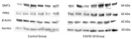
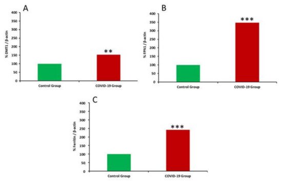
COVID-19, the disease caused by SARS-CoV-2 has reached pandemic proportions worldwide, with considerable consequences for both health and the economy. In pregnant women, COVID-19 can alter the metabolic environment, iron metabolism, and oxygen supply of trophoblastic cells, and therefore have a negative influence on pregnant women and mechanisms of fetal development, with implications in the postnatal life. The purpose of this study was to investigate, for the first time, the effects of COVID-19 infection during pregnancy with regard to the oxidative/antioxidant status in mothers’ serum and placenta, together with placental iron metabolism. Results showed no differences in superoxide dismutase activity and placental antioxidant capacity. However, antioxidant capacity decreased in the serum of infected mothers. Catalase activity decreased in the COVID-19 group, while an increase in 8-hydroxy-2’-deoxyguanosine, hydroperoxides, 15-FT-isoprostanes, and carbonyl groups were recorded in this group. Placental vitamin D, E, and Coenzyme-Q10 also showed to be increased in the COVID-19 group. As for iron-related proteins, an up-regulation of placental DMT1, ferroportin-1, and ferritin expression was recorded in infected women. Due to the potential role of iron metabolism and oxidative stress in placental function and complications, further research is needed to explain the pathogenic mechanism of COVID-19 that may affect pregnancy, so as to assess the short-term and long-term outcomes in mothers’ and infants’ health.

COVID-19 placenta pregnancy antioxidant system oxidative stress iron metabolism

1. Introduction

Novel coronavirus infection (COVID-19) is caused by SARS-CoV-2. The first case was reported in December 2019, and since then the disease has quickly spread worldwide reaching pandemic dimensions. The infection typically occurs in most patients with manifestations that include fever, cough, fatigue, shortness of breath, and frequent pneumonia. Estimates from the World Health Organization (WHO) indicate that global mortality rate ranges between 3–4%, with a high proportion of patients requiring admission to intensive care units. This results in high economic costs, related to increased investment in health care and restrictions that affect circulation and trade [1].
Pregnancy presents characteristics that make pregnant women more susceptible to respiratory diseases and the pneumonia development, this being one of the most prevalent non-obstetric infections in pregnant women [2]. These characteristics are associated with some changes that include: increased oxygen consumption, elevated diaphragm, and airway edema, which makes pregnant women much more intolerant to hypoxia. This elevated susceptibility was observed during the H1N1 (Influenza A) outbreak in 2009, in which pregnant women were four times more likely to be admitted to hospital than the rest of the population [3]. In the event that a pregnant woman gets a serious respiratory infection, the most frequent complications are: premature rupture of membranes, intrauterine growth restriction, premature delivery, and fetal death [4].
Due to the rapid expansion of the pandemic, its health and economic implications, and the increased susceptibility for pregnant women to suffer its consequences, it is necessary to conduct deeper research on the effects of the virus during pregnancy and the prediction of possible adverse outcomes in maternal and fetal health. To date, although there have been several studies on COVID-19 during pregnancy, most of the clinical studies available only include a few cases (due to the low cumulative incidence among this group of population), and all of them report on routine parameters, which shows a lack of knowledge of the implications of infection during the gestational process and its sequels in maternal-fetal health [5][6].
The placenta constitutes the active interface between the maternal and fetal blood circulations, regulating physiological changes in mother and fetus, and playing a key role in the development of many pregnancy complications. This organ maintains fetal homeostasis by performing a wide range of physiological functions, which after birth, are carried out by the kidney, gastrointestinal tract, lungs, and endocrine glands of the newborn. In pregnant women, COVID-19 can alter the oxygen supply of trophoblastic cells [7], thus affecting placental function. Moreover, oxidative stress caused by this infection is able to induce direct tissue damage, including in placenta, which would negatively affect the gestation process [8][9], especially in situations of great oxidative aggression such as pregnancy and childbirth [10].
On the other hand, iron plays a crucial role in pregnancy and fetal development [11] and can be altered by the hepcidin-like activity of SARS-CoV-2. This virus induces significant dysregulation on iron metabolism, with high levels of ferritin and ferroptosis [12], which could compromise the oxygen transport to trophoblasts and induce several cell impairments [13]. In addition, hepcidin-induced hypoferremia induced by SARS-CoV-2 impairs primary and memory responses to immunization, and the high levels of hepcidin inhibits adaptive immune responses to pathogens, indicating that serum iron, regulated by hepcidin, is an important and potentially targetable control point for immunity [14]. The pro-inflammatory state associated to iron homeostasis dysregulation plays a key role in pathogenesis of disease and the hyper-ferritinemia due to SARS-CoV-2 being associated with iron toxicity because of ferritin leakage and free iron released by damaged tissues. Therefore, iron metabolism should be investigated in COVID-19 patients to monitor the clinical course of the disease, to predict negative prognosis [15].
Nevertheless, in spite of the importance of oxidative stress and iron metabolism in fetal development, there are just a few studies available in the scientific literature about COVID-19 in pregnant women, and they record just some clinical manifestations generally near late preterm and delivery [16][17], without elucidating the crucial role of both factors during pregnancy and its maternal-fetal implications. Taking into account all that mentioned above, the purpose of this study was to investigate, for the first time, the effects of COVID-19 during pregnancy on the oxidative/antioxidant status and iron metabolism, an unexplored scientific field with noteworthy clinical and social implications.

2. Results

Table 1 shows the clinical characteristics (including the haematological and biochemical parameters measured in the hospital) of the mothers participating in this study. No statistically significant differences were observed between groups for age, weight, height, BMI, parity, or delivery method. In the biochemical parameters shown, differences were only found in serum Fe concentrations with lower values in the COVID-19 group compared to the control group (p < 0.01).
Table 1. Clinical characteristics of healthy mothers (n = 61) and those who suffered COVID-19 (n = 63).
  Control COVID-19
Age (years) 31.58 ± 1.09 31.96 ± 0.78
Weight (kg) 73.69 ± 2.55 74.12 ± 2.67
Length (cm) 166.83 ± 1.03 163.97 ± 0.68
BMI (kg/m2) 26.31 ± 1 27.3 ± 0.8
Parity Uni (%): 52.22 53.14
Multi (%): 47.28 46.86
Delivery method V (%): 56.2 58.7
A (%): 21.8 19.9
C (%): 21.8 23.8
Hemoglobin
2nd T (g/L)
11.88 ± 0.23 11.53 ± 0.13
Hemoglobin
3rd T (g/L)
11.96 ± 0.23 11.72 ± 0.17
Hematocrit
2nd T (%)
35.32 ± 0.61 34.11 ± 0.37
Hematocrit
3rd T (%)
35.70 ± 0.63 34.82 ± 0.47
Serum Iron
3rd T (µg/dL)
97.05 ± 14.35 60.14 ± 9.87 **
BMI: Body mass Index; T: Term; V: Vaginal; A: Assisted Vaginal Delivery; C: Cesarean. ** Significantly different from the control group (p < 0.01, Student’s t test).
With regard to the antioxidant systems studied (Table 2), results showed no differences in SOD and antioxidant capacity of placenta, while total antioxidant capacity decreased in serum of mothers suffering from COVID-19 (p < 0.05). On the other hand, CAT activity decreased in the COVID-19 group (p < 0.01 in placenta and p < 0.05 in serum), while an increase in 8-hydroxy-2’-deoxyguanosine (8-OHdG) (p < 0.01 for placenta and serum), hydroperoxides (p < 0.001 for placenta), 15-FT-isoprostanes (p < 0.01 for placenta and serum), and carbonyl groups (p < 0.001 for placenta and p < 0.05 for serum) were recorded in the COVID-19 group.
Table 2. Oxidative/antioxidant parameters in placenta and serum of healthy mothers (n = 61) and those who have suffered COVID-19 (n = 63).
  Placenta Serum
  Control COVID-19 Control COVID-19
ABTS (mmol/L Trolox) 3.09 ± 0.15 3.11 ± 0.11 3.34 ± 0.175 2.78 ± 0.161 *
SOD (mU/mg protein) 440.02 ± 40.14 430.25 ± 30.69 4.45 ± 0.43 3.92 ± 0.29
CAT (mU/mg protein) 186.18 × 10³ ± 9.28 168.72 × 10³ ± 5.10 ** 77.01 ± 5.90 62.86 ± 2.86 *
8-OhdG (ng/mL) 283.70 ± 7.98 316.12 ± 6.95 ** 75.82 ± 2.85 82.38 ± 1.51 **
Hydroperoxides (µM) 40.73 ± 2.47 48.02 ± 1.90 *** 1.51± 0.27 1.12 ± 0.20
Isoprostanes (ng/mL) 35.30 ± 1.31 39.85 ± 0.57 ** 4.35 ± 0.87 8.85 ± 0.22 **
Carbonyl groups
(nmol/mg protein)
12.74 ± 0.67 16.37 ± 0.84 *** 1.385 ± 0.026 1.601 ± 0.054 *
Data are shown as the mean values ± SEM. Significantly different from the control group (* p < 0.01, ** p < 0.01, *** p < 0.001, Student’s t test).
Table 3 summarizes antioxidant vitamins in placenta and serum. In placenta, Vitamin D, E, and Coenzyme Q10 (CoQ10) showed to be increased in the COVID-19 group (p < 0.01 for vitamin D and E; p < 0.05 for CoQ10. In serum, only vitamin E levels showed an increase (p < 0.05), but no differences were found for vitamin D, CoQ10, and A.
Table 3. Antioxidant vitamins in placenta and serum of healthy mothers (n = 61) and those who have suffered COVID-19 (n = 63).
    Control COVID-19
Placenta
(µg/mg protein)
Vitamin D 12.45 ± 0.58 14.06 ± 0.51 **
Vitamin E 64.80 ± 6.87 93.47 ± 7.27 **
Coenzyme Q10 82.09 ± 4.06 92.13 ± 2.84 *
Serum
(µmol/L)
Vitamin D 46.35 ± 2.59 53.22 ± 3.00
Vitamin E 23.10 ± 2.35 29.47 ± 1.59 *
Coenzyme Q10 0.44 ± 0.02 0.48 ± 0.03
Vitamin A 2.29 ± 0.20 2.72 ± 0.28
Data are shown as the mean values ± SEM. Significantly different from the control group (* p < 0.05, ** p < 0.01, Student’s t test).
Mineral levels in placenta are shown in Table 4. Mn and Fe were increased in the COVID-19 group (p < 0.01), while no differences between groups were found for Se, Ba, Cu, and Zn.
Table 4. Minerals in placenta of healthy mothers (n = 61) and those who have suffered COVID-19 (n = 63).
  Control COVID-19
Mn (µg/g DM) 3.93 ± 0.92 7.47 ± 1.07 **
Se (µg/g DM) 7.47 ± 0.63 7.40 ± 0.64
Ba (µg/g DM) 0.47 ± 0.07 0.36 ± 0.03
Cu (µg/g DM) 8.66 ± 0.57 8.72 ± 0.38
Zn (µg/g DM) 49.18 ± 2.18 49.62 ± 1.51
Fe (mg/g DM) 3.54 ± 0.40 5.39 ± 0.45 **
DM: dry matter; Data are shown as the mean values ± SEM. Significantly different from the control group (** p < 0.01, Student’s t test).
Expression of iron-regulatory proteins in placenta is shown in Figure 2 and Figure 3. An up-regulation of placental DMT1, FPN1, and ferritin expression was recorded in placentas of women who suffered the infection compared to healthy women (p < 0.01 for DMT1 and p < 0.001 for FPN1 and ferritin).
Figure 2. Inmunoblots representing placental protein expression of DMT1, FPN1, β-Actin y Ferritin in healthy and COVID-19 groups of mothers.
Figure 3. Placental iron metabolism protein expression in healthy and COVID-19 group of mothers. (A) Divalent Metal Transporter 1 (DMT1); (B): Ferroportin 1; (C): Ferritin. Values are expressed as % vs. β-actin. Significantly different from the control group (** p < 0.01, *** p < 0.001, Student’s t test).

References

  1. Huang, C.; Wang, Y.; Li, X.; Ren, L.; Zhao, J.; Hu, Y.; Zhang, L.; Fan, G.; Xu, J.; Gu, X.; et al. Clinical features of patients infected with 2019 novel coronavirus in Wuhan, China. Lancet 2020, 395, 497–506.
  2. Madinger, N.E.; Greenspoon, J.S.; Ellrodt, A.G. Pneumonia during pregnancy: Has modern technology improved maternal and fetal outcome? Am. J. Obstet. Gynecol. 1989, 161, 657–662.
  3. Jamieson, D.J.; Honein, M.A.; Rasmussen, S.A.; Williams, J.L.; Swerdlow, D.L.; Biggerstaff, M.S.; Lindstrom, S.; Louie, J.K.; Christ, C.M.; Bohm, S.R.; et al. H1N1 2009 influenza virus infection during pregnancy in the USA. Lancet 2009, 374, 451–458.
  4. Benedetti, T.J.; Valle, R.; Ledger, W.J. Antepartum pneumonia in pregnancy. Am. J. Obstet. Gynecol. 1982, 144, 413–417.
  5. Yu, N.; Li, W.; Kang, Q.; Xiong, Z.; Wang, S.; Lin, X.; Liu, Y.; Xiao, J.; Liu, H.; Deng, D.; et al. Clinical features and obstetric and neonatal outcomes of pregnant patients with COVID-19 in Wuhan, China: A retrospective, single-centre, descriptive study. Lancet Infect. Dis. 2020, 20, 559–564.
  6. Di Toro, F.; Gjoka, M.; Di Lorenzo, G.; De Santo, D.; De Seta, F.; Maso, G.; Risso, F.M.; Romano, F.; Wiesenfeld, U.; Levi-D’Ancona, R.; et al. Impact of COVID-19 on maternal and neonatal outcomes: A systematic review and meta-analysis. Clin. Microbiol. Infect. 2021, 27, 36–46.
  7. Liu, H.; Wang, L.-L.; Zhao, S.-J.; Kwak-Kim, J.; Mor, G.; Liao, A.-H. Why are pregnant women susceptible to COVID-19? An immunological viewpoint. J. Reprod. Immunol. 2020, 139, 103122.
  8. Kagan, V.E.; Tyurina, Y.Y.; Bayir, H.; Chu, C.T.; Kapralov, A.A.; Vlasova, I.I.; Belikova, N.A.; Tyurin, V.A.; Amoscato, A.; Epperly, M.; et al. The “pro-apoptotic genies” get out of mitochondria: Oxidative lipidomics and redox activity of cytochrome c/cardiolipin complexes. Chem.-Biol. Interact. 2006, 163, 15–28.
  9. Khairallah, R.J.; Kim, J.; O’Shea, K.M.; O’Connell, K.A.; Brown, B.H.; Galvão, T.; Daneault, C.; Rosiers, C.D.; Polster, B.; Hoppel, C.L.; et al. Improved Mitochondrial Function with Diet-Induced Increase in Either Docosahexaenoic Acid or Arachidonic Acid in Membrane Phospholipids. PLoS ONE 2012, 7, e34402.
  10. Díaz-Castro, J.; Pulido-Moran, M.; Moreno-Fernandez, J.; Kajarabille, N.; De Paco, C.; Garrido-Sanchez, M.; Prados, S.; Ochoa, J. Gender specific differences in oxidative stress and inflammatory signaling in healthy term neonates and their mothers. Pediatr. Res. 2016, 80, 595–601.
  11. Means, R.T. Iron Deficiency and Iron Deficiency Anemia: Implications and Impact in Pregnancy, Fetal Development, and Early Childhood Parameters. Nutrients 2020, 12, 447.
  12. Hirschhorn, T.; Stockwell, B.R. The development of the concept of ferroptosis. Free Radic. Biol. Med. 2019, 133, 130–143.
  13. Cavezzi, A.; Troiani, E.; Corrao, S. COVID-19: Hemoglobin, Iron, and Hypoxia beyond Inflammation. A Narrative Review. Clin. Pract. 2020, 10, 24–30.
  14. Frost, J.N.; Tan, T.K.; Abbas, M.; Wideman, S.K.; Bonadonna, M.; Stoffel, N.U.; Wray, K.; Kronsteiner, B.; Smits, G.; Campagna, D.R.; et al. Hepcidin-Mediated Hypoferremia Disrupts Immune Responses to Vaccination and Infection. Med 2020, 2, 164–179.e12.
  15. Lorena, D.; Isabella, N.; Daniele, V.; Giovanni Battista, V.; Andrea, M.; Andrea, V.; Patrizio, C.; Giovanna, G.; Filippo, B. COVID-19, inflammatory response, iron homeostasis and toxicity: A prospective cohort study in the Emergency Department of Piacenza (Italy). Res. Sq. 2022.
  16. Mirbeyk, M.; Saghazadeh, A.; Rezaei, N. A systematic review of pregnant women with COVID-19 and their neonates. Arch. Gynecol. Obstet. 2021, 304, 5–38.
  17. Capobianco, G.; Saderi, L.; Aliberti, S.; Mondoni, M.; Piana, A.; Dessole, F.; Dessole, M.; Cherchi, P.L.; Dessole, S.; Sotgiu, G. COVID-19 in pregnant women: A systematic review and meta-analysis. Eur. J. Obstet. Gynecol. Reprod. Biol. 2020, 252, 543–558.
More
Information
Contributors MDPI registered users' name will be linked to their SciProfiles pages. To register with us, please refer to https://encyclopedia.pub/register : ,
View Times: 508
Entry Collection: COVID-19
Revisions: 3 times (View History)
Update Date: 28 Jan 2022
1000/1000
Video Production Service