Submitted Successfully!
To reward your contribution, here is a gift for you: A free trial for our video production service.
Thank you for your contribution! You can also upload a video entry or images related to this topic.
Version Summary Created by Modification Content Size Created at Operation
1 + 2674 word(s) 2674 2021-12-15 10:21:09 |
2 Revision for minor details + 4 word(s) 2678 2022-01-25 23:58:16 | |
3 Format correct Meta information modification 2678 2022-01-26 07:03:44 | |
4 Format correct + 97 word(s) 2775 2022-01-28 06:53:15 | |
5 Format correct -3 word(s) 2772 2022-02-07 04:54:34 |

Video Upload Options

Do you have a full video?

Confirm

Are you sure to Delete?
Cite
If you have any further questions, please contact Encyclopedia Editorial Office.
Ventura, P.; Ricci, A. Mechanisms of Kidney Damage in Acute Hepatic Porphyrias. Encyclopedia. Available online: https://encyclopedia.pub/entry/18794 (accessed on 02 July 2024).
Ventura P, Ricci A. Mechanisms of Kidney Damage in Acute Hepatic Porphyrias. Encyclopedia. Available at: https://encyclopedia.pub/entry/18794. Accessed July 02, 2024.
Ventura, Paolo, Andrea Ricci. "Mechanisms of Kidney Damage in Acute Hepatic Porphyrias" Encyclopedia, https://encyclopedia.pub/entry/18794 (accessed July 02, 2024).
Ventura, P., & Ricci, A. (2022, January 25). Mechanisms of Kidney Damage in Acute Hepatic Porphyrias. In Encyclopedia. https://encyclopedia.pub/entry/18794
Ventura, Paolo and Andrea Ricci. "Mechanisms of Kidney Damage in Acute Hepatic Porphyrias." Encyclopedia. Web. 25 January, 2022.
Mechanisms of Kidney Damage in Acute Hepatic Porphyrias
Edit

Porphyrias are a group of rare disorders originating from an enzyme dysfunction in the pathway of heme biosynthesis. Depending on the specific enzyme involved, porphyrias manifest under drastically different clinical pictures. The most dramatic presentation of the four congenital acute hepatic porphyrias (AHPs: acute intermittent porphyria—AIP, ALAD deficiency, hereditary coproporphyria—HCP, and porphyria variegata—VP) consists of potentially life-threatening neurovisceral attacks, for which givosiran, a novel and effective siRNA-based therapeutic, has recently been licensed. Nonetheless, the clinical manifestations of acute porphyrias are multifaceted and do not limit themselves to acute attacks. In particular, porphyria-associated kidney disease (PAKD) is a distinct, long-term degenerating condition with specific pathological and clinical features, for which a satisfactory treatment is not available yet. In PAKD, chronic tubule-interstitial damage has been most commonly reported, though other pathologic features (e.g., chronic fibrous intimal hyperplasia) are consistent findings. Given the relevant role of the kidney in porphyrin metabolism, the mechanisms possibly intervening in causing renal damage in AHPs are different: among others, δ-aminolevulinic acid (ALA)-induced oxidative damage on mitochondria, intracellular toxic aggregation of porphyrins in proximal tubular cells, and derangements in the delicate microcirculatory balances of the kidney might be implicated. The presence of a variant of the human peptide transporter 2 (PEPT2), with a greater affinity to its substrates (including ALA), might confer a greater susceptibility to kidney damage in patients with AHPs. Furthermore, a possible effect of givosiran in worsening kidney function has been observed. In sum, the diagnostic workup of AHPs should always include a baseline evaluation of renal function, and periodic monitoring of the progression of kidney disease in patients with AHPs is strongly recommended.

porphyria kidney nephropathy chronic kidney disease kidney transplantation acute hepatic porphyrias porphyrin

1. Introduction

Porphyrias are a group of rare disorders originating from an enzyme dysfunction in the metabolic pathway of heme biosynthesis [1]. According to the specific enzyme involved, porphyrias manifest under dramatically different clinical pictures [2]: acute hepatic porphyrias (AHPs: acute intermittent porphyria—AIP, aminolevulinic acid (ALA) dehydratase deficiency porphyria—ALADp, hereditary coproporphyria—HCP, and variegate porphyria—VP) present with potentially life-threatening acute neurovisceral attacks (or acute porphyric attacks—APAs), whereas nonacute porphyrias (porphyria cutanea tarda—PCT and erythropoietic protoporphyria—EPP, among others) mainly display a range of debilitating dermatologic manifestations and—for EPP—a higher risk of developing a chronic hepatic disease (among acute porphyrias, HCP and VP may also present with cutaneous symptoms [3][4]).
Each of the porphyrias is characterized by a specific pattern of accumulation of heme precursors (δ-aminolevulinic acid, porphobilinogen—PBG, or porphyrins) in plasma, urine, and/or feces, depending on the hydrophobicity of the different compounds. Since porphyrins are highly reactive to ultraviolet rays, when found in urine they cause it to turn to a reddish hue under sunlight exposure. At the same time, when porphyrins deposit in the skin, they are responsible for the painful phototoxic reactions of cutaneous porphyrias, mainly due to the light-dependent release of cytotoxic reactive oxygen species (ROS) in the course of type I/II photosensitized reactions [5][6].
Due to their heterogeneous and often not specific presentation, added to their utmost rarity, porphyrias represent a notoriously difficult diagnostic challenge for the clinician [7]. Nonetheless, clinical awareness of this group of diseases among physicians is paramount, since patients with porphyrias are heavily burdened not only by their condition, but also by diagnostic delays in the range of months to years, with all the subsequent risks of mistreatment or suboptimal management.
Until recently, treatment options for AHPs were limited to avoiding those environmental stimuli (e.g., fasting, alcohol, “porphyrinogenic” drugs) which, by putatively increasing the metabolic demand for heme, could trigger an APA. In the absence of randomized controlled trials or a shared consensus, periodic infusions of heme arginate have been implemented as a prophylactic therapy for APAs [8], whereas acute attacks are currently managed with heme arginate, glucose infusions, and supportive therapy [8]. It should be underlined that liver transplantation is currently deemed the only curative option for patients with AHPs [9].
In recent years, a novel siRNA-based drug, givosiran, has been approved for the treatment of acute hepatic porphyrias [10][11]: by specifically inhibiting the liver isoform of ALA synthase (ALAS1), the first and rate-limiting enzyme of the heme biosynthetic pathway, givosiran has shown to be highly efficacious in reducing the frequency of porphyric attacks and improving the quality of life of patients with AHPs [12].
While givosiran has represented a breakthrough in the management of acute porphyrias, it must not be overlooked that, other than acute attacks, patients with AHPs develop long-term complications such as chronic neuropathy, hepatocellular carcinoma, and chronic kidney disease (CKD) [13][14]. In fact, porphyria-associated kidney disease (PAKD) has been recognized as a distinct entity with specific pathological and clinical features [15], for instance, most patients with AIP suffer a progressive impairment of kidney function, with an estimated decline in glomerular filtration rate (eGFR) of 1 mL/min per 1.73 m2 per year [16].
The purpose is to outline the known features of renal involvement in acute hepatic porphyrias: most of the knowledge on this subject is provided by studies on the most common AHP, acute intermittent porphyria, even though at least a few early observations on the South African cluster of variegate porphyria are available [17][18].

2. Role of the Kidney in Porphyrin Metabolism

It is generally assumed that the kidney contributes to heme production as the third major synthesizing organ, after the bone marrow and the liver—which account, respectively, for 80% and 15% of total heme biosynthesis [19]. In fact, several biochemical, ultrastructural, and fluorescence microscopy studies have suggested that the kidney is overall abundant in heme. At variance with the liver, though, the capacity for heme biosynthesis in the kidney is heterogeneously distributed and highly compartmentalized, and parallels the activity of detoxifying cytochromes and other heme-dependent functions 
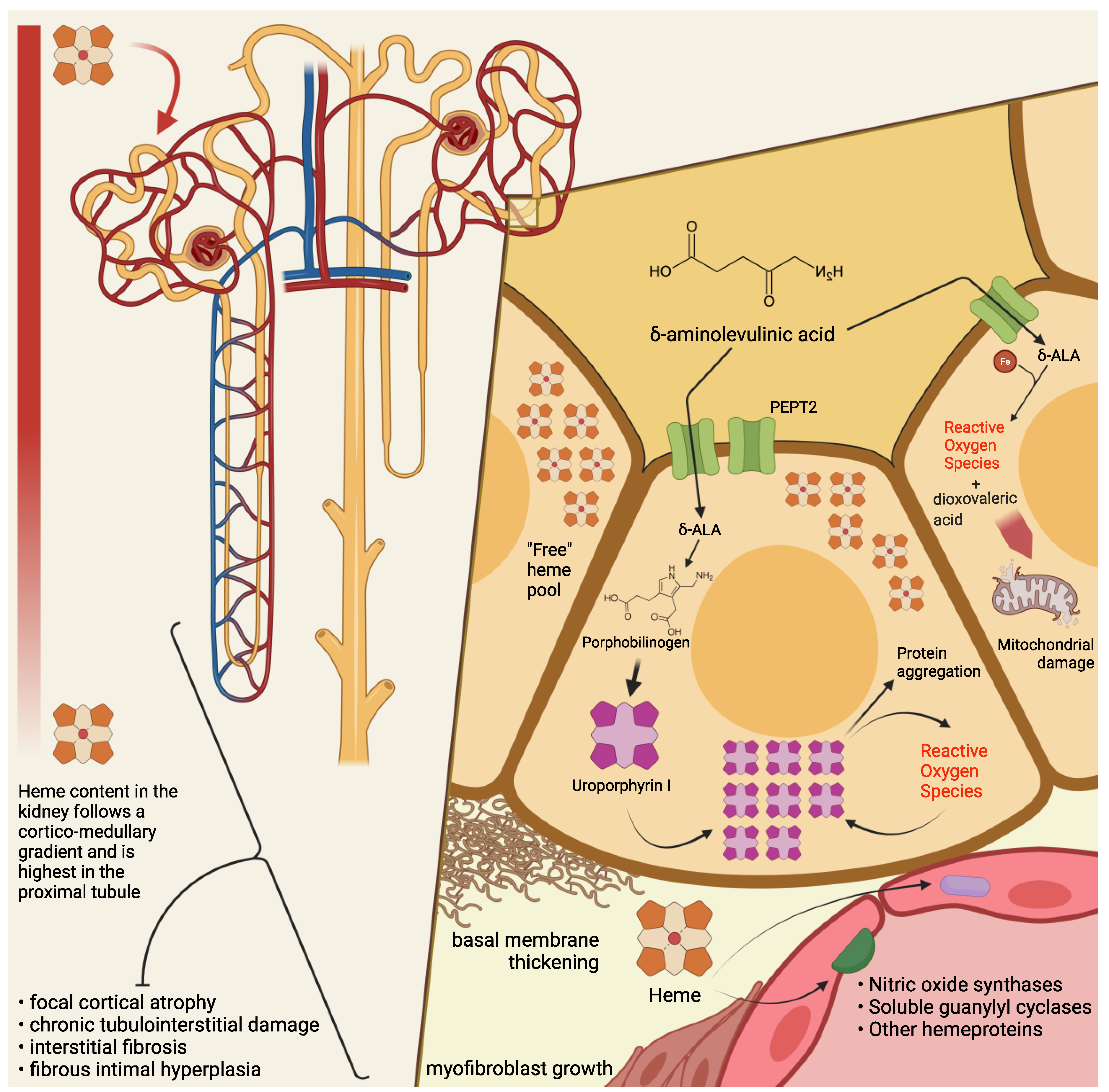
[20]. Thus, it has been demonstrated that heme biosynthetic activity and porphyrin concentration in the kidney follow a corticomedullary gradient: both are highest in the cortical proximal tubules, a metabolically active region particularly exposed to xenobiotics or other endogenously produced compounds [20].
Compared to liver cells, ALAS in the kidney is somewhat more refractory to induction by porphyrinogenic stimuli. The induction process—an initial increase in the enzyme’s activity in the cytosol and a subsequent shift into the mitochondrial matrix- seems qualitatively similar to what has been observed in the liver, but its kinetics are much slower (i.e., hours instead of minutes) [20]. By contrast, renal ALAS activity is promptly inhibited by heme, similarly to the liver isoform. Finally, a greater ratio of ferrochelatase-to-ALAS activity has been detected in renal compared to liver cells. Together with other pieces of evidence, these observations have led to the hypothesis that the kidney could benefit from a higher content of intracellular, regulatory “free” heme, which could also function as a protective buffer to acute heme-depleting stimuli [20].
This being considered, it might be of interest to estimate the amount of the “free” heme pool reserves in liver cells. Liver transplantation is deemed curative in AHPs [9]: therefore, it may be conjectured that the damage in AHP might derive, at least partially, from the tissue-specific lower concentration of intracellular unbound heme in the liver and the subsequent greater susceptibility to induction of hepatic ALAS [20].
Several observations have supported the idea that porphyrin excess in urine, e.g., during attacks, is of renal origin [17][20][21][22][23]. In particular, studies on the kidney’s porphyrin clearance, as well as observations on lead intoxication [24] and on patients with variegate porphyria [24][25], point to a renal endogenous production of coproporphyrin.

3. Pathogenesis of Kidney Damage in PAKD

Among several mechanisms by which ALA is thought to cause cytotoxic damage, the kidney may be particularly susceptible—at least in its most metabolically active segments—to mitochondrial ALA-induced oxidation. At the intracellular level, ALA undergoes a phosphate-catalyzed auto-enolization, and becomes an oxidizing agent; it reacts with iron and O2 to produce superoxide anion (O2), HO radical, and ALA radical (ALA); ALA, in the presence of oxygen, reduces iron and yields dioxo valeric acid (DOVA), a highly reactive oxidant [26][27]. Several pieces of evidence have been gathered concerning ALA toxicity on mitochondrial morphology, loss of transmembrane potential, and protein expression [28][29][30].
Renal histopathological findings in patients with PAKD point toward chronic tubulointerstitial damage [16][18][31][32][33][34] and chronic fibrous intimal hyperplasia associated with focal cortical atrophy [16]. Early autopsy reports in a South African series of patients with variegate porphyria evidenced renal tubular degeneration, more marked in distal tubules, with calcified casts [18]. More recently, Pallet et al. [16] described tubular atrophy, basal membrane thickening, and interstitial fibrosis; nonspecific arteriosclerotic lesions [16] have also been observed, with arterial fibrous intimal hyperplasia in the cortex, consisting of myofibroblast growth, sclero-fibrotic tissue production and endothelial lumen narrowing. Remarkably, glomeruli seem spared from direct damage [34], since only unspecific sclerotic and ischemic lesions have been reported [16][31]. Markers of ongoing fibrogenesis, such as cytoplasmic accumulation of ß-catenin and vimentin expression, [16] have been detected in tubular sections, and mitochondrial abnormalities have been reported anecdotally [18][33].
Cell culture studies have shown that human endothelial cells (HUVECs), when incubated with ALA and PBG, do not appear to suffer direct damage from the porphyrin precursors [16]. In contrast, human renal epithelial cells (HRECs) display a wide range of alterations in the presence of ALA and PBG in vitro, i.e.: activation of apoptosis, with signs of autophagy and endoplasmic reticulum stress; evidence of a proinflammatory and fibrogenic secretory milieu; morphologic and molecular changes suggestive of epithelial-to-mesenchymal transition (loss of the cuboid morphology, cell-to-cell contact, E-cadherin expression; nuclear translocation of β-catenin; increased expression of SLUG).
On electron microscopy, HRECs incubated with PBG showed accumulation of electron-dense cytosolic granules, whereas light microscopy detected yellow-brown granular aggregates, negative for Perl’s stain, and numerous cytoplasmic osmiophilic granules within the proximal tubular cells [16]. Intriguingly, when proximal tubular cells are incubated with PBG, the latter is completely metabolized into uroporphyrinogen I and III [16]: therefore, it has been conjectured that the observed intracellular inclusions could be aggregates of uroporphyrin obtained by the uncatalyzed polymerization and cyclisation of four PBG molecules.
It is then interesting, from a historical as well as a scientific perspective, that a few studies on acute porphyrias from the past century have reported histopathological findings suggestive of tubular deposition of porphyrins [18][35][36][37]; for instance, a case series of autopsies from patients with variegate porphyria mentioned the presence of a brown autofluorescent pigment, not staining as iron, in both casts and renal tubular cells, and detected a red-orange autofluorescence in the lumen and epithelial cells of Henle’s loop, which in the author’s experience could be possibly attributed to porphyrin deposits [18].
As a matter of fact, consistent pieces of evidence have been gathered concerning the cell-damaging effects of light-independent porphyrin-mediated toxicity [38]: in particular, intracellular, extralysosomal porphyrin accumulation engenders protein aggregation through noncovalent, oxygen-dependent, reversible mechanisms [39][40]. A particular susceptibility has been demonstrated, chiefly in hepatocytes, for intermediate filaments (nuclear laminins and cytoplasmatic keratins) [38][41], proteins in the endoplasmic reticulum (e.g., protein disulfide isomerase and calnexin) [42], proteasome regulatory particles, and key glycolytic enzymes, including glyceraldehyde 3-phosphate dehydrogenase [42]. This process could both trigger and be accelerated by the activity of other oxidizing agents (inflammation, redox reactions) [39], so that porphyrins could precipitate the production of reactive oxygen species (ROS) and intracellular protein aggregation without prior photosensitization. Of note, uroporphyrin I is reduced by the P450 cytochrome’s family and by nicotinamide adenine dinucleotide phosphate (NADPH) in a reaction that yields a superoxide radical (O2) [43][44]. It may be tempting to speculate that similar mechanisms might take place in the cytochrome-rich renal parenchyma, contributing to the renal toxicity of high concentrations of ALA and PBG.
It must be remarked that when a mouse model of AIP was employed to investigate the effects of repeated phenobarbital-induced acute attacks on renal tissues [45] relatively mild unspecific alterations were undisclosed, even in near-total (that is, 5/6) nephrectomized animals. No granular inclusions or signs of tubule-interstitial damage were evidenced, even though the same authors underscore the differences between the experimental setting and the patients’ condition with years of exposure to abnormal levels of porphyrin precursors [45].
From a clinical perspective, signs of proximal tubulointerstitial insufficiency (i.e., proteinuria, impaired erythropoietin production) and of oxidative damage (increased urinary excretion of lipoperoxides), have been anecdotally signaled in porphyric patients [32][34]. A pattern consistent with sodium losses of tubular origin has been detected in patients with variegate porphyria [17]. A case series reported that, during remission from acute attacks, patients with AHPs displayed signs of tubulointerstitial and hypertensive damage, such as poorly concentrated urines (hyposthenuria), and an impairment of the tubular excretory phase, as disclosed by isotopic renography. In this population, four patients had low serum erythropoietin levels, while all of them (11 with AIP, 1 with VP) had low plasma and erythrocyte vitamin B6 (pyridoxal phosphate, PLP) levels. Interestingly, all patients had significant hyperoxalaemia and hyperoxaluria, and an inverse relationship between plasma oxalic acid and erythrocyte vitamin B6 levels was found in AIP patients [32]. Oxalic acid is a product of glyoxylic acid metabolism, whose conversion to glycine is effected by PLP-dependent transaminases [46][47]. Inherited excessive urinary excretion of oxalic acid (primary hyperoxaluria) is linked to an increased risk of urolithiasis (formation of calcium oxalate kidney stones) and kidney damage [48]. Even though the efficacy of PLP supplementation in reducing oxaluria is debated [47][48][49][50][51], AHPs patients are known to suffer from a poorer vitamin B6 status [52][53] compared to the general population

4. Conclusions

The clinical manifestations of acute porphyrias are multifaceted and do not limit themselves to acute attacks. In particular, porphyria-associated kidney disease is a long-term, degenerating condition, for which a satisfactory treatment is still not available. A deeper understanding of the mechanisms of kidney damage in AHPs (Figure 1) is crucial for tailoring a treatment aimed at preventing progression to ESRD in these patients.
Diagnostics 11 02324 g001
Figure 1. Mechanisms of kidney damage in acute hepatic porphyrias. The capacity for heme biosynthesis in the kidney parallels the activity of detoxifying cytochromes and other heme-dependent functions. Compared to the liver, the kidney could benefit from a higher content of intracellular regulatory “free” heme, serving as a protective buffer to acute heme-depleting stimuli. Histopathological findings in patients with porphyria-associated kidney disease have shown chronic tubulointerstitial damage and chronic fibrous intimal hyperplasia associated with focal cortical atrophy. PEPT2 is a proton-coupled symporter expressed on the brush border of tubular proximal cells: the PEPT2*1 variant has a greater affinity to its substrates (including δ-aminolevulinic acid—ALA) compared to PEPT2*2, and its presence has been independently associated with a worse decline in renal function in patients with HMBS mutation. At the intracellular level, ALA undergoes a multistep reaction in the presence of iron and O2 to produce dioxovaleric acid, a highly reactive oxidant: among others, this is a mechanism by which ALA could exert its mitochondrial toxicity. When proximal tubular cells are incubated with porphobilinogen (PBG), the latter is completely metabolized into uroporphyrinogen I and III: thus, the uncatalyzed polymerization and cyclisation of four PBG molecules may lead to the intracellular accumulation of (uro)porphyrin aggregates. Porphyrins have been shown to produce reactive oxygen species (ROS) and cause intracellular protein aggregation without prior photosensitization; this process could both trigger and be accelerated by an oxidizing milieu. Relative heme depletion, as well as hemodynamic rearrangements due to sudden alterations in the levels of circulating ALA, could impact the delicate microcirculatory balances of the kidney regulated by nitric oxide synthases (NOSs), soluble guanylyl cyclases (sGC) or other hemeproteins with vasoactive effects. Other possible mechanisms of kidney damage are discussed in the text. Created with BioRender.com (last accessed date: 5 December 2021).
In fact, a baseline evaluation of kidney function should always be included in the diagnostic workup of AHPs, and herein strongly recommend a periodic monitoring of the progression of kidney damage in these patients, whether they are under siRNA-based therapy or not.
Since acute attacks are the most dramatic manifestation of AHPs, a great deal of the research in this field has focused on the pathogenesis and treatment of neurovisceral damage [54]. In this regard, givosiran has truly represented a game-changer in decreasing the rate of attacks and improving the patients’ quality of life. Notwithstanding the concern for some possibly drug-related adverse events, suspension of treatment should be weighed against the heavy burden of the reoccurrence of potentially life-threatening APAs.

Acronyms

AHP Acute hepatic porphyrias
AIP Acute intermittent porphyria
ALA Aminolevulinic acid
ALAD Aminolevulinic acid dehydratase
APA Acute porphyric attack
CKD Chronic kidney disease
DOVA Dioxovaleric acid
eGFR Estimated Glomerular Filtration Rate
ESRD End-Stage Renal Disease
HCP Hereditary coproporphyria
HMBS Porphobilinogen-deaminase or hydroxymethylbilane-synthase
HREC Human Renal Epithelial Cells
HUVEC Human Umbilical Vein Endothelial Cells
mRNA Messenger RNA
NADPH Nicotinamide adenine dinucleotide phosphate
NOS Nitric oxide synthase
PAKD Porphyria-associated kidney disease
PBG Porphobilinogen
PEPT2 Human Peptide Transporter 2
PLP Pyridoxal phosphate
RNA Ribonucleic Acid
ROS Reactive Oxygen Species
sGC Soluble guanylyl cyclase
siRNA Small interfering RNA
VP Variegate porphyria

References

  1. Bissell, D.M.; Anderson, K.E.; Bonkovsky, H.L. Porphyria. N. Engl. J. Med. 2017, 377, 862–872.
  2. Souza, P.V.S.; Badia, B.M.L.; Farias, I.B.; Gonçalves, E.A.; Pinto, W.B.V.R.; Oliveira, A.S.B. Acute hepatic porphyrias for the neurologist: Current concepts and perspectives. Arq. Neuropsiquiatr. 2021, 79, 68–80.
  3. Martinez, M.D.C.; Cerbino, G.N.; Granata, B.X.; Batlle, A.; Parera, V.E.; Rossetti, M.V. Clinical, biochemical, and genetic characterization of acute hepatic porphyrias in a cohort of Argentine patients. Mol. Genet. Genom. Med. 2021, 9, e1059.
  4. Kaftory, R.; Edel, Y.; Snast, I.; Lapidoth, M.; Mamet, R.; Elis, A.; Hodak, E.; Levi, A. Greater disease burden of variegate porphyria than hereditary coproporphyria: An Israeli nationwide study of neurocutaneous porphyrias. Mol. Genet. Metab. Rep. 2021, 26, 100707.
  5. Foote, C.S. Definition of type I and type II photosensitized oxidation. Photochem. Photobiol. 1991, 54, 659.
  6. Brun, A.; Sandberg, S. Mechanisms of photosensitivity in porphyric patients with special emphasis on erythropoietic protoporphyria. J. Photochem. Photobiol. B 1991, 10, 285–302.
  7. Ventura, P.; Cappellini, M.D.; Rocchi, E. The acute porphyrias: A diagnostic and therapeutic challenge in internal and emergency medicine. Intern. Emerg. Med. 2009, 4, 297–308.
  8. Ventura, P. When awareness makes the difference: Diagnosing and treating the acute hepatic porphyrias. Intern. Emerg. Med. 2021, 16, 25–27.
  9. Lissing, M.; Nowak, G.; Adam, R.; Karam, V.; Boyd, A.; Gouya, L.; Meersseman, W.; Melum, E.; Oldakowska-Jedynak, U.; Reiter, F.P.; et al. Liver transplantation for acute intermittent porphyria. Liver Transplant. 2021, 27, 491–501.
  10. Balwani, M.; Sardh, E.; Ventura, P.; Peiró, P.A.; Rees, D.C.; St¨olzel, U.; Bissell, D.M.; Bonkovsky, H.L.; Windyga, J.; Anderson, K.E.; et al. Phase 3 trial of RNAi therapeutic givosiran for acute intermittent porphyria. N. Eng. J. Med. 2020, 382, 2289–2301.
  11. Ventura, P.; Bonkovsky, H.L.; Gouya, L.; Aguilera-Peiró, P.; Montgomery Bissell, D.; Stein, P.E.; Balwani, M.; Anderson, D.K.E.; Parker, C.; Kuter, D.J.; et al. Efficacy and safety of givosiran for acute hepatic porphyria: 24-month interim analysis of the randomized phase 3 ENVISION study. Liver Int. 2021.
  12. Souza, P.V.S.; Badia, B.M.L.; Farias, I.B.; Pinto, W.B.V.R.; Oliveira, A.S.B. Acute Hepatic Porphyria: Pathophysiological Basis of Neuromuscular Manifestations. Front. Neurosci. 2021, 15, 715523.
  13. Stewart, M.F. Review of hepatocellular cancer, hypertension and renal impairment as late complications of acute porphyria and recommendations for patient follow-up. J. Clin. Pathol. 2012, 65, 976–980.
  14. Buendía-Martínez, J.; Barreda-Sánchez, M.; Rodríguez-Peña, L.; Ballesta-Martínez, M.J.; López-González, V.; Sánchez-Soler, M.J.; Serrano-Antón, A.T.; Pérez-Tomás, M.E.; Gil-Ferrer, R.; Avilés-Plaza, F.; et al. Health impact of acute intermittent porphyria in latent and non-recurrent attacks patients. Orphanet. J. Rare Dis. 2021, 16, 1–8.
  15. Pallet, N.; Karras, A.; Thervet, E.; Gouya, L.; Karim, Z.; Puy, H. Porphyria and kidney diseases. Clin. Kidney J. 2018, 11, 191–197.
  16. Pallet, N.; Mami, I.; Schmitt, C.; Karim, Z.; Fran¸cois, A.; Rabant, M.; Nochy, D.; Gouya, L.; Deybach, J.C.; Xu-Dubois, Y.; et al. High prevalence of and potential mechanisms for chronic kidney disease in patients with acute intermittent porphyria. Kidney Int. 2015, 88, 386–395.
  17. Bales, L.; Day, R.; Blekkenhorst, G. The clinical and biochemical features of variegate porphyria: An analysis of 300 cases studied at Groote Schuur hospital, Cape Town. Int. J. Biochem. 1980, 12, 837–853.
  18. Campbell, J.A. The pathology of South African genetic porphyria. S. Afr. J. Lab. Clin. Med. 1963, 14, 197–203.
  19. Szlendak, U.; Bykowska, K.; Lipniacka, A. Clinical, biochemical and molecular characteristics of the main types of porphyria. Adv. Clin. Exp. Med. 2016, 25, 361–368.
  20. Woods, J. Regulation of porphyrin and heme metabolism in the kidney. Semin Hematol. 1988, 25, 336–348.
  21. Day, R.; Eales, L.; Disler, P. Porphyrias and the kidney. Nephron 1981, 28, 261–267.
  22. Day, R.; Eales, L. Porphyrins in chronic renal failure. Nephron 1980, 26, 90–95.
  23. Day, R.; Eales, L. Porphyrins in renal transplantation. Nephron 1982, 30, 22–27.
  24. Martásek, P.; Jirsa, M.; Kordaˇc, V. Role of the kidneys in porphyrias. Nephron 1982, 32, 277–278.
  25. Day, R.; Blekkenhorst, G.; Eales, L. Hepatic porphyrins in variegate porphyria. N. Eng. J. Med. 1980, 303, 1368–1369.
  26. Rocha, M.E.M.; Dutra, F.; Bandy, B.; Baldini, R.L.; Gomes, S.L.; Faljoni-Alário, A.; Liria, C.W.; Miranda, M.T.M.; Bechara, E.J.H. Oxidative damage to ferritin by 5-aminolevulinic acid. Arch. Biochem. Biophys. 2003, 409, 349–356.
  27. Oteiza, P.I.; Bechara, E.J. 5-Aminolevulinic acid induces lipid peroxidation in cardiolipin-rich liposomes. Arch. Biochem. Biophys. 1993, 305, 282–287.
  28. Hermes-Lima, M.; Castilho, R.F.; Valle, V.G.; Bechara, E.J.; Vercesi, A.E. Calcium-dependent mitochondrial oxidative damage promoted by 5-aminolevulinic acid. Biochim. Biophys. Acta Mol. Basis Dis. 1992, 1180, 201–206.
  29. Vercesi, A.E.; Castilho, R.F.; Meinicke, A.R.; Valle, V.G.; Hermes-Lima, M.; Bechara, E.J. Oxidative damage of mitochondria induced by 5-aminolevulinic acid: Role of Ca2+ and membrane protein thiols. Biochim. Biophys. Acta 1994, 1188, 86–92.
  30. Laafi, J.; Homedan, C.; Jacques, C.; Gueguen, N.; Schmitt, C.; Puy, H.; Reynier, P.; Carmen Martinez, M.; Malthièery, Y. Pro-oxidant effect of ALA is implicated in mitochondrial dysfunction of HepG2 cells. Biochimie 2014, 106, 157–166.
  31. Andersson, C.; Wikberg, A.; Stegmayr, B.; Lithner, F. Renal symptomatology in patients with acute intermittent porphyria. a population-based study. J. Intern. Med. 2000, 248, 319–325.
  32. Mydlík, M.; Derzsiová, K. Kidney damage in acute intermittent porphyria. Prz. Lek. 2011, 68, 610–613.
  33. Onozato, M.; Tojo, A.; Kamijo, A.; Taniguchi, S.; Kimura, K.; Goto, A.; Fujita, T. Tubulointerstitial nephritis associated with acute intermittent porphyria. Clin. Nephrol. 2001, 55, 171–174.
  34. Marsden, J.; Chowdhury, P.; Wang, J.; Deacon, A.; Dutt, N.; Peters, T.; Macdougall, I. Acute intermittent porphyria and chronic renal failure. Clin. Nephrol. 2008, 69, 339–346.
  35. Schley, G.; Bock, K.; Debusmann, E.; Hocevar, V.; Merguet, P.; Paar, D.; Rausch-Stroomann, J. Untersuchungen über die nierenfunktion bei der akuten intermittierenden porphyrie. Klin. Wochenschr. 1970, 48, 616–623.
  36. Schmid, R.; Schwartz, S.; Watson, C.J. Porphyrin content of bone marrow and liver in the various forms of porphyria. Arch. Intern. Med. 1954, 93, 167–190.
  37. Prunty, F. Sodium and chloride depletion in acute porphyria with reference to the status of adrenal cortical function. J. Clin. Investig. 1949, 28, 690–699.
  38. Maitra, D.; Cunha, J.B.; Elenbaas, J.S.; Bonkovsky, H.L.; Shavit, J.A.; Omary, M.B. Porphyrin-induced protein oxidation and aggregation as a mechanism of porphyria-associated cell injury. Cell. Mol. Gastroenterol. Hepatol. 2019, 8, 535–548.
  39. Maitra, D.; Carter, E.L.; Richardson, R.; Rittié, L.; Basrur, V.; Zhang, H.; Nesvizhskii, A.I.; Osawa, Y.; Wolf, M.W.; Ragsdale, S.W.; et al. Oxygen and conformation dependent protein oxidation and aggregation by porphyrins in hepatocytes and light-exposed cells. Cell. Mol. Gastroenterol. Hepatol. 2019, 8, 659–682.
  40. Maitra, D.; Pinsky, B.M.; Soherwardy, A.; Zheng, H.; Banerjee, R.; Omary, B. Protoporphyrin-IX nanostructures modulate their protein aggregation ability via differential oxidation and protein binding. bioRxiv 2021.
  41. Singla, A.; Griggs, N.W.; Kwan, R.; Snider, N.T.; Maitra, D.; Ernst, S.A.; Herrmann, H.; Omary, M.B. Lamin aggregation is an early sensor of porphyria-induced liver injury. J. Cell Sci. 2013, 126, 3105–3112.
  42. Maitra, D.; Elenbaas, J.S.; Whitesall, S.E.; Basrur, V.; D’Alecy, L.G.; Omary, M.B. Ambient light promotes selective subcellular proteotoxicity after endogenous and exogenous porphyrinogenic stress. J. Biol. Chem. 2015, 290, 23711–23724.
  43. Morehouse, K.M.; Moreno, S.N.; Mason, R.P. The one-electron reduction of uroporphyrin I by rat hepatic microsomes. Arch. Biochem. Biophys. 1987, 257, 276–284.
  44. Morehouse, K.M.; Mason, R.P. The enzymatic one-electron reduction of por phyrins to their anion free radicals. Arch. Biochem. Biophys. 1990, 283, 306–310.
  45. Unzu, C.; Sampedro, A.; Sardh, E.; Mauleón, I.; De Salamanca, R.E.; Prieto, J.; Salido, E.; Harper, P.; Fontanellas, A. Renal failure affects the enzymatic activities of the three first steps in hepatic heme biosynthesis in the acute intermittent porphyria mouse. PLoS ONE 2012, 7, e32978.
  46. Williams, H.E.; Smith, L.H., Jr. Disorders of oxalate metabolism. Am. J. Med. 1968, 45, 715–735.
  47. Balcke, P. Pyridoxine therapy in patients with renal calcium oxalate calculi. Proc. Eur. Dial. Transplant. Assoc. 1983, 20, 417–421.
  48. Hoppe, B. An update on primary hyperoxaluria. Nat. Rev. Nephrol. 2012, 8, 467–475.
  49. Ferraro, P.M.; Taylor, E.N.; Gambaro, G.; Curhan, G.C. Vitamin B6 intake and the risk of incident kidney stones. Urolithiasis 2018, 46, 265–270.
  50. Curhan, G.C.; Willett, W.C.; Rimm, E.B.; Stampfer, M.J. A prospective study of the intake of vitamins C and B6, and the risk of kidney stones in men. J. Urol. 1996, 155, 1847–1851.
  51. Curhan, G.C.; Willett, W.C.; Speizer, F.E.; Stampfer, M.J. Intake of vitamins B6 and C and the risk of kidney stones in women. J. Am. Soc. Nephrol. 1999, 10, 840–845.
  52. Hamfelt, A.; Wetterberg, L. Pyridoxal phosphate in acute intermittent porphyria. Ann. N. Y. Acad. Sci. 1969, 166, 361–364.
  53. Ventura, P.; Marcacci, M.; Marchini, S.; Cuoghi, C.; Vaccari, D.; Pietrangelo, A. Is poor vitamin status a reliable target for treatment of symptomatic patients with hepatic acute porphyrias? Dig. Liver Dis. 2019, 51, e23–e24.
  54. Ricci, A.; Di Pierro, E.; Marcacci, M.; Ventura, P. Mechanisms of Neuronal Damage in Acute Hepatic Porphyrias. Diagnostics 2021, 11, 2205.
More
Information
Contributors MDPI registered users' name will be linked to their SciProfiles pages. To register with us, please refer to https://encyclopedia.pub/register : ,
View Times: 577
Revisions: 5 times (View History)
Update Date: 07 Feb 2022
1000/1000
Video Production Service