Submitted Successfully!
To reward your contribution, here is a gift for you: A free trial for our video production service.
Thank you for your contribution! You can also upload a video entry or images related to this topic.
Version Summary Created by Modification Content Size Created at Operation
1 + 2422 word(s) 2422 2022-01-17 10:11:18 |
2 format correct Meta information modification 2422 2022-01-18 02:47:01 |

Video Upload Options

We provide professional Video Production Services to translate complex research into visually appealing presentations. Would you like to try it?

Confirm

Are you sure to Delete?
Cite
If you have any further questions, please contact Encyclopedia Editorial Office.
Cardoso, A. Decellularized Colorectal Cancer Matrices as Bioactive Scaffolds. Encyclopedia. Available online: https://encyclopedia.pub/entry/18356 (accessed on 15 November 2024).
Cardoso A. Decellularized Colorectal Cancer Matrices as Bioactive Scaffolds. Encyclopedia. Available at: https://encyclopedia.pub/entry/18356. Accessed November 15, 2024.
Cardoso, Ana. "Decellularized Colorectal Cancer Matrices as Bioactive Scaffolds" Encyclopedia, https://encyclopedia.pub/entry/18356 (accessed November 15, 2024).
Cardoso, A. (2022, January 17). Decellularized Colorectal Cancer Matrices as Bioactive Scaffolds. In Encyclopedia. https://encyclopedia.pub/entry/18356
Cardoso, Ana. "Decellularized Colorectal Cancer Matrices as Bioactive Scaffolds." Encyclopedia. Web. 17 January, 2022.
Decellularized Colorectal Cancer Matrices as Bioactive Scaffolds
Edit

More than a physical structure providing support to tissues, the extracellular matrix (ECM) is a complex and dynamic network of macromolecules that modulates the behavior of both cancer cells and associated stromal cells of the tumor microenvironment (TME). Over the last few years, several efforts have been made to develop new models that accurately mimic the interconnections within the TME and specifically the biomechanical and biomolecular complexity of the tumor ECM. Particularly in colorectal cancer, the ECM is highly remodeled and disorganized and constitutes a key component that affects cancer hallmarks, such as cell differentiation, proliferation, angiogenesis, invasion and metastasis. Therefore, several scaffolds produced from natural and/or synthetic polymers and ceramics have been used in 3D biomimetic strategies for colorectal cancer research. Nevertheless, decellularized ECM from colorectal tumors is a unique model that offers the maintenance of native ECM architecture and molecular composition.

colorectal cancer extracellular matrix patient-derived scaffolds

1. Introduction

Colorectal cancer (CRC) is an increasingly prevalent disease that accounts for substantial mortality and morbidity and is responsible for an impaired quality of life and high financial resource consumption [1]. Despite advances in the development of less invasive screening and diagnostic approaches, approximately 25% of CRC patients are still diagnosed with a distant metastatic disease [2]. Currently, available therapies have not only limited the curative impact but also developed resistance, leading to poor prognosis and increased mortality rates [3]. In particular, immunotherapy has a limited application in CRC, being only recommended to patients with high microsatellite instable (MSI) tumors, which correspond to less than 15% of all CRC cases [4]. This scenario highlights the urgent need to better understand the biological mechanisms underlying CRC onset, progression and spread to improve CRC diagnosis and establish tailored therapeutic strategies. For that, a detailed understanding of the tumor microenvironment is fundamental, since it is where the tumorigenic process begins and evolves under the heavy influence of the complex crosstalk between the cellular component (cancer cells and the non-malignant stromal cells), the non-cellular component (extracellular matrix—ECM) and the interstitial fluids [5].
Over the last few years, the ECM has become a hot topic of research since this complex network of macromolecules is much more than a physical and stable structure providing support to tissues. The ECM is an extremely dynamic component of the TME [6] that modulates the behavior of both tumor and cancer-associated stromal cells through its particular biochemical and biomechanical properties [7]. During tumor development, the ECM is significantly altered, both structurally and in terms of composition, usually enabling cellular transformation, angiogenesis, inflammation, invasion and metastasis [8][9]. These tumor ECM alterations translate into dysfunctional biomechanical tissue properties with increased stiffness activating several cellular pathways, such as YAP/TAZ [10], TXNIP [11], Rho/Rock-PTEN [12], PI3K-AKT [13], GSK3β [14] and AMPK [15][16].
Considering the relevant role of this cellular–acellular communication, several efforts have been made to develop new CRC models that accurately mimic the interconnections within the TME to understand the disease [17][18][19][20][21][22][23]. Until now, most cancer research has been performed with in vitro two-dimensional (2D) cell culture. However, it is known that cells behave differently in 2D and three-dimensional (3D) cultures, and that animal models do not truly represent the human tumor architecture [17]. Current 3D cancer models are now managing to bridge the gap between 2D monolayer cell lines, animal models and clinical research. There is an increasingly growing field for the development of 3D cell culture models that are able to closely recapitulate the TME landscape and screen anti-cancer drugs in CRC, such as bio-fabricated tissues [18], organotypic 3D-bioactive models [19] and cancer tissue-originated spheroids [20]. Among these, several reports have described interesting strategies using decellularized ECM from native tissues where the cellular component is removed and the tissue physiology is maintained [24][25][26].

2. Decellularized Colorectal Cancer Matrices as Bioactive Scaffolds for Modeling the Tumor Microenvironment

Decellularized ECM from malignant tissues is gaining attention in the field of organotypic modeling of tumor-stroma interactions by successfully incorporating key biochemical and biophysical characteristics of the native TME [27][28][29]. Particularly, patient-derived scaffolds allow comparisons between the tumor and the normal adjacent tissues, as well as deliver the potential of a preclinical platform to test patient-specific responses to treatment therapies [30][31]. However, decellularized ECM as a biomimetic model for CRC research is just beginning to be explored (Table 1) [24][25].
Table 1. Methods used for the decellularization and evaluation of biochemical/biomechanical properties of decellularized ECM from colorectal tissues.
ECM Sources Decellularization Method Biochemical Evaluation Biomechanical Evaluation REF
Cell-derived matrix
HT-29
SW480
CCD-841-Com
-CHEMICAL
0.5% Triton X-100
20 mM NH4OH
Ionic and nonionic surfactants
n/a n/a [32][33][34]
Human-derived tissue CHEMICAL
5 mM EDTA
10% DMSO
1% Triton X-100
10 mM sodium
cholate hydrate
50 mM Tris-HCl
Centrifugal rotation
Ionic and nonionic surfactants
Mechanical mixing
-Cellular proteins (cytokeratin, vimentin) and stromal components (collagen IV, fibrinogen, hyaluronic acid): Immunohistochemistry
-Actin: Western Blot
-DNA content: SYBR agarose gel
-Architecture: HE
-3D structure: FITC staining of ECMs
[35]
CHEMICAL/ENZYMATIC
4% sodium deoxycholate
2000 kU DNase-I
-DNA content: DNeasy Blood & Tissue kit
-Stromal components (GAGs, Col IV): PAS and Immunohistochemistry
-Cellular proteins (Ki67, vimentin, E-cadherin, DAPI): Immunofluorescence
-Architecture: HE and Laminin
-3D structure: SEM
-Permeability: In-house developed permeability device
[36]
-DNA content: DNeasy Blood & Tissue kit and 1% SYBRsafe agarose gel
-Stromal components (GAGs, Col IV): PAS, Masson’s Trichrome, Immunohistochemistry and Alcian blue
-Architecture: HE,
Gieson and Silver stains
-3D structure: SEM
[19]
PHYSICAL/CHEMICAL
Freezing
2% SDC
1% Triton X-100
Physical disruption
Ionic and nonionic surfactants
-Nucleic acids: HE
-Collagens: SHG
-Stiffness: AMR
-Topography: SHG
[37]
CHEMICAL/ENZYMATIC
0.1% SDS
50 U/mL DNase-I
Ionic surfactant
-Nucleic acids: DAPI
-DNA content: PureLink Genomic DNA Mini Kit
-Histomorphological analysis: HE and Masson’s Trichrome
-Major ECM proteins (Collagens I and IV, Laminin, Fibronectin and Hyaluronic acid): Immunofluorescence
-Stiffness: Rheology
-3D structure: SEM
[26]
CHEMICAL
1% SDS
1% Triton X-100
-DNA content: Nanodrop
-Major ECM proteins (GAGs, Collagen I, Laminin and fibronectin): Immunostaining
-Cellular proteins: F-actin (cytoskeleton), DAPI and HE (nuclei acid)
-Structure and architecture: SEM and TEM [38]
SISmuc
(small intestine submucosa + mucosa from decellularized porcine jejunum)
CHEMICAL
4% SDS
200 U/mL DNase I-
n/e n/e [39]
Mice-derived tissue CHEMICAL/ENZYMATIC
4% sodium deoxycholate
2000 kU DNase-I
-DNA content: Roche’s DNA isolation Kit and Quant-It PicoGreen dsDNA Assay
-Nucleic acids: DAPI and HE
-Major ECM proteins (Collagens I and IV, Fibronectin and Laminin): Immunofluorescence and Masson’s Trichrome
-Tensile testing: RSA-G2 solids analyzer [40]
AMR: Active microrheology; AFM: Atomic force microscopy; SHG: Second harmonic generation; n/a: not applicable; HE: Hematoxylin & Eosin; PAS: Periodic acid-Schiff; SEM: Scanning electron microscopy; SDS: sodium dodecyl sulfate; GAG: Glycosaminoglycan; n/e: not evaluated.
Overall, decellularization protocols aim to eliminate all cell material while maintaining ECM architecture and biochemical components and are mainly a combination of physical, chemical and enzymatic methodologies [41]. The removal of cellular components without impairing the original ECM architecture, protein and glycoprotein distribution, and viscoelastic properties renders decellularization a reliable and attractive technique for studying ECM [19][26][36][37]. Several decellularization protocols and methodologies for evaluating the biochemical and biomechanical features of decellularized CRC ECM have been reported (Table 1). The lack of uniformity in these approaches obviously results in differences in decellularization efficiency, ECM characterization and ECM-mediated biological effects. Therefore, the uniformization of methodologies concerning CRC tissue decellularization would be essential for the reproducibility of results. Decellularized ECM can be cell-, animal-, or human- or patient-derived and have been applied as scaffolds for 3D cell cultures [42], bioprinting techniques for creating adaptable 3D structures, and as components in synthetic and natural cell culture platforms [43], providing bioactive ECM components in tunable constructs [44]. For example, the use of porcine jejunum decellularized matrix, derived from the patented Biological Vascularized Scaffold (DE:302014007893; BioVaSc®, proprietary knowhow Fraunhofer IGB) [45], is an interesting 3D in vitro colon model. This scaffold functions as an alternative biological colon-like structure with a mucosal tissue layer, including crypt, villi and a basement membrane, allowing the investigation of tumor cancer cell growth, EMT processes, cell invasion across the basement membrane, and metastization. Importantly, once this dynamic model is maintained in a bioreactor, the repopulation with CRC cells (i.e., SW480) and fibroblasts results in the formation of tumor-like clusters. Such data highlight the relevance of mimicking the tumor milieu to support malignant and stroma cell interactions [39]. Another exciting CRC biomimetic model was described by Alabi et al., who repurposed the use of mouse decellularized colon matrices, recellularized with HT-29 and HCT-116 human CRC cell lines, to investigate the crosstalk between ECM and cancer cell traits. Interestingly, mouse colon scaffolds were a better support for CRC cell HT-29 proliferation and differentiation than MatrigelTM. Moreover, HCT-116 cells displayed a higher rate of cell invasion when cultured in mouse colon cancer decellularized matrices than in cells cultured in wild-type mouse colon scaffolds or MatrigelTM [40].
Three-dimensional ECM-hydrogels from decellularized human normal and tumor colon tissues have already been prepared through lyophilization, powdering and solubilization techniques and were useful for showing that tumor ECM components induced faster growth of HT-29 cells and their shift toward a glycolytic metabolism [37]. In an elegant study by Tian et al., organ-specific metastases were obtained by seeding CRC cells in a biomatrix coating composed of mouse lung and liver decellularized ECM [46]. In this system, 3D colonies were spontaneously formed and mimicked in vivo metastasis in terms of histological, molecular and phenotypic characteristics. Remarkably, these conditioned cancer cells exhibited tissue-specific tropism when injected into Nu/Nu mice.
Despite the undeniable utility and potential of these works that clearly show the impact of the matrix in CRC progression and metastasis, these approaches lack the native ECM architecture and mechanical properties that exhibit an active role in cell behavior [7][19].
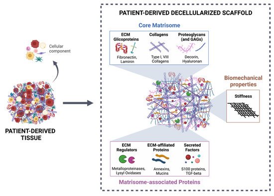
In human CRC, strategies using decellularized ECM to study TME dynamics have been broadly focused on patient-derived scaffolds. Several protocols have been developed with the aim of efficiently removing cellular components from intestinal tissue while maintaining the architecture and biomechanical/biochemical features (Figure 2). These scaffolds have been successfully recellularized with different types of cells [19][26][36]. In 2016, Chen and Shuler described detailed procedures for establishing an organotypic human colon model from decellularized biopsy specimens, which were then recellularized with primary colon epithelial cells, endothelial cells, and fibroblasts [38]. This methodology allowed the identification of several genes that drive invasion in APC and KRAS mutated cells and demonstrated this model’s success in studying cancer biology under physiologically relevant conditions that include cell–matrix interactions and the spatial localization of multiple cell types, crucial in the TME context.
Figure 2. Patient-derived decellularized scaffold as a suitable tool for studying tumor–stroma interactions. The decellularization process of patient-derived tissue allows the efficient removal of the cellular component of the ECM, maintaining the 3D structure, as well as the biochemical categories (core matrisome and matrisome-associated proteins), according to Naba et al. [47], and biomechanical properties (stiffness). Created with BioRender.com (accessed on 10 November 2021).
In this field, approaches that consider paired CRC and normal adjacent tissue benefit from the direct comparison of samples from the same individual and allow the consideration of the role of tumor versus normal ECM on various cancer-associated activities and interactions with other TME components. Nevertheless, studies with access to this type of exceptionally valuable sample have been mainly restricted to recellularization with only one type of cell and require a further complex to create a structure that most trustworthily resembles the TME. Beyond proteomic and structural characterization of the decellularized ECM, reports showed that tumor ECM modulates IL-8 expression by HT29 cells [19] and that both HT-29 and HCT-116 cells reduced the sensitivity to 5-fluoracil when in a 3D ECM-setting [36][48]. In comparison with 2D cultures, HT-29 cells grown in CRC patient-derived scaffolds also displayed changes in the expression of genes and proteins related to proliferation and an increase in those concerning pluripotency and stemness [49].
From a different perspective, Pinto et al. [26] implemented a novel approach by studying the effect of human decellularized normal and tumor matrices derived from CRC patients’ surgical resections on the macrophage inflammatory signature. This work showed for the first time that, although derived from the same patient, normal and tumor matrices differently modulated the macrophage phenotype, with the last inducing an anti-inflammatory polarization, mimicking the immunosuppressive tumor microenvironment. Additionally, macrophages differentiated within tumor decellularized matrices stimulated CRC cell invasion through the expression of CCL18, an immunosuppressive chemokine identified as a key molecule in this process.
Decellularized tissues have also been applied in the study of the CRC metastatic process. D’Angelo and colleagues created a model with decellularized normal and primary tumor CRC tissue, as well as matched CRC liver metastasis with the aim of recapitulating this specific microenvironment in vitro [48]. This system demonstrated that HT-29 cells cultured in scaffolds derived from liver metastasis exhibit a higher EMT transition, a loss of E-cadherin and a higher vimentin expression, among other biological processes.
One of the major drawbacks of CRC patient-derived scaffolds is the limited amount of tumor tissue available from each individual, since it derives from biopsies or surgical resections, from which most tissue is required for further diagnostic molecular and histological characterization. Another question to keep in mind is that normal mucosa adjacent to the tumor, while often considered a healthy control from the same individual, in fact represents an intermediate state between normal and tumor tissues [50]. Despite collecting from at least 10 cm away from the tumor, a normal adjacent mucosa has a large number of differentially expressed genes in comparison with normal mucosa from healthy donors. This genetic disparity is essentially related to functions concerning the inhibition of matrix metalloproteinases (MMPs), cell adhesion molecules, TGF-β and integrin signaling pathways, inflammation, and cytokine–receptor interaction [51]. Interestingly, an integrative analysis of TCGA and GTEx RNA-seq data showed that normal adjacent tissue from the sigmoid colon is more similar to the tumor, while normal adjacent tissue from the transverse colon is more comparable to healthy tissue [50].
The recellularization of decellularized ECM also presents a few challenges, namely the choice of cell(s), the cells’ distribution within the scaffold and the reproducibility of recellularization efficiencies, even in samples from the same patient [52]. Concerning the specific case of CRC, strategies have been focused on small decellularized tissue fragments, static culture conditions, and recellularization with cancer cell lines. However, more sophisticated systems for mimicking the TME will require the inclusion of immune and stromal cells under dynamic cultures that will allow the flow of nutrients and molecules between the different compartments. To surpass the issue of spatial heterogeneity of the matrisome [53], tissue samples have to be representative of distinct tumor regions to avoid biasing experimental outcomes.
These are relevant topics to be considered when establishing an organotypic 3D model for CRC cancer with decellularized tissues, as well as for previously determining if there was previous neoadjuvant therapy. Still, the possibilities of these kinds of systems to incorporate ECM, cancer, stromal and immune cells will allow the study of the dynamic and complex crosstalks between the different components and recapitulate more closely the TME and, eventually, design strategies with potential for predicting clinical outcomes.

References

  1. Dubé, C. Tackling colorectal cancer as a public health issue: What can the gastroenterologist do? Can. J. Gastroenterol. 2012, 26, 417–418.
  2. Vatandoust, S.; Price, T.J.; Karapetis, C.S. Colorectal cancer: Metastases to a single organ. World J. Gastroenterol. 2015, 21, 11767–11776.
  3. Choi, Y.; Sateia, H.F.; Peairs, K.S.; Stewart, R.W. Screening for colorectal cancer. Semin. Oncol. 2017, 44, 34–44.
  4. Ganesh, K.; Stadler, Z.K.; Cercek, A.; Mendelsohn, R.B.; Shia, J.; Segal, N.H.; Diaz, L.A., Jr. Immunotherapy in colorectal cancer: Rationale, challenges and potential. Nat. Rev. Gastroenterol. Hepatol. 2019, 16, 361–375.
  5. Balkwill, F.R.; Capasso, M.; Hagemann, T. The tumor microenvironment at a glance. J. Cell Sci. 2012, 125, 5591–5596.
  6. Hynes, R.O. The extracellular matrix: Not just pretty fibrils. Science 2009, 326, 1216–1219.
  7. Pickup, M.W.; Mouw, J.K.; Weaver, V.M. The extracellular matrix modulates the hallmarks of cancer. EMBO Rep. 2014, 15, 1243–1253.
  8. Hanahan, D.; Weinberg, R.A. Hallmarks of Cancer: The Next Generation. Cell 2011, 144, 646–674.
  9. Winkler, J.; Abisoye-Ogunniyan, A.; Metcalf, K.J.; Werb, Z. Concepts of extracellular matrix remodelling in tumour progression and metastasis. Nat. Commun. 2020, 11, 5120.
  10. Scott, K.E.; Fraley, S.I.; Rangamani, P. A spatial model of YAP/TAZ signaling reveals how stiffness, dimensionality, and shape contribute to emergent outcomes. Proc. Natl. Acad. Sci. USA 2021, 118, e2021571118.
  11. Sullivan, W.J.; Mullen, P.J.; Schmid, E.W.; Flores, A.; Momcilovic, M.; Sharpley, M.S.; Jelinek, D.; Whiteley, A.E.; Maxwell, M.B.; Wilde, B.R.; et al. Extracellular Matrix Remodeling Regulates Glucose Metabolism through TXNIP Destabilization. Cell 2018, 175, 117–132.e21.
  12. Park, J.S.; Burckhardt, C.J.; Lazcano, R.; Solis, L.M.; Isogai, T.; Li, L.; Chen, C.S.; Gao, B.; Minna, J.D.; Bachoo, R.; et al. Mechanical regulation of glycolysis via cytoskeleton architecture. Nature 2020, 578, 621–626.
  13. Peyrou, M.; Clément, S.; Maier, C.; Bourgoin, L.; Branche, E.; Conzelmann, S.; Kaddai, V.; Foti, M.; Negro, F. PTEN protein phosphatase activity regulates hepatitis C virus secretion through modulation of cholesterol metabolism. J. Hepatol. 2013, 59, 420–426.
  14. You, Y.; Zheng, Q.; Dong, Y.; Wang, Y.; Zhang, L.; Xue, T.; Xie, X.; Hu, C.; Wang, Z.; Chen, R.; et al. Higher Matrix Stiffness Upregulates Osteopontin Expression in Hepatocellular Carcinoma Cells Mediated by Integrin β1/GSK3β/β-Catenin Signaling Pathway. PLoS ONE 2015, 10, e0134243.
  15. Levental, K.R.; Yu, H.; Kass, L.; Lakins, J.N.; Egeblad, M.; Erler, J.T.; Fong, S.F.T.; Csiszar, K.; Giaccia, A.; Weninger, W.; et al. Matrix Crosslinking Forces Tumor Progression by Enhancing Integrin Signaling. Cell 2009, 139, 891–906.
  16. Bays, J.L.; Campbell, H.K.; Heidema, C.; Sebbagh, M.; DeMali, K.A. Linking E-cadherin mechanotransduction to cell metabolism through force-mediated activation of AMPK. Nat. Cell Biol. 2017, 19, 724–731.
  17. Baker, B.M.; Chen, C.S. Deconstructing the third dimension—How 3D culture microenvironments alter cellular cues. J. Cell Sci. 2012, 125, 3015–3024.
  18. Devarasetty, M.; Dominijanni, A.; Herberg, S.; Shelkey, E.; Skardal, A.; Soker, S. Simulating the human colorectal cancer microenvironment in 3D tumor-stroma co-cultures in vitro and in vivo. Sci. Rep. 2020, 10, 9832.
  19. Piccoli, M.; D’Angelo, E.; Crotti, S.; Sensi, F.; Urbani, L.; Maghin, E.; Burns, A.; De Coppi, P.; Fassan, M.; Rugge, M.; et al. Decellularized colorectal cancer matrix as bioactive microenvironment for in vitro 3D cancer research. J. Cell. Physiol. 2018, 233, 5937–5948.
  20. Kondo, J.; Ekawa, T.; Endo, H.; Yamazaki, K.; Tanaka, N.; Kukita, Y.; Okuyama, H.; Okami, J.; Imamura, F.; Ohue, M.; et al. High-throughput screening in colorectal cancer tissue-originated spheroids. Cancer Sci. 2019, 110, 345–355.
  21. Di Modugno, F.; Colosi, C.; Trono, P.; Antonacci, G.; Ruocco, G.; Nisticò, P. 3D models in the new era of immune oncology: Focus on T cells, CAF and ECM. J. Exp. Clin. Cancer Res. 2019, 38, 7.
  22. Sensi, F.; D’Angelo, E.; D’Aronco, S.; Molinaro, R.; Agostini, M. Preclinical three-dimensional colorectal cancer model: The next generation of in vitro drug efficacy evaluation. J. Cell. Physiol. 2018, 234, 181–191.
  23. Xu, R.; Zhou, X.; Wang, S.; Trinkle, C. Tumor organoid models in precision medicine and investigating cancer-stromal interactions. Pharmacol. Ther. 2021, 218, 107668.
  24. Hoshiba, T. Decellularized Extracellular Matrix for Cancer Research. Materials 2019, 12, 1311.
  25. Ferreira, L.P.; Gaspar, V.M.; Mano, J.F. Decellularized Extracellular Matrix for Bioengineering Physiomimetic 3D in Vitro Tumor Models. Trends Biotechnol. 2020, 38, 1397–1414.
  26. Pinto, M.L.; Rios, E.; Silva, A.C.; Neves, S.C.; Caires, H.R.; Pinto, A.T.; Durães, C.; Carvalho, F.A.; Cardoso, A.P.; Santos, N.C.; et al. Decellularized human colorectal cancer matrices polarize macrophages towards an anti-inflammatory phenotype promoting cancer cell invasion via CCL18. Biomaterials 2017, 124, 211–224.
  27. Wishart, A.L.; Conner, S.J.; Guarin, J.R.; Fatherree, J.P.; Peng, Y.; McGinn, R.A.; Crews, R.; Naber, S.P.; Hunter, M.; Greenberg, A.S.; et al. Decellularized extracellular matrix scaffolds identify full-length collagen VI as a driver of breast cancer cell invasion in obesity and metastasis. Sci. Adv. 2020, 6, eabc3175.
  28. Lv, Y.; Wang, H.; Li, G.; Zhao, B. Three-dimensional decellularized tumor extracellular matrices with different stiffness as bioengineered tumor scaffolds. Bioact. Mater. 2021, 6, 2767–2782.
  29. Liu, G.; Wang, B.; Li, S.; Jin, Q.; Dai, Y. Human breast cancer decellularized scaffolds promote epithelial-to-mesenchymal transitions and stemness of breast cancer cells in vitro. J. Cell. Physiol. 2019, 234, 9447–9456.
  30. Leiva, M.C.; Garre, E.; Gustafsson, A.; Svanström, A.; Bogestål, Y.; Håkansson, J.; Ståhlberg, A.; Landberg, G. Breast cancer patient-derived scaffolds as a tool to monitor chemotherapy responses in human tumor microenvironments. J. Cell. Physiol. 2021, 236, 4709–4724.
  31. Landberg, G.; Fitzpatrick, P.; Isakson, P.; Jonasson, E.; Karlsson, J.; Larsson, E.; Svanström, A.; Rafnsdottir, S.; Persson, E.; Gustafsson, A.; et al. Patient-Derived Scaffolds Uncover Breast Cancer Promoting Properties of the Microenvironment. Biomaterials 2020, 235, 119705.
  32. Hoshiba, T.; Tanaka, M. Decellularized matrices as in vitro models of extracellular matrix in tumor tissues at different malignant levels: Mechanism of 5-fluorouracil resistance in colorectal tumor cells. Biochim. Biophys. Acta (BBA)-Bioenerg. 2016, 1863, 2749–2757.
  33. Hoshiba, T.; Tanaka, M. Optimization of the tissue source, malignancy, and initial substrate of tumor cell-derived matrices to increase cancer cell chemoresistance against 5-fluorouracil. Biochem. Biophys. Res. Commun. 2015, 457, 353–357.
  34. Hoshiba, T. An extracellular matrix (ECM) model at high malignant colorectal tumor increases chondroitin sulfate chains to promote epithelial-mesenchymal transition and chemoresistance acquisition. Exp. Cell Res. 2018, 370, 571–578.
  35. Genovese, L.; Zawada, L.; Tosoni, A.L.; Ferri, A.; Zerbi, P.; Allevi, R.; Nebuloni, M.; Alfano, M. Cellular Localization, Invasion, and Turnover Are Differently Influenced by Healthy and Tumor-Derived Extracellular Matrix. Tissue Eng. Part A 2014, 20, 2005–2018.
  36. Sensi, F.; D’Angelo, E.; Piccoli, M.; Pavan, P.; Mastrotto, F.; Caliceti, P.; Biccari, A.; Corallo, D.; Urbani, L.; Fassan, M.; et al. Recellularized Colorectal Cancer Patient-Derived Scaffolds as In Vitro Pre-Clinical 3D Model for Drug Screening. Cancers 2020, 12, 681.
  37. Romero-López, M.; Trinh, A.; Sobrino, A.; Hatch, M.M.; Keating, M.T.; Fimbres, C.; Lewis, D.E.; Gershon, P.D.; Botvinick, E.L.; Digman, M.; et al. Recapitulating the human tumor microenvironment: Colon tumor-derived extracellular matrix promotes angiogenesis and tumor cell growth. Biomaterials 2017, 116, 118–129.
  38. Chen, H.J.; Wei, Z.; Sun, J.; Bhattacharya, A.; Savage, D.J.; Serda, R.; Mackeyev, Y.; Curley, S.A.; Bu, P.; Wang, L.; et al. A recellularized human colon model identifies cancer driver genes. Nat. Biotechnol. 2016, 34, 845–851.
  39. Nietzer, S.; Baur, F.; Sieber, S.; Hansmann, J.; Schwarz, T.; Stoffer, C.; Häfner, H.; Gasser, M.; Waaga-Gasser, A.M.; Walles, H.; et al. Mimicking Metastases Including Tumor Stroma: A New Technique to Generate a Three-Dimensional Colorectal Cancer Model Based on a Biological Decellularized Intestinal Scaffold. Tissue Eng. Part C Methods 2016, 22, 621–635.
  40. Alabi, B.R.; LaRanger, R.; Shay, J.W. Decellularized mice colons as models to study the contribution of the extracellular matrix to cell behavior and colon cancer progression. Acta Biomater. 2019, 100, 213–222.
  41. Taylor, D.A.; Sampaio, L.C.; Ferdous, Z.; Gobin, A.S.; Taite, L.J. Decellularized matrices in regenerative medicine. Acta Biomater. 2018, 74, 74–89.
  42. Park, Y.; Huh, K.M.; Kang, S.-W. Applications of Biomaterials in 3D Cell Culture and Contributions of 3D Cell Culture to Drug Development and Basic Biomedical Research. Int. J. Mol. Sci. 2021, 22, 2491.
  43. Dzobo, K.; Motaung, K.; Adesida, A. Recent Trends in Decellularized Extracellular Matrix Bioinks for 3D Printing: An Updated Review. Int. J. Mol. Sci. 2019, 20, 4628.
  44. Zhang, W.; Du, A.; Liu, S.; Lv, M.; Chen, S. Research progress in decellularized extracellular matrix-derived hydrogels. Regen. Ther. 2021, 18, 88–96.
  45. Schanz, J.; Pusch, J.; Hansmann, J.; Walles, H. Vascularised human tissue models: A new approach for the refinement of biomedical research. J. Biotechnol. 2010, 148, 56–63.
  46. Tian, X.; Werner, M.E.; Roche, K.C.; Hanson, A.D.; Foote, H.P.; Yu, S.K.; Warner, S.B.; Copp, J.A.; Lara, H.; Wauthier, E.L.; et al. Organ-specific metastases obtained by culturing colorectal cancer cells on tissue-specific decellularized scaffolds. Nat. Biomed. Eng. 2018, 2, 443–452.
  47. Naba, A.; Clauser, K.R.; Ding, H.; Whittaker, C.A.; Carr, S.A.; Hynes, R.O. The extracellular matrix: Tools and insights for the “omics” era. Matrix Biol. 2016, 49, 10–24.
  48. D’Angelo, E.; Natarajan, D.; Sensi, F.; Ajayi, O.; Fassan, M.; Mammano, E.; Pilati, P.; Pavan, P.; Bresolin, S.; Preziosi, M.; et al. Patient-Derived Scaffolds of Colorectal Cancer Metastases as an Organotypic 3D Model of the Liver Metastatic Microenvironment. Cancers 2020, 12, 364.
  49. Parkinson, G.T.; Salerno, S.; Ranji, P.; Håkansson, J.; Bogestål, Y.; Wettergren, Y.; Ståhlberg, A.; Bexe Lindskog, E.B.; Landberg, G. Patient-derived scaffolds as a model of colorectal cancer. Cancer Med. 2021, 10, 867–882.
  50. Aran, D.; Camarda, R.; Odegaard, J.; Paik, H.; Oskotsky, B.; Krings, G.; Goga, A.; Sirota, M.; Butte, A.J. Comprehensive analysis of normal adjacent to tumor transcriptomes. Nat. Commun. 2017, 8, 077.
  51. Sanz-Pamplona, R.; Berenguer, A.; Cordero, D.; Molleví, D.G.; Crous-Bou, M.; Sole, X.; Paré-Brunet, L.; Guino, E.; Salazar, R.; Santos, C.; et al. Aberrant gene expression in mucosa adjacent to tumor reveals a molecular crosstalk in colon cancer. Mol. Cancer 2014, 13, 46.
  52. Ahmed, E.; Saleh, T.; Xu, M. Recellularization of Native Tissue Derived Acellular Scaffolds with Mesenchymal Stem Cells. Cells 2021, 10, 1787.
  53. Gustafsson, A.; Garre, E.; Leiva, M.C.; Salerno, S.; Ståhlberg, A.; Landberg, G. Patient-derived scaffolds as a drug-testing platform for endocrine therapies in breast cancer. Sci. Rep. 2021, 11, 3334.
More
Information
Contributor MDPI registered users' name will be linked to their SciProfiles pages. To register with us, please refer to https://encyclopedia.pub/register :
View Times: 989
Entry Collection: Gastrointestinal Disease
Revisions: 2 times (View History)
Update Date: 18 Jan 2022
1000/1000
ScholarVision Creations