Submitted Successfully!
To reward your contribution, here is a gift for you: A free trial for our video production service.
Thank you for your contribution! You can also upload a video entry or images related to this topic.
Version Summary Created by Modification Content Size Created at Operation
1 + 2061 word(s) 2061 2021-12-29 07:50:47 |
2 format corrected. Meta information modification 2061 2022-01-13 03:06:18 |

Video Upload Options

Do you have a full video?

Confirm

Are you sure to Delete?
Cite
If you have any further questions, please contact Encyclopedia Editorial Office.
Chen, W. Secondary Metabolites of Purpureocillium lilacinum. Encyclopedia. Available online: https://encyclopedia.pub/entry/18170 (accessed on 26 July 2024).
Chen W. Secondary Metabolites of Purpureocillium lilacinum. Encyclopedia. Available at: https://encyclopedia.pub/entry/18170. Accessed July 26, 2024.
Chen, Wei. "Secondary Metabolites of Purpureocillium lilacinum" Encyclopedia, https://encyclopedia.pub/entry/18170 (accessed July 26, 2024).
Chen, W. (2022, January 13). Secondary Metabolites of Purpureocillium lilacinum. In Encyclopedia. https://encyclopedia.pub/entry/18170
Chen, Wei. "Secondary Metabolites of Purpureocillium lilacinum." Encyclopedia. Web. 13 January, 2022.
Secondary Metabolites of Purpureocillium lilacinum
Edit

Fungi can synthesize a wealth of secondary metabolites, which are widely used in the exploration of lead compounds of pharmaceutical or agricultural importance. Beauveria, Metarhizium, and Cordyceps are the most extensively studied fungi in which a large number of biologically active metabolites have been identified. However, relatively little attention has been paid to Purpureocillium lilacinum. P. lilacinum are soil-habituated fungi that are widely distributed in nature and are very important biocontrol fungi in agriculture, providing good biological control of plant parasitic nematodes and having a significant effect on Aphidoidea, Tetranychus cinnbarinus, and Aleyrodidae. At the same time, it produces secondary metabolites with various biological activities such as anticancer, antimicrobial, and insecticidal. 

entomogenous fungi biosynthesis biocontrol nematodes

1. Introduction

The genus Purpureocillium in the Ophiocordycipitaceae family was structured by Luangsa-Ard et al. In 2011, based on the medical importance, the Purpureocillium lilacinum was designated as the type species of the Paecilomyces genus [1]. This species was nominated as Penicillium lilacinum by Thom in 1901, and then it was revised as Paecilomyces lilacinus by Samson in 1974 [2]. After comparing the 18S rRNA gene, internal transcribed spacer, and partial translation elongation factor 1-a sequences with P. lilacinus, Luangsa-Ard proposed a new genus name Purpureocillium and made the new combination P. lilacinum in 2011. The fungus was found in a wide range of land and marine environments [3][4][5]. They are often isolated from insects, nematodes, and the rhizosphere of many crops [6][7][8]. The species can grow in a wide range of temperatures from 8 to 38 °C with optimal temperatures of 26–30 °C [3]. It also has a wide pH tolerance and can grow on a variety of substrates [9]. This fungus has promising potential as a biocontrol agent to control crops‘ root-knot nematodes [10][11]. The parasitism of nematodes is that the hyphae directly invade the surface of nematodes’ eggs and then produce appressoria on the surface, which infects the nematodes’ eggs after adsorption. In the process of infection, P. lilacinum secretes a variety of enzymes, such as serine protease and chitinase, which can lead to the degradation of protein and chitin components of the nematode epidermis, which is conducive to the invasion of fungi and destruction of cell components. It has been shown that the fermentation filtrate of P. lilacinum can inhibit the mycelial growth of the pathogenic fungi Helminthosporium maydis and Fusarium graminearum, and has a significant inhibitory effect on the spore germination of Fusarium oxysporum [12][13]. Currently, there are eight registered pesticide products of P. lilacinum in China used to control root-knot nematodes (http://www.chinapesticide.org.cn/hysj/index.jhtml, accessed on 23 February 2021); similar pesticides are also registered in the USA (https://iaspub.epa.gov/apex/pesticides, accessed on 23 February 2021) and European Union (http://www.efsa.europa.eu/, accessed on 23 February 2021). In addition, P. lilacinum has been shown to be effective against Phyllotreta striolata, Thrips palmi, and predatory mite [14][15]. However, the P. lilacinum strains used for biocontrol agents have a high identity with those strains causing infections in humans [16][17].
Secondary metabolites are produced in a certain growth period of plants and microorganisms. They are small molecules with complex chemical structures that are not necessary for growth and reproduction, such as pigments, hormones, toxins, and antibiotics [18]. Fungi are important organisms that produce active secondary metabolites. Different kinds of fungi produce different secondary metabolites. The discovery of fungal secondary metabolites has become an important source of new drugs and pesticides [19]. Fumosorinone was isolated from the Isaria fumosorosea, and it is a potential medicine for the treatment of type II diabetes and other associated metabolic syndromes [20]. Diorcinol K, D, and I were isolated from Aspergillus, displaying significant antibacterial activities against Staphylococcus aureus and methicillin-resistant S. aureus [21]. Pyrenocine A was produced by Paecilomyces and showed a significant antitrypanosomal activity against Trypanosoma brucei [22]. Paeciloxanthone was isolated from the extracts of Paecilomyces sp. and showed significant cytotoxicity against HepG2 cell lines [23]. The research on the synthesis and regulation of secondary metabolites is helpful to develop new active compounds and increase the output of active compounds. The common secondary metabolites of fungi are polyketones, nonribosome peptides, sterols, alkaloids, and terpenes.

2. Biosynthesis of Secondary Metabolites in Purpureocillium lilacinum

In 2015, Prasad sequenced the TERIBC-1 strain of P. lilacinus with a genome size of 40.02 Mb by using Illumina Hiseq technology, and predicted 30 secondary metabolite synthesis genes: 12 polyketide syntheses (PKs, details of all abbreviations are in Table S1), 2 PKs-like, 7 nonribosome peptide synthetases, 7 NRPSs-like, 1 PK-NRPS, and 1 dimethylallyl tryptophan synthases (DMATs) gene [24]. In 2016, Wang sequenced P. lilacinus PLBJ-1 and PLFJ-1 strains. The genome sizes of the two strains were 38.14 Mb and 38.53 Mb, respectively [18]. Using SMURF [25] and anti-SMASH [26] software to predict the secondary metabolite synthesis gene cluster, PLBJ-1 and PLFJ-1 strains were found to encode 13 PKs, 2 PKs-like, 10 NRPSs, 10 NRPSs-like, 1 PK-NRPS, 4 terpene synthases (TSs), and 1 DMAT genes. It can be seen that the secondary metabolites produced by different species of P. lilacinum are not identical, but in general, P. lilacinum has great potential in the synthesis of secondary metabolites.
We know that the typical NRPS modules have adenylation (A), thiolation (T) or peptidyl carrier (PCP), condensation (C), and thioesterase (TE) domains [27][28], which are, respectively, responsible for the activation of amino acids, the extension of peptide chains, the formation of amide bonds, and the release of peptide chains [29][30]. The synthesis mechanism of NRPSs is shown in Figure 1. Generally speaking, the A domain combines with the amino acid substrate under the action of ATP to form the corresponding aminoacyl AMP, and the aminoacyl AMP combines with the sulfhydryl group of the T domain to form the aminoacyl-s-carrier complex. Finally, the carriers carrying the aminoacyl group and the peptide acyl group combine with the specific region of the C domain, and the amino group on the aminoacyl-s-carrier complex attacks the acyl group on the peptidyl-s-carrier complex, forming a new peptide bond, and finally forming a complete peptide chain, through the action of multiple modules, wherein the amino acids in the peptide chain correspond to the modules in the NRPS one by one. Some NRPS modules also contain epimerization (E), formylation (F), methylation (M), heterocyclization (CY) [31], reduction (R), and oxidation (OX) domains, which are involved in the structural modification of peptide chains. Finally, mature peptide chains are released from the NRP assembly line under the action of the TE domain [32].
Figure 1. The biosynthesis of NRPSs [33].
In 2016, Wang sequenced and analyzed the whole genome of P. lilacinum, and predicted the knock-out of the NRPS synthetic gene (LcsA), PK synthetase (LcsC), Acyl CoA ligase (LcsD), and thioesterase (LcsE), using high-performance liquid chromatography (HPLC) to compare the crude extracts of wild-type and mutant strains of P. lilacinum. It was found that the crude extracts of ΔLcsA, ΔLcsC, ΔLcsD, and ΔLcsE had a lack of Leucinostatin A and Leucinostatin B, and then these enzymes were found to play a key role in the synthesis of Leucinostatin, and the synthesis of leucinostatin of P. lilacinum was suggested. This hypothetical biosynthesis is initiated by the assembly of 4-methylhex-2-enoic acid via reductive PKs. However, they were unable to estimate which PKs were responsible for 4-methylhex-2-enoic acid [18].
In microorganisms, PK comes from the independent hypothesis of a variety of compounds. Polyketide compounds are assembled by repeated Claisen condensations between the activated acyl initiation unit and the chain extender unit derived from malonyl-CoA. This process is catalyzed by the synergistic action of keto synthase (KS), acyltransferase (AT), and phosphopan ethylation acyl carrier protein (ACP) or CoA linked to the primary chain. After each extension step, the functionality of β-keto can be reduced by further involved enzymes [33]. This general PK catalytic mechanism is realized by different enzyme mechanisms (Figure 2). Three types of PKs are described below, which are responsible for the biosynthesis of polyketide chains.
Figure 2. The biosynthesis of PKs [34].
PKs can be divided into three types: type I PKs are modular enzymes composed of several functional domains, which are arranged linearly and covalently. Any functional domain is not reused in the process of chain synthesis and extension. They mainly synthesize polyether, polyene, and macrolides. PKs of type II are aromatic, starting from acetyl CoA. Polyketones with an aromatic ring structure are synthesized with malonyl coenzyme A as an extension unit. Type III PKs are chalcone synthetases, a kind of homologous dimer enzyme that can be reused. It catalyzes the condensation of acetyl-CoA molecules to synthesize one ring or multi-ring aromatic polyketones [31].
It is generally believed that most α-pyrones are synthesized through the polyketide pathway [35]. Terpenoids are a kind of chain or cyclic secondary metabolites, which are composed of isoprene as the basic unit. Terpenoids are synthesized by terpene synthase and can be divided into: monoterpenes, with geranyl diphosphate as the synthetic precursor; sesquiterpenes, with farnesyl diphosphate as the synthetic precursor; diterpenes, with geranyl pyrophosphate as the precursor. According to the degree of reduction, it can be divided into reduced terpenoids and nonreduced terpenoids [36].

3. Problems and Perspectives

Among the more than 40 metabolites reviewed in this paper, we can see that most SMs of P. lilacinus that have been reported so far have the functions of anticancer activity, antimicrobial activity, insecticidal activity, cytotoxicity, drug carriers, and so on. Most importantly, some of the compounds showed potent activities compared to those of the positive controls, which indicates that they could be used to develop new medicines. These include the anticancer lead compound leucinostatins, ergosterol peroxide, (22E,24R)-5α, 6α-epoxy-3β-hydroxyergosta-22-ene-7-one, and paecilaminol. Leucinostatins is cytotoxic to HeLa cells, Ehrlich subcutaneous solid tumors, and prostate cancer. However, it was found to be toxic to rats by intraperitoneal injection, so more attention should be paid to its safety assessment when developing the drug. The other three compounds have the ability to inhibit human cancer K562, MCF-7, HL-60, and BGC-823 cells, but their safety for other species is still unknown. Acremoxanthone and acremonidin were both calmodulin inhibitors; paecilomide is an acetylcholinesterase inhibitor and kojic acid showed tyrosinase inhibitory activity, indicating their potential as insecticides. These remarkable activities make many of these compounds suitable candidates for new drugs and insecticides discovery and may lead to future synthesis studies. However, some of the SMs of P. lilacinus are toxic to animals and humanity. Hocquette, Dr. Qian, Pastor, and others have reported infections caused by P. lilacinus in immunocompromised patients [37][38].
With the development of society, more and more attention has been paid to biological control, more and more fungal products will come out, and the safety of related products has also received great attention. Therefore, how to ensure the safety of fungal products has become particularly important.
Generally, in production and in life, there are six destinations (i.e., target organisms, nontarget organisms, soil, water, atmosphere, and humans) involved in the production and application of P. lilacinum pesticide formulations. The most important destination is target organisms, including pests and crops when preparations are released in fields. Soil is another important destination, especially when it is released through soil treatments for nematodes. Water and the atmosphere are the destinations of the drifting formulations. Humans contact P. lilacinum through direct and indirect pathways. There is no doubt that the biosafety risks of P. lilacinum are closely related to the sources and fates of the SMs produced by entomopathogenic fungi [39].
Surveying the SMs will be beneficial to improving the safety of P. lilacinum fungal products. Thus, developing the discovery, structure, function, and synthesis pathway of secondary metabolites of P. lilacinum are of great significance to biomedicine, human health, and agricultural disease control. For a long time, due to the gene silencing or low expression of most gene clusters in common culture medium, the research of fungal secondary metabolites has been hindered to some extent. There are only a few kinds of research on SMs of P. lilacinum, which are leucinostatins, acremoxanthones, and paecilomides, and their synthetic pathway and regulatory mechanism are still unclear. Therefore, it is necessary to use a super-expressing transcription factor, to replace the promoter in the synthetic gene cluster with an inducible strong promoter, to modify the histone, to heterologously express the gene cluster to activate the silent gene cluster, and to further discover that the structure is novel and biologically active. The SMs production yield of P. lilacinum needs to be improved by changing the culture conditions. First, gene knockout methods need to be used to further clarify the synthesis mechanism of secondary metabolites. In addition, it is necessary to continuously improve the efficiency and precision of chemical separation detection, in order to be more conducive to the separation of secondary metabolites and the identification of structural functions.

References

  1. Luangsa-Ard, J.; Houbraken, J.; van Doorn, T.; Hong, S.B.; Borman, A.M.; Hywel-Jones, N.L.; Samson, R.A. Purpureocillium, a new genus for the medically important Paecilomyces lilacinus. FEMS Microbiol. Lett. 2011, 321, 141–149.
  2. Sampson, R.A. Paecilomyces and Some Allied Hyphomycetes. Cent. Voor Schimmelcultures 1975, 64, 174.
  3. Srilakshmi, A.; Sai Gopal, D.V.R.; Narasimha, G. Impact of bioprocess parameters on cellulase production by Purpureocillium lilacinum isolated from forest soil. Int. J. Pharma Bio Sci. 2017, 8, 157–165.
  4. Zhu, Y.; Ai, D.; Zhang, W. Difference of soil microbiota in perennial ryegrass turf before and after turning green using high-throughput sequencing technology. Res. J. BioTechnol. 2017, 12, 50–60.
  5. Redou, V.; Navarri, M.; Meslet-Cladiere, L.; Barbier, G.; Burgaud, G. Species richness and adaptation of marine fungi from deep-subseafloor sediments. Appl. Environ. Microbiol. 2015, 81, 3571–3583.
  6. Liu, L.; Zhang, C.; Fan, H.; Guo, Z.; Yang, H.; Chen, M.; Han, J.; Cao, Y.; Xu, J.; Zhang, K.; et al. An efficient gene disruption system for the nematophagous fungus Purpureocillium lavendulum. Fungal. Biol. 2019, 123, 274–282.
  7. Silva, S.D.; Carneiro, R.M.D.G.; Faria, M.; Souza, D.A.; Monnerat, R.G.; Lopes, R.B. Evaluation of Pochonia chlamydosporia and Purpureocillium lilacinum for suppression of Meloidogyne enterolobii on tomato and banana. J. Nematol. 2017, 49, 77–85.
  8. Gine, A.; Sorribas, F.J. Effect of plant resistance and BioAct WG (Purpureocillium lilacinum strain 251) on Meloidogyne incognita in a tomato-cucumber rotation in a greenhouse. Pest Manag. Sci. 2017, 73, 880–887.
  9. Cavello, I.A.; Hours, R.A.; Rojas, N.L.; Cavalitto, S.F. Purification and characterization of a keratinolytic serine protease from Purpureocillium lilacinum LPS # 876. Process. Biochem. 2013, 48, 972–978.
  10. Desaeger, J.A.; Watson, T.T. Evaluation of new chemical and biological nematicides for managing Meloidogyne javanica in tomato production and associated double-crops in Florida. Pest Manag. Sci. 2019, 75, 3363–3370.
  11. Jiao, Y.; Li, Y.; Li, Y.; Cao, H.; Mao, Z.; Ling, J.; Yang, Y.; Xie, B. Functional genetic analysis of the leucinostatin biosynthesis transcription regulator lcsL in Purpureocillium lilacinum using CRISPR-Cas9 technology. Appl. Microbiol. Biotechnol. 2019, 103, 6187–6194.
  12. Wang, M.; Zhou, H.; Fu, Y.; Wang, C. The Antifungal Activities of the Fungus 36–1 to Several Plant Pathogens. Chin. J. Biol. Control. 1996, 12, 20–23.
  13. Li, F.; Chen, J.; Shi, H.; Liu, B. Anatgoinstic effect of biocontrol fungus, Paecilomyces lilacinus strain NH-PL-3 and its mechainsm against Fusairum oxyspourm. J. Plant Prot. 2005, 32, 373–378.
  14. Hotaka, D.; Amnuaykanjanasin, A.; Maketon, C.; Siritutsoontorn, S.; Maketon, M. Efficacy of Purpureocillium lilacinum CKPL-053 in controlling Thrips palmi (Thysanoptera: Thripidae) in orchid farms in Thailand. Appl. Entomol. Zool. 2015, 50, 317–329.
  15. Yoder, J.A.; Fisher, K.A.; Dobrotka, C.J. A report on Purpureocillium lilacinum found naturally infecting the predatory mite, Balaustium murorum (Parasitengona: Erythraeidae). Int. J. Acarol. 2018, 44, 139–145.
  16. Deng, J.X.; Paul, N.C.; Sang, H.K.; Lee, J.H.; Hwang, Y.S.; Yu, S.H. First Report on Isolation of Penicillium adametzioides and Purpureocillium lilacinum from Decayed Fruit of Cheongsoo Grapes in Korea. Mycobiology 2012, 40, 66–70.
  17. Guo, L.-N.; Wang, H.; Hsueh, P.-R.; Meis, J.F.; Chen, H.; Xu, Y.-C. Endophthalmitis caused by Purpureocillium lilacinum. J. Microbiol. Immunol. Infect. 2019, 52, 170–171.
  18. Wang, G.; Liu, Z.; Lin, R.; Li, E.; Mao, Z.; Ling, J.; Yang, Y.; Yin, W.-B.; Xie, B. Biosynthesis of antibiotic leucinostatins in bio-control fungus Purpureocillium lilacinum and their inhibition on Phytophthora revealed by genome mining. PLoS Pathog. 2016, 12, e1005685.
  19. Bode, H.B.; Bethe, B.; Hofs, R.; Zeeck, A. Big effects from small changes: Possible ways to explore nature’s chemical diversity. Chembiochem 2002, 3, 619–627.
  20. Liu, L.; Zhang, J.; Chen, C.; Teng, J.; Wang, C.; Luo, D. Structure and biosynthesis of fumosorinone, a new protein tyrosine phosphatase 1B inhibitor firstly isolated from the entomogenous fungus Isaria fumosorosea. Fungal Genet. Biol. 2015, 81, 191–200.
  21. Yurchenko, A.N.; Girich, E.V.; Yurchenko, E.A. Metabolites of Marine Sediment-Derived Fungi: Actual Trends of Biological Activity Studies. Mar. Drugs 2021, 19, 88.
  22. Li, X.-Q.; Xu, K.; Liu, X.-M.; Zhang, P. A Systematic Review on Secondary Metabolites of Paecilomyces Species: Chemical Diversity and Biological Activity. Planta Medica 2020, 86, 805–821.
  23. Wen, L.; Lin, Y.C.; She, Z.G.; Du, D.S.; Chan, W.L.; Zheng, Z.H. Paeciloxanthone, a new cytotoxic xanthone from the marine mangrove fungus Paecilomyces sp. (Tree1–7). J. Asian Nat. Prod. Res. 2008, 10, 133–137.
  24. Prasad, P.; Varshney, D.; Adholeya, A. Whole genome annotation and comparative genomic analyses of bio-control fungus Purpureocillium lilacinum. BMC Genom. 2015, 16, 1004.
  25. Khaldi, N.; Seifuddin, F.T.; Turner, G.; Haft, D.; Nierman, W.C.; Wolfe, K.H.; Fedorova, N.D. SMURF: Genomic mapping of fungal secondary metabolite clusters. Fungal Genet. Biol. 2010, 47, 736–741.
  26. Weber, T.; Blin, K.; Duddela, S.; Krug, D.; Kim, H.U.; Bruccoleri, R.; Lee, S.Y.; Fischbach, M.A.; Muller, R.; Wohlleben, W.; et al. antiSMASH 3.0-a comprehensive resource for the genome mining of biosynthetic gene clusters. Nucleic Acids Res. 2015, 43, W237–W243.
  27. Takeda, K.; Kemmoku, K.; Satoh, Y.; Ogasawara, Y.; Shinya, K.; Dairi, T.J.A.C.B. N-Phenylacetylation and Nonribosomal Peptide Synthetases with Substrate Promiscuity for Biosynthesis of Heptapeptide Variants, JBIR-78 and JBIR-95. ACS Chem. Biol. 2017, 12, 1813.
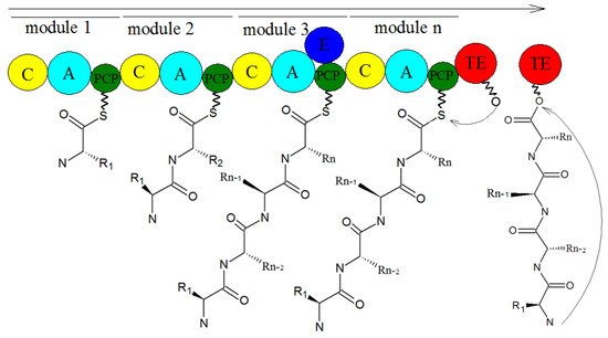
  28. Han, M.; Chen, J.; Qiao, Y.; Zhu, P. Advances in the nonribosomal peptide synthetases. Yaoxue Xuebao 2018, 53, 1080–1089.
  29. Sung, C.T.; Chang, S.; Entwistle, R.; Ahn, G.; Lin, T.; Petrova, V.; Yeh, H.; Praseuth, M.B.; Chiang, Y.; Oakley, B.R.; et al. Overexpression of a three-gene conidial pigment biosynthetic pathway in Aspergillus nidulans reveals the first NRPS known to acetylate tryptophan. Fungal Genet. Biol. 2017, 101, 1–6.
  30. Payne, J.A.E.; Schoppet, M.; Hansen, M.H.; Cryle, M.J. Diversity of nature’s assembly lines—recent discoveries in non-ribosomal peptide synthesis. Mol. Biosyst. 2017, 13, 9–22.
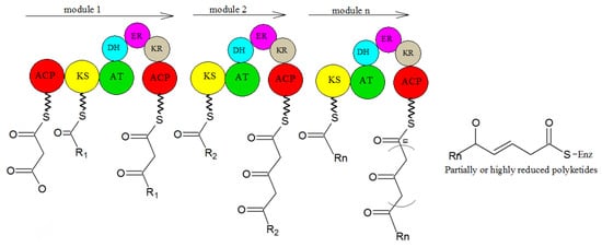
  31. Sun, Y.-H.; Deng, Z.-X. Polyketides and combinatorial biosynthetic approaches. Zhongguo Kangshengsu Zazhi 2006, 31, 6.
  32. Bloudoff, K.; Fage, C.D.; Marahiel, M.A.; Schmeing, T.M. Structural and mutational analysis of the nonribosomal peptide synthetase heterocyclization domain provides insight into catalysis. Proc. Natl. Acad. Sci. USA 2017, 114, 95–100.
  33. Strieker, M.; Tanovic, A.; Marahiel, M.A. Nonribosomal peptide synthetases: Structures and dynamics. Curr. Opin. Struct. Biol. 2010, 20, 234–240.
  34. Hertweck, C. The Biosynthetic Logic of Polyketide Diversity. Angew. Chem. Int. Ed. 2009, 48, 4688–4716.
  35. Schaeberle, T.F. Biosynthesis of alpha-pyrones. Beilstein J. Org. Chem. 2016, 12, 571–588.
  36. Sun, J.; Bu, J.; Cui, G.; Ma, Y.; Zhao, H.; Mao, Y.; Zeng, W.; Guo, J.; Huang, L. Accumulation and biosynthetic of curcuminoids and terpenoids in turmeric rhizome in different development periods. Zhongguo Zhong Yao Za Zhi 2019, 44, 927–934.
  37. Hocquette, A.; Grondin, M.; Bertout, S.; Mallié, M. Les champignons des genres Acremonium, Beauveria, Chrysosporium, Fusarium, Onychocola, Paecilomyces, Penicillium, Scedosporium et Scopulariopsis responsables de hyalohyphomycoses. J. De Mycol. Médicale 2005, 15, 136–149.
  38. Okhravi, N.; Lightman, S. Clinical manifestations, treatment and outcome of Paecilomyces lilacinus infections. Clin. Microbiol. Infect. 2007, 13, 554.
  39. Weng, Q.; Zhang, X.; Chen, W.; Hu, Q. Secondary Metabolites and the Risks of Isaria fumosorosea and Isaria farinosa. Molecules 2019, 24, 664.
More
Information
Subjects: Chemistry, Organic
Contributor MDPI registered users' name will be linked to their SciProfiles pages. To register with us, please refer to https://encyclopedia.pub/register :
View Times: 643
Revisions: 2 times (View History)
Update Date: 13 Jan 2022
1000/1000
Video Production Service