Submitted Successfully!
To reward your contribution, here is a gift for you: A free trial for our video production service.
Thank you for your contribution! You can also upload a video entry or images related to this topic.
Version Summary Created by Modification Content Size Created at Operation
1 + 1711 word(s) 1711 2021-12-06 07:26:04 |
2 Format Change Meta information modification 1711 2021-12-07 02:32:23 | |
3 Format Change Meta information modification 1711 2021-12-07 02:34:02 | |
4 Format change + 60 word(s) 1771 2021-12-07 02:38:48 | |
5 Format Change + 60 word(s) 1771 2021-12-07 02:39:30 | |
6 Format change + 60 word(s) 1771 2021-12-07 02:40:06 | |
7 format correct + 60 word(s) 1771 2021-12-07 02:45:19 | |
8 format correct Meta information modification 1771 2021-12-07 02:46:54 |

Video Upload Options

Do you have a full video?

Confirm

Are you sure to Delete?
Cite
If you have any further questions, please contact Encyclopedia Editorial Office.
Zhang, Y. Cytokine-Induced Killer Cells on Lymphoma Therapy. Encyclopedia. Available online: https://encyclopedia.pub/entry/16798 (accessed on 27 July 2024).
Zhang Y. Cytokine-Induced Killer Cells on Lymphoma Therapy. Encyclopedia. Available at: https://encyclopedia.pub/entry/16798. Accessed July 27, 2024.
Zhang, Ying. "Cytokine-Induced Killer Cells on Lymphoma Therapy" Encyclopedia, https://encyclopedia.pub/entry/16798 (accessed July 27, 2024).
Zhang, Y. (2021, December 06). Cytokine-Induced Killer Cells on Lymphoma Therapy. In Encyclopedia. https://encyclopedia.pub/entry/16798
Zhang, Ying. "Cytokine-Induced Killer Cells on Lymphoma Therapy." Encyclopedia. Web. 06 December, 2021.
Cytokine-Induced Killer Cells on Lymphoma Therapy
Edit

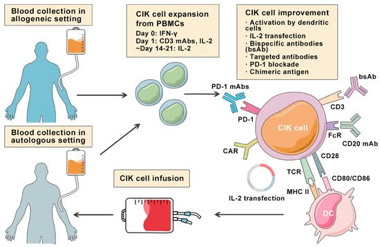
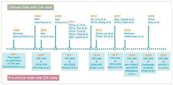
Lymphoma is a heterogeneous group of neoplasms including over 70 different subtypes. Its biological characteristic of deriving from lymphoid tissues makes it ideal for immunotherapy. Cytokine-induced killer (CIK) cells therapy is one of the adoptive immunotherapy which eliminates cancer cells by transfusing immune cells that have been expanded and activated in vitro. CIK cells are heterogeneous in nature and composed of CD3+CD56- (T cells), CD3-CD56+ (NK cells), and CD3+CD56+ (NKT cells) cell populations. The CD3+CD56+ subset is considered as the major effector cells that exert potent anti-tumor cytotoxicity in a major histocompatibility complex (MHC)-unrestricted manner. From 1991 to 2020, there are nearly 20 clinical trials conducted for the treatment of lymphoma. Moreover, a number of pre-clinical approaches are being investigated to improve CIK cell therapy to enhance its anti-tumor activity.

lymphoma cytokine-induced killer cells adoptive cell transfer immunotherapy

1. Introduction

Contrary to solid cancers, hematologic malignancies (blood cancers) have some unique characteristics and often require different treatments. Importantly, it is now well established that the hematologic malignancies (lymphoma, leukemia, and myeloma) despite sharing some common clinical features are also biologically distinct entities. Among them, lymphoma, a cancer of lymphocytes, is a very heterogeneous group of neoplasms exhibiting diverse clinical presentations, prognoses, and therapeutic responses and includes more than 70 different subtypes. Lymphomas are conventionally divided into two main groups, Hodgkin’s lymphoma (HL) and non-Hodgkin’s lymphoma (NHL), with both types having multiple early clinical manifestations, often delaying the diagnosis. By comparison, HLs are rare and less heterogeneous, whereas NHLs are more a common and highly heterogeneous group of B-, T-, or NK-cell neoplasms. HL only accounts for approximately 10% of newly diagnosed lymphoma [1]. The complex heterogeneity can also be evident from the fact that in HL alone, the distinctions can be made between two subtypes, classic HL (cHL) and rare nodular lymphocyte-predominant HL (nLPHL) [2]. Besides, cHL is further categorized into nodular sclerosis (NS), mixed cellularity (MC), lymphocyte-rich (LR) and lymphocyte-depleted (LD) cHL. The cHL subtype is composed of Hodgkin (H) and Reed-Sternberg (RS) cells, collectively referred to as Hodgkin and Reed-Sternberg cells (HRS). The scenario is more complex in NHL, which is classified according to the type of lymphocytes involved: B lymphocytes (B-cells) or T lymphocytes (T-cells). Diffuse large B-cell lymphoma (DLBCL), follicular lymphoma (FL), and Burkitt’s lymphoma (BL) are common NHL types that are usually classified according to their aggressive or slow growing characteristics.
As per reports from the SEER database (https://seer.cancer.gov/, accessed on 3 August 2021), HL affected approximately 219,128 people in the United States in 2018. To mention, HL is quite curable, but there are differences in incidence rates, sex and age distribution (young and adults), mainly based on the socioeconomic background [3]. The most common age group of diagnosis is generally between 20 and 34 years old. Contrarily, NHL pose a challenge to the treatment response. According to the German Center for Cancer Registry Data (ZfKD), about 19,200 people in Germany were diagnosed with NHL in 2017, mostly in people of advanced age (average: women 72 years, men 70 years). In 2020, the number of incident cases is increased to 18,549 in Germany from the data of GLOBOCAN 2020 (http://gco.iarc.fr/today, accessed on 25 November 2021). Similarly, approximately 81,560 patients are expected to be diagnosed with NHL in the United States in 2021 according to data from Cancer Statistics Center of American Cancer Society (http://cancerstatisticscenter.cancer.org/, accessed on 3 August 2021).

2. Cytokine-Induced Killer Cells

Since cancer immunotherapy focuses on enhancing the anti-cancer capabilities of immune cells rather than killing cancer cells, one such potential approach of adoptive immunotherapy is cytokine-induced killer (CIK) therapy, which eliminates cancer cells by transfusing immune cells that have been expanded and activated in vitro. Phenotypically, CIK cells are heterogeneous in nature and composed of CD3+CD56− (T cells), CD3−CD56+ (NK cells), and CD3+CD56+ (NKT cells) cell populations [4]. In fact, the CD3+CD56+ subset is considered as the major effector cells that exert potent anti-tumor cytotoxicity in a major histocompatibility complex (MHC)-unrestricted manner. The generation of CIK cells was first reported in 1991 by sequential incubation of peripheral blood mononuclear cells (PBMCs) with interferon-γ (IFN-γ), anti-CD3 monoclonal antibody (OKT3), and recombinant human interleukin-2 (rhIL-2) [5]. The manufacturing process of autologous and allogeneic CIK cells is shown in Figure 1.
Cancers 13 06007 g001 550
Figure 1. Overview of multiple approaches aiming at improving cytokine-induced killer (CIK)cell therapy in lymphoma. Illustration showing the expansion of autologous or allogeneic CIK cells from PBMCs collected from the blood of lymphoma patients under incubation of IFN-γ, CD3 mAbs, and IL-2 for 2–3 weeks. To increase the cytotoxicity and specificity of CIK cells against lymphoma cells, several modifications are implemented.
Particularly, apparent differences can be drawn between CIK cells and the impressive immunologic approaches against lymphoma, including CAR-T and BiTE therapies (Table 1). Unlike CAR-T and BiTE that recognize tumor cells by targeting the tumor-associated antigens (TAAs), the majority of the CIK cell cytotoxicity results from the natural killer group 2 member D (NKG2D) interactions [6][7][8][9]. NKG2D is expressed on all NK cells and recognizes at least six ligands that are relatively restricted on malignant tissues, including MHC class I-related molecules A and B (MICA and MICB) and members of the UL16-binding protein family (ULBP1-4) [10]. The manufacturing of CAR-T cells requires genetic modification and cell expansion ex vivo, which is time-consuming and brings some financial considerations. However, the relative easiness of CIK cell preparation facilitates the fast growing of CIK cell therapy. Besides, cytokine release syndrome (CRS) is the most severe toxicity in CAR-T and BiTE therapies. It is associated with the rapid and extensive activation of T cells and the release of high-level proinflammatory cytokines (TNFα and IL-6) [11]. Nonetheless, the crucial advantages of CIK cells are the relatively low toxicity and lack of graft-versus-host diseases (GVHD) response. The most common side effects in CIK cell therapy are fever, chills, fatigue, headache, and skin rash, and patients can usually recover with simple treatment [12].
Table 1. Similarities and differences between CIK cells, CAR-T cells and BiTEs in lymphoma.
  CIK Cells CAR-T Cells BiTEs
Manufacturing Process PBMCs sequentially activated with 1000 IU/m IFN-γ on day 0, and 50 ng/mL anti-CD3 mAb and 600 IU/mL IL-2 on the following day; IL-2 supplemented every 2–3 days T cells with CAR gene transduction primarily by lentiviruses; CARs consist of an scFv based ectodomain for antigen-binding, a transmembrane domain, and an endodomain containing TCR CD3ζ chain with or without costimulatory signaling components Antibodies designed to bind to a selected TAA and CD3 on T cells simultaneously; produced in bioreactors by mammalian cell lines as secreted polypeptides
Effector Cells CD3+CD56+ cells Mostly αβ-TCR+ T cells Endogenous CD8+ or CD4+ T cells
Cell Source Autologous/allogeneic Autologous/allogeneic Autologous
Target Antigen MIC A/B and ULBP1–4 CD19, CD20, CD22, CD30, BCMA, etc. CD19
MHC Restriction Dual-functional capability (non-MHC-restricted and TCR-mediated lysis) TAA recognition by CARs is MHC-unrestricted MHC-unrestricted
Mechanism Release of perforin and granzyme B from CIK cells Release of perforin and granzyme B from CAR-T cells Activating T cells to release perforin and granzyme B by linking TAAs to CD3 on T cells
Toxicities and Side effects Low-grade toxicities including fever, chills, fatigue, headache, and skin rash; grade 3 and 4 toxicities are rare; limited GVHD response in the allogeneic setting CRS, ICANS, and MAS/HLH; potentially life-threatening CRS and ICANS; severe toxicities are one of the major concerns
Clinical Efficacy Varied due to heterogeneity of expension method and study design Axicabtagene ciloleucel: 83% ORR, 58% CR; Tisagenlecleucel: 54% ORR, 40% CR; Lisocabtagene maraleucel: 73% ORR, 53% CR [13][14][15] Blinatumomab: 36% to 69% ORR in relapsed/refractory NHL [16][17]
CIK cells, cytokine-induced killer cells; MIC A/B, MHC class I-related molecules A and B; ULBP1–4, UL16-binding protein; GVHD, graft-versus-host diseases; CAR, chimeric antigen receptors; scFv, single-chain variable fragment; TCR, T cell receptor; TAA, tumor-associated antigen; CRS, cytokine release syndrome; ICANS, immune effector cell-associated neurotoxicity syndrome; MAS/HLH, Macrophage activation syndrome/hemophagocytic lymphohistiocytosis; CR, complete remission; ORR, objective response rate; BiTE, bispecific T cell engager.

3. Improving CIK Cell Therapy on Lymphoma

Despite the positive clinical benefit observed in trials, it is difficult to achieve long-lasting response and complete cancer eradication in patients with refractory or relapsed lymphoma. Therefore, a number of pre-clinical approaches are being investigated to improve CIK cell therapy to enhance its anti-tumor activity (Figure 2). Among these innovative methods, bispecific antibodies (bsAb) represent a promising development bringing targeted antigen on tumor cells into close proximity to receptors on cytotoxic T-cells, and thereby triggering T-cell receptor signaling and anti-tumor immune response [18]. In a particular study, it was reported that the bsAb CD19 × CD5 (HD37 × T5.16) enhanced the cytotoxicity activity of CIK cells against B lymphoma cells [19]. Moore et al. used a bispecific antibody platform known as dual affinity retargeting (DART) to eradicate B-cell lymphomas by targeting the B-cell-specific antigen CD19 and the TCR/CD3 complex to effector T cells [20]. Subsequently, it was shown that CIK cells and CD19xCD3 DART can control and/or eradicate patient-derived tumor xenografts from chemo-refractory B-ALL and diffuse large B-cell lymphoma (DLBCL) patients [21].
Cancers 13 06007 g002 550
Figure 2. Highlights in the development of CIK cell immunotherapy. In 1991, the development of CIK therapy began (in Germany) and continued in several countries, with 2011 being the year with the most clinical trial reports. The supporting pre-clinical models that are used to improve the therapeutic activity of CIK cells are also highlighted.
Likewise, studies also have shown an enhancement of CIK cell activity when combined with anti-CD20 antibodies. For instance, Pievani et al. showed that the addition of the anti-CD20 antibodies GA101 or rituximab in B-NHL increased the cytotoxicity of CIK cells by 35% and 15%, respectively [22]. The authors also suggested that activation of the MAPK pathway may be a possible mechanism for the anti-apoptosis effect on CIK cell proliferation. Esser et al., investigated the effect of CIK cells in combination with brentuximab vedotin (SGN-35) on three different CD30+ lymphoma cell lines (Daudi, KI-JK, and L-540), and was found that the combined approach led to better results in vitro [23]. Recently, a novel antibody-cell conjugation method for the enhancement and characterization of cytokine-induced killer cells has been presented [24]. The authors demonstrated that CIK cells conjugated with rituximab exhibited increased cytotoxic activity against CD20+ lymphoma cell lines and suggested that without any genetic modification, CIK cells can be rapidly equipped with monoclonal antibodies to target tumor cells.
Biederbick et al. recently contributed to raise concerns about synergistic molecular mechanisms of CIK cells by using a combination of anti-CD40 and anti-GITR mAb in the human lymphoma cell lines SU-DHL-4 and Daudi (both CD40-positive) [25]. More recently, Li et al. reported an increase in IFN-γ secretion in B-NHL cell lines treated with CIK alone or with the PD-1 antibody, yet this trend was not observed for PD-L1, raising the question of whether PD-1 and PD-L1 are comparable and interchangeable in the clinical practice [26].

References

  1. Armitage, J.O.; Gascoyne, R.D.; Lunning, M.A.; Cavalli, F. Non-Hodgkin lymphoma. Lancet 2017, 390, 298–310.
  2. Swerdlow, S.H.; Campo, E.; Pileri, S.A.; Harris, N.L.; Stein, H.; Siebert, R.; Advani, R.; Ghielmini, M.; Salles, G.A.; Zelenetz, A.D.; et al. The 2016 revision of the World Health Organization classification of lymphoid neoplasms. Blood 2016, 127, 2375–2390.
  3. Shanbhag, S.; Ambinder, R.F. Hodgkin lymphoma: A review and update on recent progress. CA A Cancer J. Clin. 2017, 68, 116–132.
  4. Introna, M. CIK as therapeutic agents against tumors. J. Autoimmun. 2017, 85, 32–44.
  5. Schmidt-Wolf, I.G.; Negrin, R.S.; Kiem, H.P.; Blume, K.G.; Weissman, I.L. Use of a SCID mouse/human lymphoma model to evaluate cytokine-induced killer cells with potent antitumor cell activity. J. Exp. Med. 1991, 174, 139–149.
  6. Verneris, M.R.; Karami, M.; Baker, J.; Jayaswal, A.; Negrin, R.S. Role of NKG2D signaling in the cytotoxicity of activated and expanded CD8+ T cells. Blood 2004, 103, 3065–3072.
  7. Karimi, M.; Cao, T.M.; Baker, J.A.; Verneris, M.R.; Soares, L.; Negrin, R.S. Silencing Human NKG2D, DAP10, and DAP12 Reduces Cytotoxicity of Activated CD8+T Cells and NK Cells. J. Immunol. 2005, 175, 7819–7828.
  8. Frigault, M.J.; Maus, M.V. State of the art in CAR T cell therapy for CD19+ B cell malignancies. J. Clin. Investig. 2020, 130, 1586–1594.
  9. Goebeler, M.-E.; Bargou, R.C. T cell-engaging therapies—BiTEs and beyond. Nat. Rev. Clin. Oncol. 2020, 17, 418–434.
  10. Diefenbach, A.; Jamieson, A.; Liu, S.D.; Shastri, N.; Raulet, D. Ligands for the murine NKG2D receptor: Expression by tumor cells and activation of NK cells and macrophages. Nat. Immunol. 2000, 1, 119–126.
  11. Ramos, C.A.; Heslop, H.E.; Brenner, M.K. CAR-T Cell Therapy for Lymphoma. Annu. Rev. Med. 2016, 67, 165–183.
  12. Zhang, Y.; Schmidt-Wolf, I.G.H. Ten-year update of the international registry on cytokine-induced killer cells in cancer immunotherapy. J. Cell. Physiol. 2020, 235, 9291–93033.
  13. Locke, F.L.; Ghobadi, A.; Jacobson, C.A.; Miklos, D.B.; Lekakis, L.J.; Oluwole, O.O.; Lin, Y.; Braunschweig, I.; Hill, B.T.; Timmerman, J.M.; et al. Long-term safety and activity of axicabtagene ciloleucel in refractory large B-cell lymphoma (ZUMA-1): A single-arm, multicentre, phase 1–2 trial. Lancet Oncol. 2019, 20, 31–42.
  14. Abramson, M.J.S.; Palomba, M.L.; Gordon, L.I.; Lunning, D.M.A.; Wang, M.L.; Arnason, J.E.; Mehta, A.; Purev, E.; Maloney, D.G.; Andreadis, M.C.; et al. Pivotal Safety and Efficacy Results from Transcend NHL 001, a Multicenter Phase 1 Study of Lisocabtagene Maraleucel (liso-cel) in Relapsed/Refractory (R/R) Large B Cell Lymphomas. Blood 2019, 134, 241.
  15. Bachanova, V.; Westin, J.; Tam, C.; Borchmann, P.; Jaeger, U.; McGuirk, J.; Holte, H.; Waller, E.; Jaglowski, S.; Bishop, M.; et al. Correlative Analyses of Cytokine Release Syndrome and Neurological Events in Tisagenlecleucel-Treated Relapsed/Refractory Diffuse Large B-Cell Lymphoma Patients. Clin. Lymphoma Myeloma Leuk. 2019, 19, S251–S252.
  16. Viardot, A.; Goebeler, M.-E.; Hess, G.; Neumann, S.; Pfreundschuh, M.; Adrian, N.; Zettl, F.; Libicher, M.; Sayehli, C.; Stieglmaier, J.; et al. Phase 2 study of the bispecific T-cell engager (BiTE) antibody blinatumomab in relapsed/refractory diffuse large B-cell lymphoma. Blood 2016, 127, 1410–1416.
  17. Goebeler, M.-E.; Knop, S.; Viardot, A.; Kufer, P.; Topp, M.S.; Einsele, H.; Noppeney, R.; Hess, G.; Kallert, S.; Mackensen, A.; et al. Bispecific T-Cell Engager (BiTE) Antibody Construct Blinatumomab for the Treatment of Patients with Relapsed/Refractory Non-Hodgkin Lymphoma: Final Results from a Phase I Study. J. Clin. Oncol. 2016, 34, 1104–1111.
  18. Ansell, S.M. Fundamentals of immunology for understanding immunotherapy for lymphoma. Hematol. Am. Soc. Hematol. Educ. Program 2020, 2020, 585–589.
  19. Tita-Nwa, F.; Moldenhauer, G.; Herbst, M.; Kleist, C.; Ho, A.D.; Kornacker, M. Cytokine-induced killer cells targeted by the novel bispecific antibody CD19xCD5 (HD37xT5.16) efficiently lyse B-lymphoma cells. Cancer Immunol. Immunother. 2007, 56, 1911–1920.
  20. Moore, P.A.; Zhang, W.; Rainey, G.J.; Burke, S.; Li, H.; Huang, L.; Gorlatov, S.; Veri, M.C.; Aggarwal, S.; Yang, Y.; et al. Ap-plication of dual affinity retargeting molecules to achieve optimal redirected T-cell killing of B-cell lymphoma. Blood 2011, 117, 4542–4551.
  21. Circosta, P.; Elia, A.R.; Landra, I.; Machiorlatti, R.; Todaro, M.; Aliberti, S.; Brusa, D.; Deaglio, S.; Chiaretti, S.; Bruna, R.; et al. Tailoring CD19xCD3-DART exposure enhances T-cells to eradication of B-cell neoplasms. OncoImmunology 2018, 7, e1341032.
  22. Pievani, A.; Belussi, C.; Klein, C.; Rambaldi, A.; Golay, J.T.; Introna, M. Enhanced killing of human B-cell lymphoma targets by combined use of cytokine-induced killer cell (CIK) cultures and anti-CD20 antibodies. Blood 2011, 117, 510–518.
  23. Esser, L.; Weiher, H.; Schmidt-Wolf, I. Increased Efficacy of Brentuximab Vedotin (SGN-35) in Combination with Cytokine-Induced Killer Cells in Lymphoma. Int. J. Mol. Sci. 2016, 17, 1056.
  24. Frank, M.J.; Olsson, N.; Huang, A.; Tang, S.-W.; Negrin, R.S.; Elias, J.E.; Meyer, E.H. A novel antibody-cell conjugation method to enhance and characterize cytokine-induced killer cells. Cytotherapy 2020, 22, 135–143.
  25. Biederbick, K.D.; Schmidt-Wolf, I.G. Efficacy of cytokine-induced killer cells targeting CD40 and GITR. Oncol. Lett. 2019, 17, 2425–2430.
  26. Li, Y.; Sharma, A.; Bloemendal, M.; Schmidt-Wolf, R.; Kornek, M.; Schmidt-Wolf, I.G.H. PD-1 blockade enhances cytokine-induced killer cell-mediated cytotoxicity in B-cell non-Hodgkin lymphoma cell lines. Oncol. Lett. 2021, 22, 613.
More
Information
Subjects: Oncology
Contributor MDPI registered users' name will be linked to their SciProfiles pages. To register with us, please refer to https://encyclopedia.pub/register :
View Times: 349
Revisions: 8 times (View History)
Update Date: 07 Dec 2021
1000/1000
Video Production Service