Submitted Successfully!
To reward your contribution, here is a gift for you: A free trial for our video production service.
Thank you for your contribution! You can also upload a video entry or images related to this topic.
Version Summary Created by Modification Content Size Created at Operation
1 + 2046 word(s) 2046 2021-11-03 08:01:59 |
2 Format change Meta information modification 2046 2021-11-09 02:35:52 | |
3 There were no changes introduced Meta information modification 2046 2021-11-09 11:44:07 |

Video Upload Options

Do you have a full video?

Confirm

Are you sure to Delete?
Cite
If you have any further questions, please contact Encyclopedia Editorial Office.
Garaschuk, O.; Lushchak, V.; Gospodaryov, D. Reactive Oxygen Species in the Brain. Encyclopedia. Available online: https://encyclopedia.pub/entry/15803 (accessed on 27 July 2024).
Garaschuk O, Lushchak V, Gospodaryov D. Reactive Oxygen Species in the Brain. Encyclopedia. Available at: https://encyclopedia.pub/entry/15803. Accessed July 27, 2024.
Garaschuk, Olga, Volodymyr Lushchak, Dmytro Gospodaryov. "Reactive Oxygen Species in the Brain" Encyclopedia, https://encyclopedia.pub/entry/15803 (accessed July 27, 2024).
Garaschuk, O., Lushchak, V., & Gospodaryov, D. (2021, November 08). Reactive Oxygen Species in the Brain. In Encyclopedia. https://encyclopedia.pub/entry/15803
Garaschuk, Olga, et al. "Reactive Oxygen Species in the Brain." Encyclopedia. Web. 08 November, 2021.
Reactive Oxygen Species in the Brain
Edit

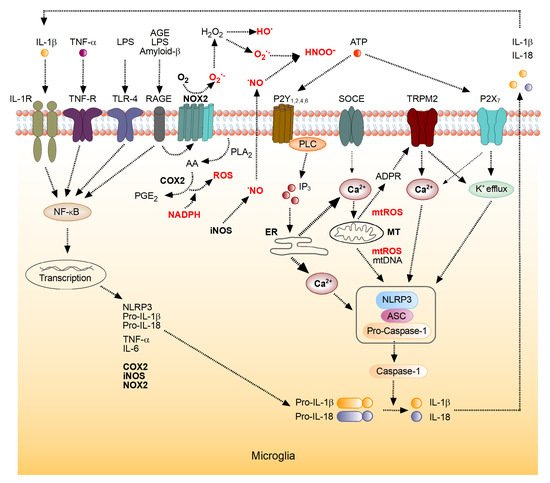
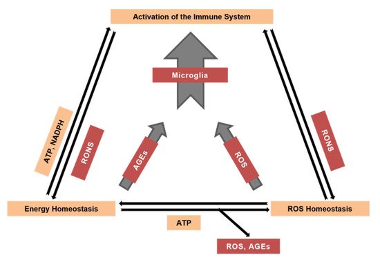
Reactive oxygen and nitrogen species are crucial contributors to the age-dependent decline in all tissues. Neural tissue, one of the main oxygen consumers in the mammalian body, is especially prone to reactive species-mediated damage. Brain cells, including neurons, astrocytes, and microglia, produce reactive oxygen species (ROS) by specific enzymatic systems, including complexes of the mitochondrial respiratory chain, multienzyme flavin-containing complexes, monoamine and xanthine oxidases, microglial and endothelial NADPH oxidases and cyclooxygenases in addition to non-enzymatic and potentially uncontrolled mechanisms of ROS production, such as autooxidation of quinones or other aromatic compounds. Nitric oxide produced by nitric oxide synthases powers the conversion of ROS into reactive nitrogen species (RNS). Both ROS and RNS play important signaling roles and are also capable of modifying other molecules such as proteins, nucleic and fatty acids, lipids and carbohydrates. The antioxidant system, comprising low molecular mass antioxidants (e.g., tocopherol, ascorbic acid and glutathione) and high molecular mass antioxidants such as enzymes (e.g., catalases, peroxidases, superoxide dismutases) and others, protects cells from potential damage caused by ROS or RNS. Powering antioxidant systems by NADPH provides neural tissue with defense against ROS but may also trigger ROS production by NADPH oxidases and cyclooxygenases. In turn, mitochondria start using ketone bodies as an energy source under certain conditions. Increased steady-state levels of ROS and RNS, along with the aforementioned ROS-modified molecules, activate the organisms’ immune system including brain’s microglia.

Reactive Oxygen Species Reactive Nitrogen Species Oxidative Stress Advanced Glycation End Products Brain Aging Microglia

1. Introduction

Living organisms are open thermodynamic systems, critically relying on their energy metabolism for the maintenance of structural integrity and function. Energy homeostasis includes catabolic processes that start with the degradation of polysaccharides, lipids and proteins and lead to the production of macroergic compounds such as adenosine triphosphate (ATP). This universal macroergic compound used by living organisms, is produced via multistep anaerobic or aerobic oxidation processes.
Aerobic ATP production is accompanied by the generation of reactive oxygen species (ROS) as side products of operation of the mitochondrial electron transport chain [1][2]. Thus, besides producing energy-rich substances, the energy homeostasis generates potentially damaging side products such as ROS in the respiratory chain and methylglyoxal in glycolysis. Methylglyoxal and other α-dicarbonyl compounds can react with arginine and lysine residues of proteins, yielding glycated derivatives called advanced glycation end products (AGEs), while ROS can interact with virtually all components of living organisms, causing their modification [3][4][5]. ROS are also produced by the metabolic systems not connected with energy production. In particular, monoamine oxidases, cytochromes P450, peroxisomal oxidases and plasma membrane-bound Nicotinamide adenine dinucleotide phosphate (NADPH) oxidases (NOX) can produce ROS [6]. Since ROS discovery in living organisms in the early 1950th, they have been considered as damaging species [7]. Therefore, most ROS studies focused on the investigation of their negative effects and protection against them [8]. However, besides the damaging effects, at physiological concentrations mitochondrial and cytosolic ROS play important signaling roles in multiple cellular processes, including inflammation, cellular growth and differentiation [9][10][11][12]. Whether ROS effects are beneficial or detrimental depends on the balance between ROS generation and elimination as well as on the targets attacked [11]. Usually, the antioxidant system of a young and adult organism copes with oxidative modifications of biomolecules but gradually loses this ability during aging. Therefore, aging is accompanied by an intensification of oxidative stress, a decreased efficiency of ATP production and the concomitant activation of the immune system [3][4][13]. That is because both ROS and AGEs activate key proinflammatory molecules nuclear factor kappa-light-chain-enhancer of activated B cells (NF-κB) and NLR Family Pyrin Domain Containing 3 (NLRP3) (Figure 1), causing enhanced production of proinflammatory cytokines [14][15][16]. Inflammation, triggered in this way, leads to the production of different reactive species, particularly ROS (Figure 1), thus feeding back to energy and ROS homeostasis (Figure 2) [17][18]. While in a young organism byproducts generated by the energy metabolism are efficiently neutralized by the respective defense systems, these processes become imbalanced with advancing age [4][19].
Figure 1. Molecular mechanisms underlying microglial reactive oxygen species (ROS) metabolism. Mechanisms involved in the interplay between the inflammation-mediated ROS production, increases in [Ca2+]i and cytokine production by microglia (see text for detailed description). IL-1β, interleukin 1β; TNF-α, tumor necrosis factor α; LPS, lipopolysaccharide; AGE, advanced glycation endproducts; IL-1R, IL-1β receptor; TNF-R, TNF-α receptor; TLR-4, Toll-Like Receptor 4; RAGE, a receptor for AGE; NADPH, a reduced form of nicotinamide adenine dinucleotide phosphate; NOX2, NADPH oxidase 2; P2Y1,2,4,6, metabotropic ATP receptors, SOCE, store-operated Ca2+ entry channel; TRPM2, Transient receptor potential cation channel, subfamily M, member 2; P2X7, ionotropic ATP receptor, PLA2, phospholipase A2; AA, arachidonic acid; COX2, cyclooxygenase 2; PGE2, prostaglandin E2; iNOS, inducible NO-Synthase; PLC, Phospholipase C; IP3, inositol 1,4,5-trisphosphate; ER, endoplasmic reticulum; MT, mitochondrion; mtROS, reactive oxygen species of mitochondrial origin; mtDNA, mitochondrial DNA; ADPR, ADP-ribose. The boxed structure consisting of NLRP3, adaptor protein ASC and Pro-Caspase-1 represents the NLRP3 inflammasome (modified from [14][16]).
The brain is especially prone to ROS-mediated toxicity for several reasons: (i) the intense oxidative metabolism, (ii) the high levels of polyunsaturated fatty acids, serving as primary substrates for ROS-promoted oxidation and (iii) a rather high number of resident immune cells [20].

2. Mechanisms of ROS Generation

The bulk ROS amount in the organisms is generated by poorly controlled non-enzymatic processes such as autoxidation of small molecules (e.g., epinephrine, quinones), by better controlled enzymatic processes (e.g., operation of different oxidases) and by the escape of active electrons from electron transport chains [11]. Under normoxic conditions, more than 90% ROS in eukaryotic cells are generated by mitochondria [21], where above 95% of consumed oxygen is reduced via four-electron reduction of molecular oxygen by cytochrome oxidase [22]. During the operation of mitochondria, however, some of the transported electrons escape ETC and join molecular oxygen giving rise to ROS. Besides the respiratory chain, ROS can be generated by multi-enzyme flavin-containing complexes such as alpha-ketoglutarate dehydrogenase and pyruvate dehydrogenase. Other enzymatic generators of ROS are monoamine oxidases located in the mitochondrial outer membrane. The latter way of ROS formation is especially important for neurons that produce and secrete biogenic amines [6][23]. Brain cyclooxygenases, lipoxygenases and NADPH oxidases also generate ROS. NADPH oxidases (NOXs) are often associated with immune cells like neutrophils and macrophages. However, they are also expressed in brain vascular endothelium and microglia and may contribute to age-related changes [12][14][24]. These enzymes specifically produce either superoxide anion radical (O2•−) or hydrogen peroxide (H2O2) or both, transferring electrons from NADPH to flavin adenine dinucleotide (FAD) first, and then—to heme of cytochrome b558, and—from cytochrome b558 to oxygen [25]. Cyclooxygenases are heme-containing enzymes that convert arachidonic acid to prostaglandin H2. These enzymes can produce O2•− since the catalysis occurs via the formation of transient carbon-centered radical species that, in turn, can react with molecular oxygen. Importantly, the formation of O2•− by cyclooxygenases requires reducing co-substrates such as NADH or NADPH [26][27]. Superoxide is also produced in the brain endothelial cells by xanthine oxidase, a peroxisomal flavin-containing enzyme [28].
In most cases where ROS are formed by enzymatic systems, sequential multistep one-electron O2 reduction takes place. Interaction of one electron with molecular oxygen results in the formation of O2•−. The latter can interact with one more electron and two protons forming H2O2. At the next reduction step, H2O2 can accept one more electron, resulting in the production of hydroxyl radical (HO) and hydroxyl anion (OH). Finally, HO may accept another electron and one proton, while OH may associate with a proton; in both cases, water molecules are formed. All three partially reduced oxygen intermediates, namely free radicals O2•− and HO as well as H2O2 are called reactive oxygen species because they are more active than molecular triplet oxygen [2][29]. ROS-induced reactions occur spontaneously and are poorly controlled by living organisms.
All cells with aerobic metabolism possess a set of low and high molecular mass antioxidants [11]. Low molecular mass antioxidants such as vitamins C and E, carotenoids, anthocyans and glutathione interact directly with any ROS type at low specificity. However, biological protection against HO is difficult, probably due to its high chemical activity, short lifespan and small diffusion distance, and thus organisms try to prevent HO production. In terms of high molecular mass antioxidants, levels of O2•− and H2O2 are also controlled by enzymatic systems. Antioxidant enzymes form the so-called primary line, which directly deals with ROS, and a secondary line, which assists to the primary one and converts ROS-related components to less dangerous products. In addition, several enzymes regenerate low molecular mass antioxidants and repair certain types of oxidative damage [6][11]. Superoxide dismutases accelerate the conversion of O2•− in H2O2 with concomitant formation of molecular oxygen, i.e., they dismutate one type of substrate molecules in two different products. Hydrogen peroxide can be eliminated by two types of enzymes: catalase, which dismutates H2O2 to water and molecular oxygen, and peroxidases, which use diverse co-substrates to reduce H2O2 to water and the respective oxidized co-substrate. Manganese-containing superoxide dismutase (Mn-SOD), specific glutaredoxins (Grx5), thioredoxins and peroxiredoxins (Prdx3 and Prdx5) scavenge ROS released to both, mitochondrial matrix and intermembrane space [30]. In turn, O2•− released by non-mitochondrial sources, such as NOX, cytochromes P450 and peroxisomal oxidases (for instance, xanthine oxidase) is scavenged by cytosolic copper-zinc-containing superoxide dismutase (Cu,Zn-SOD). Hydrogen peroxide, which is small and uncharged molecule, can easily cross lipid membranes. Whatever the place of H2O2 formation, it has a great chance to occur in the cytosol where it can be converted to water by peroxidases. Peroxidases use different cofactors and glutathione peroxidases are likely the best-studied enzymes of this group [31]. They use the reductive power of glutathione that is oxidized to a dimeric form and may be further reduced by glutathione reductase at the expense of NADPH. The formed NADP+ is then reduced by glucose-6-phosphate dehydrogenase, the key enzyme of the pentose phosphate pathway, that oxidizes glucose-6-phosphate to 6-phosphoglucolactone. This reaction finally connects ROS homeostasis with the catabolism of carbohydrates and overall energy-providing processes (Figure 2). An imbalance between ROS generation and detoxification will eventually enhance the oxidation of important biomolecules and induce a loss of their functions. The systems responsible for the antioxidant defense, i.e., the elimination of oxidized molecules either by repair or biosynthesis wear out with cell’s age. This is especially relevant for neural tissue, which is predominantly composed of cells that rarely divide or regenerate.
Figure 2. The interplay between the energy/ROS homeostasis and the activation of the brain’s immune system during aging. RONS, reactive oxygen/nitrogen species.

3. The Role of the Brain’s Immune System in the Generation of ROS

Microglia, the main immune cells of the brain, vividly utilize ROS-mediated signaling under (patho)physiological conditions, and, consequently, possess several mechanisms for the generation of both intra- and extracellular ROS [12][14][15]. As illustrated in Figure 1, microglia express NADPH oxidases, capable of generating O2•− and H2O2. Recent single-cell RNA sequencing analyses identified NADPH oxidase NOX2 as an isotype with the highest expression level in both human and mouse microglia [12][14]. In both species, the robust expression of NOX2 was seen not only during adulthood but also during development. NOX4 was also expressed in microglia, albeit at a much lower level than NOX2 [12]. Besides NOX, NO synthases and cyclooxygenases are relevant sources of microglial ROS [14]. For example, activation of microglia is associated with an NF-κB-dependent upregulation of iNOS and COX2 expression and a concomitant overproduction of intracellular ROS (Figure 1). The intracellularly generated NO diffuses out of the cell and, besides acting as a secondary messenger, reacts with superoxide anion, generated by NOX2, forming peroxynitrite (ONOO). The latter is a highly reactive nitrogen species often causing tissue, cell and mitochondrial damage [12][32].
Like many other non-excitable cells, microglia utilize changes in the intracellular free Ca2+ concentration ([Ca2+]i) for executing their sensor and effector functions [33][34][35]. Such Ca2+ signaling, mediated, for example, by activation of a plentitude of metabotropic receptors or store-operated Ca2+ channels (Figure 1) causes a release of ROS from mitochondria [16][36]. In turn, ROS produced in cytosol or mitochondria increase the production of ADP-ribose through degradation of poly-ADP-ribose in the nucleus or degradation of NAD+ released from damaged mitochondria in the cytoplasm [36][37]. Together, ADP-ribose and Ca2+ activate Ca2+-permeant Transient Receptor Potential (TRPM2) channels (Figure 1), known for their sensitivity to endogenous ROS [37], thus further increasing [Ca2+]i. In this way, TRPM2 channels link ROS production to inflammasome activation in immune cells, where the expression of these channels is abundant.

References

  1. Adam-Vizi, V. Production of Reactive Oxygen Species in Brain Mitochondria: Contribution by Electron Transport Chain and Non-Electron Transport Chain Sources. Antioxid. Redox Signal. 2005, 7, 1140–1149.
  2. Lushchak, V.I. Free Radicals, Reactive Oxygen Species, Oxidative Stresses and their Classifications. UKR. Biochem. J. 2015, 87, 11–18.
  3. Garaschuk, O.; Semchyshyn, H.M.; Lushchak, V.I. Healthy Brain Aging: Interplay between Reactive Species, Inflammation and Energy Supply. Ageing Res. Rev. 2018, 43, 26–45.
  4. Lushchak, V.I. Interplay between Bioenergetics and Oxidative Stress at Normal Brain Aging. Aging as a Result of Increasing Disbalance in the System Oxidative Stress-Energy Provision. Pflugers Arch. 2021, 473, 713–722.
  5. Semchyshyn, H. Is Carbonyl/AGE/RAGE Stress a Hallmark of the Brain Aging? Pflugers Arch. 2021, 473, 723–734.
  6. Zorov, D.B.; Juhaszova, M.; Sollott, S.J. Mitochondrial Reactive Oxygen Species (ROS) and ROS-Induced ROS Release. Physiol. Rev. 2014, 94, 909–950.
  7. Harman, D. Aging: A Theory Based on Free Radical and Radiation Chemistry. J. Gerontol. 1956, 11, 298–300.
  8. McCord, J.M.; Fridovich, I. Superoxide Dismutase. An Enzymic Function for Erythrocuprein (Hemocuprein). J. Biol. Chem. 1969, 244, 6049–6055.
  9. Reczek, C.R.; Chandel, N.S. ROS-Dependent Signal Transduction. Curr. Opin. Cell Biol. 2015, 33, 8–13.
  10. Zandalinas, S.I.; Mittler, R. ROS-Induced ROS Release in Plant and Animal Cells. Free Radic. Biol. Med. 2018, 122, 21–27.
  11. Sies, H.; Jones, D.P. Reactive Oxygen Species (ROS) as Pleiotropic Physiological Signalling Agents. Nat. Rev. Mol. Cell Biol. 2020, 21, 363–383.
  12. Simpson, D.S.A.; Oliver, P.L. ROS Generation in Microglia: Understanding Oxidative Stress and Inflammation in Neurodegenerative Disease. Antioxidants 2020, 9, 743.
  13. Longo, V.D.; Di Tano, M.; Mattson, M.P.; Guidi, N. Intermittent and Periodic Fasting, Longevity and Disease. Nat. Aging 2021, 1, 47–59.
  14. García-Revilla, J.; Alonso-Bellido, I.M.; Burguillos, M.A.; Herrera, A.J.; Espinosa-Oliva, A.M.; Ruiz, R.; Cruz-Hernández, L.; García-Domínguez, I.; Roca-Ceballos, M.A.; Santiago, M.; et al. Reformulating Pro-Oxidant Microglia in Neurodegeneration. J. Clin. Med. 2019, 8, 1719.
  15. Cepas, V.; Collino, M.; Mayo, J.C.; Sainz, R.M. Redox Signaling and Advanced Glycation Endproducts (AGEs) in Diet-Related Diseases. Antioxidants 2020, 9, 142.
  16. Garaschuk, O. The Role of NLRP3 Inflammasome for Microglial Response to Peripheral Inflammation. Neural Regen. Res. 2021, 16, 294–295.
  17. Tschopp, J.; Schroder, K. NLRP3 Inflammasome Activation: The Convergence of Multiple Signalling Pathways on ROS Production? Nat. Rev. Immunol. 2010, 10, 210–215.
  18. Yang, S.; Qin, C.; Hu, Z.-W.; Zhou, L.-Q.; Yu, H.-H.; Chen, M.; Bosco, D.B.; Wang, W.; Wu, L.-J.; Tian, D.-S. Microglia Reprogram Metabolic Profiles for Phenotype and Function Changes in Central Nervous System. Neurobiol. Dis. 2021, 152, 105290.
  19. Metcalfe, N.B.; Alonso-Alvarez, C. Oxidative Stress as a Life-History Constraint: The Role of Reactive Oxygen Species in Shaping Phenotypes from Conception to Death: Oxidative Stress as a Life-History Constraint. Funct. Ecol. 2010, 24, 984–996.
  20. Sikora, E.; Bielak-Zmijewska, A.; Dudkowska, M.; Krzystyniak, A.; Mosieniak, G.; Wesierska, M.; Wlodarczyk, J. Cellular Senescence in Brain Aging. Front. Aging Neurosci. 2021, 13, 646924.
  21. Papa, S.; Skulachev, V.P. Reactive Oxygen Species, Mitochondria, Apoptosis and Aging. Mol. Cell. Biochem. 1997, 174, 305–319.
  22. Büeler, H. Mitochondrial and Autophagic Regulation of Adult Neurogenesis in the Healthy and Diseased Brain. Int. J. Mol. Sci. 2021, 22, 3342.
  23. Mattson, M.P.; Gleichmann, M.; Cheng, A. Mitochondria in Neuroplasticity and Neurological Disorders. Neuron 2008, 60, 748–766.
  24. Fan, L.M.; Geng, L.; Cahill-Smith, S.; Liu, F.; Douglas, G.; Mckenzie, C.-A.; Smith, C.; Brooks, G.; Channon, K.M.; Li, J.-M. Nox2 Contributes to Age-Related Oxidative Damage to Neurons and the Cerebral Vasculature. J. Clin. Investig. 2019, 129, 3374–3386.
  25. Vignais, P.V. The Superoxide-Generating NADPH Oxidase: Structural Aspects and Activation Mechanism. Cell. Mol. Life Sci. CMLS 2002, 59, 1428–1459.
  26. Kontos, H.A.; George, E. Brown Memorial Lecture. Oxygen Radicals in Cerebral Vascular Injury. Circ. Res. 1985, 57, 508–516.
  27. Kukreja, R.C.; Kontos, H.A.; Hess, M.L.; Ellis, E.F. PGH Synthase and Lipoxygenase Generate Superoxide in the Presence of NADH or NADPH. Circ. Res. 1986, 59, 612–619.
  28. Terada, L.S.; Willingham, I.R.; Rosandich, M.E.; Leff, J.A.; Kindt, G.W.; Repine, J.E. Generation of Superoxide Anion by Brain Endothelial Cell Xanthine Oxidase. J. Cell. Physiol. 1991, 148, 191–196.
  29. Minaev, B.F. How Cofactor-Free Oxygenases Can Overcome Spin Prohibition in Substrates Oxygenation by Dioxygen. Chem. Phys. 2019, 521, 61–68.
  30. Hanschmann, E.-M.; Godoy, J.R.; Berndt, C.; Hudemann, C.; Lillig, C.H. Thioredoxins, Glutaredoxins, and Peroxiredoxins—Molecular Mechanisms and Health Significance: From Cofactors to Antioxidants to Redox Signaling. Antioxid. Redox Signal. 2013, 19, 1539–1605.
  31. Jones, D.P. Redox Theory of Aging. Redox Biol. 2015, 5, 71–79.
  32. Asiimwe, N.; Yeo, S.G.; Kim, M.-S.; Jung, J.; Jeong, N.Y. Nitric Oxide: Exploring the Contextual Link with Alzheimer’s Disease. Oxid. Med. Cell. Longev. 2016, 2016, 7205747.
  33. Brawek, B.; Garaschuk, O. Microglial Calcium Signaling in the Adult, Aged and Diseased Brain. Cell Calcium 2013, 53, 159–169.
  34. Brawek, B.; Garaschuk, O. Monitoring In Vivo Function of Cortical Microglia. Cell Calcium 2017, 64, 109–117.
  35. Garaschuk, O.; Verkhratsky, A. Physiology of Microglia. Methods Mol. Biol. 2019, 2034, 27–40.
  36. Brookes, P.S.; Yoon, Y.; Robotham, J.L.; Anders, M.W.; Sheu, S.-S. Calcium, ATP, and ROS: A Mitochondrial Love-Hate Triangle. Am. J. Physiol. Cell Physiol. 2004, 287, C817–C833.
  37. Wang, L.; Negro, R.; Wu, H. TRPM2, Linking Oxidative Stress and Ca2+ Permeation to NLRP3 Inflammasome Activation. Curr. Opin. Immunol. 2020, 62, 131–135.
More
Information
Contributors MDPI registered users' name will be linked to their SciProfiles pages. To register with us, please refer to https://encyclopedia.pub/register : , ,
View Times: 631
Revisions: 3 times (View History)
Update Date: 09 Nov 2021
1000/1000
Video Production Service