Submitted Successfully!
To reward your contribution, here is a gift for you: A free trial for our video production service.
Thank you for your contribution! You can also upload a video entry or images related to this topic.
Version Summary Created by Modification Content Size Created at Operation
1 + 2175 word(s) 2175 2021-09-12 07:27:35 |
2 format correct Meta information modification 2175 2021-10-08 11:03:01 |

Video Upload Options

Do you have a full video?

Confirm

Are you sure to Delete?
Cite
If you have any further questions, please contact Encyclopedia Editorial Office.
Yoon, S.K. Immune Responses Following HCC LRTs. Encyclopedia. Available online: https://encyclopedia.pub/entry/14706 (accessed on 16 April 2024).
Yoon SK. Immune Responses Following HCC LRTs. Encyclopedia. Available at: https://encyclopedia.pub/entry/14706. Accessed April 16, 2024.
Yoon, Seung Kew. "Immune Responses Following HCC LRTs" Encyclopedia, https://encyclopedia.pub/entry/14706 (accessed April 16, 2024).
Yoon, S.K. (2021, September 29). Immune Responses Following HCC LRTs. In Encyclopedia. https://encyclopedia.pub/entry/14706
Yoon, Seung Kew. "Immune Responses Following HCC LRTs." Encyclopedia. Web. 29 September, 2021.
Immune Responses Following HCC LRTs
Edit

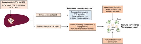
Hepatocellular carcinoma (HCC) is a common cause of cancer-related deaths worldwide. Unlike other types of cancer, HCC can be treated with locoregional treatments (LRTs) such as radiofrequency ablation (RFA) or transarterial chemoembolization (TACE). However, recurrences following LRTs are common, and strategies to improve long-term outcomes need to be developed. The exhaustion of anti-tumor immunity in HCC has been well established in many reports and the immunomodulatory effects of LRTs (enhancement of tumor antigen-specific T cell responses after RFA, reduction of effector regulatory T cells after TACE) have also been reported in several previous studies. However, a comprehensive review of previous studies and the possible roles of immunotherapy following LRTs in HCC are not known.

HCC immunotherapy locoregional treatment adjuvant treatment

1. Effects of Cell Death in Anti-Tumor Immune Responses Induced by LRTs

Several mechanisms have been suggested regarding the effects of LRTs on anti-tumor immune responses; cancer cell death caused by LRTs is closely associated with immunomodulatory effects. Both apoptosis and necrosis of tumor cells can be induced by LRTs. In general, necrosis is known to be immunogenic, because the plasma membrane is broken, and necrotic cells release cellular contents including cancer-specific antigens and DAMPs [1]. This phenomenon augments innate and adaptive immune responses via necroinflammation [2]. Conversely, apoptosis has been known to be non-immunogenic, because that is a process of programmed cell death, in which the plasma membrane is not disrupted [1]. Nevertheless, previous reports have also implicated that certain types of apoptosis could be immunogenic [1]. For example, melphalan-induced cancer cell apoptosis was associated with the secretion of proinflammatory cytokines [3]. Additionally, the exposure of cardiac glycosides induced HMGB-1, a representative DAMP, which resulted in immunogenic cancer cell apoptosis [4]. Thus, in addition to the type of cell death, whether LRT-related cell death is immunogenic or non-immunogenic has been investigated.
The release of tumor antigens due to cell death and subsequent changes in antigen-presenting cells (APCs) and effector immune cells are the main processes responsible for the changes in anti-tumor immune responses after LRTs [5]. Tumor cell death caused by RFA releases tumor antigens for DCs, which induce anti-tumor immune responses [6]. In another study, RFA induced APC infiltration and effective anti-tumor immune responses [7].
The type of cell death might also be important in inducing an anti-tumor immune response. In a previous study, the apoptosis of tumor cells due to irradiation (not injection) of necrotic tumor cells augmented the effects of tumor cell vaccines [8]. In another report, necrosis of tumor cells failed to induce CD8+ T cell responses and was associated with the release of peptidases [9]. LRTs induce tumor cell necrosis by thermal injury, but may also induce tumor cell apoptosis. For example, subtotal thermal injury in the transitional zone of RFA caused apoptosis and increased expression of heat shock protein 70, which might be associated with enhanced immunogenicity [10]. In another study, tumor cell apoptosis was shown to be activated by RFA in the early period [11]. Most of the tumor cells in HCC patients who underwent TACE before surgical resection were necrotic, and 11% of the tumor cells exhibited apoptosis [12]. These findings indicated that immunogenic cell death and apoptosis could also be induced by LRTs, as well as necrosis, which is considered as non-immunogenic cell death. However, considering the high recurrence rate even after successful LRTs, a strategy to augment the anti-tumor immune responses following LRTs might also be necessary to improve tumor surveillance.
Doxorubicin, which is the most commonly used agent in TACE, can induce immunogenic cell death. The effects on cell death can differ with the types of chemotherapeutics; doxorubicin can promote immunologic cell death, but cisplatin cannot [13]. Conversely, doxorubicin in a transplantable murine lymphoma model expanded myeloid-derived suppressor cells (MDSCs) and resulted in PD-1/PD-L1-dependent immunosuppression [14], indicating that targeting immune checkpoint molecules following TACE with doxorubicin is helpful for effective anti-tumor responses.

2. Immunological Changes Following LRTs in HCC

Evidence regarding immunological changes based on the HCC stage is lacking; however, in the analysis of the exhausted, dysfunctional immune cells within the tumor and adjacent liver, the surgical samples used in most immunological studies were from relatively early stage HCC patients. Immune dysfunction may begin in the early stage of tumor development. In a murine model, tumor-specific T cell exhaustion was initiated at the early pre-malignant phase of tumorigenesis [15]. In a previous study with human samples, PD-1+-exhausted CD8+ T cells within the tumor were also increased in patients with stage I and stage II or III HCC [16]. Furthermore, peritumoral CD8+ T cells exhibit exhausted features [16]. Although further investigation is necessary, these functional and phenotypic features of T cell exhaustion caused by chronic antigenic stimulation might be restored, but do not appear fully recovered by complete antigen clearance [17]. Thus, immune dysfunction can remain after successful LRT for HCC and LRT alone may induce limited antitumor immune responses. In the following sections, the previous studies in which the immunological changes caused by LRT in HCC were investigated are introduced and summarized in Figure 1.
Figure 1. Insufficient immune surveillance after LRTs for HCC. Image-guided locoregional treatments (LRTs) such as radiofrequency ablation (RFA), microwave ablation (MWA), percutaneous ethanol injection (PEI), cryoablation, transarterial chemoembolization (TACE), and transarterial radioembolization (TARE) cause tumor-cell necrosis or apoptosis. LRT can induce immunogenic cell death, which can be characterized by increased tumor-antigen release, antigen presenting cell (APC) activation, and decrease of regulatory T (Treg) cell frequency. These phenomena result in the augmentation of anti-tumor T cell responses and tumor surveillance. However, LRT can also induce a non-immunogenic cell death, which increases the tumor-promoting inflammation, angiogenesis, and the expression of immune checkpoint molecules. Furthermore, incomplete T cell restoration despite antigen clearance and immune-tolerant liver environment might also affect the attenuation of immune surveillance; these factors might be associated with frequent tumor recurrence even after successful LRTs for HCC.

2.1. Local Ablative Therapies

RFA is the most studied LRT in terms of immunological changes following LRTs. The immunomodulatory effect of RFA may be associated with tumor cell death caused by coagulative necrosis and apoptosis [18]. Cryoablation also showed inflammatory and coagulative responses, which may be associated with immune activation [19].
The increased number of tumor-specific T cells is associated with reduced tumor recurrence following RFA [20]. Similarly, MWA also increased the peripheral T cell number and increased the Th1/Th2 cytokine ratio [21]. A rat model of HCC showed that RFA treatment of the tumor in the left hepatic lobe enhanced T cell infiltration in the right lobe, thereby reducing tumor growth [22]. RFA enhanced T cell trafficking to the liver, which possesses an effector function against tumor antigens, but has no effect on B cell responses [23]. Notably, T cell activation caused by RFA tended to be stronger than that caused by surgical resection [24]. The activation of T cells may also be associated with DC activation. In HCC patients treated with RFA or PEI, activation of myeloid DCs and increased serum levels of tumor necrosis factor-α (TNF-α) and IL-1β were observed [25]. RFA or cryoablation also increases antigen-loaded DCs within the lymph nodes [6]. RFA also increased Th1 cytokines (interferon-γ and TNF-α) in HCC patients, which might be associated with enhanced T cell responses [26]. These results indicated that RFA can augment T cell infiltration into the liver and anti-tumor T cell responses in number and strength.
A 50–70% recurrence rate after successful RFA within 20–30 months has been reported in several studies [27]. In a previous study, tumor-specific T cell responses were enhanced by RFA in a liver cancer rabbit model, including circulating T cell activation and intrahepatic T cell infiltration; however, a high recurrence rate was also observed [28]. This finding was further validated in human subjects, which showed that immune activation following RFA was not associated with the prevention of HCC recurrence [29]. In addition, circulating PD-L1/PD-1 expression was significantly increased after cryoablation, correlating with poor clinical outcomes [30]. Increased Th17 cells after MWA have been shown to be a risk factor for tumor recurrence [31]. These findings indicate that the augmentation of immune responses following RFA is insufficient to prevent tumor recurrence.
The Toll-like receptor 9 agonist CpG increased anti-tumor T cell responses following RFA and prolonged survival and reduced new tumor growth [32]. A weak tumor-specific T cell response induced by RFA was potentiated by an anti-CTLA-4 blockade [33]. In another study, anti-CTLA-4 blockade or Treg cell depletion after RFA or cryoablation increased tumor-specific T cell number and cytokine secretion [6]. Interestingly, RFA of liver metastases increased T cell infiltration but also increased PD-L1 expression in primary tumors, which was overcome by anti-PD-1 blockade [34]. Furthermore, a mouse model showed that the administration of CC chemokine ligand 3 after RFA potentiated tumor-specific T cell responses via CD11c+cells in a CCR1-dependent manner [35]. In addition to the activation of T cell responses, RFA reduces the frequency of MDSCs, which is associated with a better recurrence-free survival [36]. These findings indicate that further immunotherapeutic strategies after local ablation treatments is helpful for improving clinical outcomes and enhancing tumor surveillance by effective immune responses, and strategies beyond targeting immune checkpoint molecules need to be investigated.

2.2. Transarterial Therapies

2.2.1. TACE

TACE is the preferred method for treating intermediate-stage, unresectable HCCs. Despite procedural advancement, the clinical outcome has not been satisfactory and the median OS, which is affected by tumor size, tumor markers, liver function, and vascular invasion, was 19.9 months based on a recent report [37]. Similar to local ablative treatments, immune dysfunction may remain even after successful treatment. Furthermore, in a previous study with human samples, PD-1+-exhausted CD8+ T cells in the tumor were more increased in stage II or III patients than in stage I patients [16], indicating that T-cell exhaustion is more advanced in patients receiving TACE than in patients receiving local ablation treatments. As TACE is not a curative treatment and a small number of tumor cells can remain in circulation and within the liver, surveillance and clearance by immune cells and their augmentation using ICBs could have clinical benefit [38] and clinical trials are ongoing.
The changes in the composition or function of immune cells after TACE remain to be elucidated, and whether the peripheral T cell population might be affected by TACE has been investigated in only a few studies. Growing evidence indicates that tumor necrosis caused by TACE induces immunological activation. For example, the inflammatory cytokine IL-6 significantly increased early after TACE [39]. However, Th2 cytokines (IL-4, IL-5, or suppressive cytokine IL-10) were also increased in the late phase after TACE, indicating that the immunological changes caused by TACE are complicated processes.
Immunologic cell death markers, such as high mobility group box 1 (HMGB1) and soluble receptor for advanced glycation end products (sRAGE), inducers of PD-L1 in tumor cells, are increased by TACE in HCC patients [40][41]. In a previous report, the peripheral CD4+/CD8+ T cell ratio was significantly lower in patients with HCC prior to TACE than in healthy volunteers and was increased 1 month after TACE [42][43], although its effect on anti-tumor immunity was not investigated in those studies. However, in another study, TACE increased the frequency of tumor-specific CD4+ T cells, which was associated with improved clinical outcomes [44]. Notably, a recent report showed that baseline PD-L1 expression within peripheral blood mononuclear cells was significantly higher in poor TACE responders, which were defined as SD or PD by RECIST criteria, and patients with high PD-L1 expression of PBMCs after TACE showed poor OS [45]. Additionally, TACE increases PD-1 expression in peripheral mononuclear cells [45]. These findings indicate that targeting the PD-1-PD-L1 axis might benefit the clinical outcome after TACE in terms of response rate and survival, although the effects of PD-1 expression on anti-tumor immunity before and after TACE remain unclear.
Several studies have indicated that TACE also affects Treg cells. Peripheral Treg cells were significantly increased in HCC patients and decreased by TACE [42][46]; their reduction was associated with improved clinical outcomes [47]. Among Treg cells, the effector Treg population, which exerts suppressive function, was also decreased by TACE, and post-TACE frequency in this population was associated with clinical outcome [46]. As Treg cells also highly express PD-1 in HCC patients [48], ICBs including PD-1 or CTLA-4 inhibitors might also be beneficial for HCC patients following TACE via further Treg reduction. Taken together, partial immune reconstitution due to TACE can occur, and targeting immune checkpoint molecules and immune-suppressive components, including Treg cells, would further augment anti-tumor immune responses and tumor surveillance even after successful TACE.

2.2.2. Transarterial Radioembolization (TARE)

Transarterial radioembolization (TARE) using yttrium-90 (Y90) is an emerging option for treating locally advanced HCC that is not eligible for surgical resection. The method delivers Y90 via tumor-supplying arteries and provides tumor-restricting effects without damaging the non-malignant liver [49], although its long-term clinical outcome has yet to be determined. Similar to TACE, TARE also activates pro-inflammatory cytokines such as IL-6 and IL-8 [50], and baseline values of these cytokines are associated with liver function and survival [51].
A recent in-depth analysis using CyTOF in resected HCC samples who underwent TARE before resection showed that TARE induces the activation of local immune cells, including CD8+ T cells, CD56+ NK cells, and CD8+CD56+ NKT cells [52]. Notably, TARE also induces peripheral T-cell function and increases APCs [52]. Furthermore, PD-1+TIM-3+CD8+ T cells were observed in TARE responders, indicating that further enhancement of T cell responses might be feasible by using ICBs following TARE.

References

  1. Tonnus, W.; Meyer, C.; Paliege, A.; Belavgeni, A.; Von Mässenhausen, A.; Bornstein, S.R.; Hugo, C.; Becker, J.U.; Linkermann, A. The pathological features of regulated necrosis. J. Pathol. 2019, 247, 697–707.
  2. Asadzadeh, Z.; Safarzadeh, E.; Safaei, S.; Baradaran, A.; Mohammadi, A.; Hajiasgharzadeh, K.; Derakhshani, A.; Argentiero, A.; Silvestris, N.; Baradaran, B. Current Approaches for Combination Therapy of Cancer: The Role of Immunogenic Cell Death. Cancers 2020, 12, 1047.
  3. Dudek-Peric, A.M.; Ferreira, G.B.; Muchowicz, A.; Wouters, J.; Prada, N.; Martin, S.; Kiviluoto, S.; Winiarska, M.; Boon, L.; Mathieu, C.; et al. Antitumor immunity triggered by melphalan is potentiated by melanoma cell surface-associated calreticulin. Cancer Res. 2015, 75, 1603–1614.
  4. Kepp, O.; Menger, L.; Vacchelli, E.; Adjemian, S.; Martins, I.; Ma, Y.; Sukkurwala, A.Q.; Michaud, M.; Galluzzi, L.; Zitvogel, L.; et al. Anticancer activity of cardiac glycosides: At the frontier between cell-autonomous and immunological effects. Oncoimmunology 2012, 1, 1640–1642.
  5. Greten, T.F.; Mauda-Havakuk, M.; Heinrich, B.; Korangy, F.; Wood, B. Combined locoregional-immunotherapy for liver cancer. J. Hepatol. 2019, 70, 999–1007.
  6. Den Brok, M.H.M.G.M.; Sutmuller, R.P.M.; Nierkens, S.; Bennink, E.J.; Frielink, C.; Toonen, L.W.J.; Boerman, O.C.; Figdor, C.; Ruers, T.J.M.; Adema, G.J. Efficient loading of dendritic cells following cryo and radiofrequency ablation in combination with immune modulation induces anti-tumour immunity. Br. J. Cancer 2006, 95, 896–905.
  7. Dromi, S.A.; Walsh, M.P.; Herby, S.; Traughber, B.; Xie, J.; Sharma, K.V.; Sekhar, K.P.; Luk, A.; Liewehr, D.J.; Dreher, M.R.; et al. Radiofrequency Ablation Induces Antigen-presenting Cell Infiltration and Amplification of Weak Tumor-induced Immunity. Radiology 2009, 251, 58–66.
  8. Scheffer, S.R.; Nave, H.; Korangy, F.; Schlote, K.; Pabst, R.; Jaffee, E.M.; Manns, M.P.; Greten, T.F. Apoptotic, but not necrotic, tumor cell vaccines induce a potent immune response in vivo. Int. J. Cancer 2003, 103, 205–211.
  9. Gamrekelashvili, J.; Kapanadze, T.; Han, M.; Wissing, J.; Ma, C.; Jaensch, L.; Manns, M.P.; Armstrong, T.; Jaffee, E.; White, A.O.; et al. Peptidases released by necrotic cells control CD8+ T cell cross-priming. J. Clin. Investig. 2013, 123, 4755–4768.
  10. Rai, R.; Richardson, C.; Flecknell, P.; Robertson, H.; Burt, A.; Manas, D. Study of Apoptosis and Heat Shock Protein (HSP) Expression in Hepatocytes Following Radiofrequency Ablation (RFA). J. Surg. Res. 2005, 129, 147–151.
  11. Vanagas, T.; Gulbinas, A.; Sadauskiene, I.; Dambrauskas, Z.; Pundzius, J.; Barauskas, G. Apoptosis is activated in an early period after radiofrequency ablation of liver tissue. Hepatogastroenterology 2009, 56, 1095–1099.
  12. Lu, W.; Li, Y.-H.; He, X.-F.; Zhao, J.-B.; Chen, Y.; Mei, Q.-L. Necrosis and Apoptosis in Hepatocellular Carcinoma Following Low-Dose Versus High-Dose Preoperative Chemoembolization. Cardiovasc. Interv. Radiol. 2008, 31, 1133–1140.
  13. Galluzzi, L.; Buqué, A.; Kepp, O.; Zitvogel, L.; Kroemer, G. Immunological Effects of Conventional Chemotherapy and Targeted Anticancer Agents. Cancer Cell 2015, 28, 690–714.
  14. Ding, Z.-C.; Lu, X.; Yu, M.; Lemos, H.D.P.; Huang, L.; Chandler, P.; Liu, K.; Walters, M.; Krasinski, A.; Mack, M.; et al. Immunosuppressive Myeloid Cells Induced by Chemotherapy Attenuate Antitumor CD4+ T-Cell Responses through the PD-1–PD-L1 Axis. Cancer Res. 2014, 74, 3441–3453.
  15. Schietinger, A.; Philip, M.; Krisnawan, V.E.; Chiu, E.Y.; Delrow, J.J.; Basom, R.S.; Lauer, P.; Brockstedt, D.G.; Knoblaugh, S.E.; Hammerling, G.J.; et al. Tumor-Specific T Cell Dysfunction Is a Dynamic Antigen-Driven Differentiation Program Initiated Early during Tumorigenesis. Immunity 2016, 45, 389–401.
  16. Ma, J.; Zheng, B.; Goswami, S.; Meng, L.; Zhang, D.; Cao, C.; Li, T.; Zhu, F.; Ma, L.; Zhang, Z.; et al. PD1(Hi) CD8(+) T cells correlate with exhausted signature and poor clinical outcome in hepatocellular carcinoma. J. Immunother. Cancer 2019, 7, 331.
  17. Han, J.W.; Sung, P.S.; Kim, K.H.; Hong, S.-H.; Shin, E.-C.; Song, M.J.; Park, S.-H. Dynamic Changes in Ex Vivo T-Cell Function After Viral Clearance in Chronic HCV Infection. J. Infect. Dis. 2019, 220, 1290–1301.
  18. Li, G.; Staveley-O’Carroll, K.F.; Kimchi, E.T. Potential of Radiofrequency Ablation in Combination with Immunotherapy in the Treatment of Hepatocellular Carcinoma. J. Clin. Trials 2016, 6, 1–5.
  19. Jansen, M.C.; van Hillegersberg, R.; Schoots, I.G.; Levi, M.; Beek, J.F.; Crezee, H.; van Gulik, T.M. Cryoablation induces greater inflammatory and coagulative responses than radiofrequency ablation or laser induced thermotherapy in a rat liver model. Surgery 2010, 147, 686–695.
  20. Mizukoshi, E.; Yamashita, T.; Arai, K.; Sunagozaka, H.; Ueda, T.; Arihara, F.; Kagaya, T.; Yamashita, T.; Fushimi, K.; Kaneko, S. Enhancement of tumor-associated antigen-specific T cell responses by radiofrequency ablation of hepatocellular carcinoma. Hepatology 2013, 57, 1448–1457.
  21. Zhang, H.; Hou, X.; Cai, H.; Zhuang, X. Effects of microwave ablation on T-cell subsets and cytokines of patients with hepatocellular carcinoma. Minim. Invasive Ther. Allied Technol. 2017, 26, 207–211.
  22. De Bethlenfalva-Hora, C.E.; Piguet, A.C.; Schmitt, J.; Mertens, J.C.; Dufour, J.-F.; Geier, A.; Kettenbach, J.; Terracciano, L.; Weimann, R. Radiofrequency ablation suppresses distant tumour growth in a novel rat model of multifocal hepatocellular carcinoma. Clin. Sci. 2014, 126, 243–252.
  23. Napoletano, C.; Taurino, F.; Biffoni, M.; De Majo, A.; Coscarella, G.; Bellati, F.; Rahimi, H.; Pauselli, S.; Pellicciotta, I.; Burchell, J.M.; et al. RFA strongly modulates the immune system and anti-tumor immune responses in metastatic liver patients. Int. J. Oncol. 2008, 32, 481–490.
  24. Nobuoka, D.; Motomura, Y.; Shirakawa, H.; Yoshikawa, T.; Kuronuma, T.; Takahashi, M.; Nakachi, K.; Ishii, H.; Furuse, J.; Gotohda, D.; et al. Radiofrequency ablation for hepatocellular carcinoma induces glypican-3 peptide-specific cytotoxic T lymphocytes. Int. J. Oncol. 2011, 40, 63–70.
  25. Ali, M.Y.; Grimm, C.F.; Ritter, M.; Mohr, L.; Allgaier, H.-P.; Weth, R.; Bocher, W.O.; Endrulat, K.; Blum, H.E.; Geissler, M. Activation of dendritic cells by local ablation of hepatocellular carcinoma. J. Hepatol. 2005, 43, 817–822.
  26. Ji, L.; Gu, J.; Chen, L.; Miao, D. Changes of Th1/Th2 cytokines in patients with primary hepatocellular carcinoma after ultrasound-guided ablation. Int. J. Clin Exp. Pathol. 2017, 10, 8715–8720.
  27. Cho, Y.K.; Kim, J.K.; Kim, M.Y.; Rhim, H.; Han, J.K. Systematic review of randomized trials for hepatocellular carcinoma treated with percutaneous ablation therapies. Hepatology 2009, 49, 453–459.
  28. Wissniowski, T.T.; Hänsler, J.; Neureiter, D.; Frieser, M.; Schaber, S.; Esslinger, B.; Voll, R.; Strobel, D.; Hahn, E.G.; Schuppan, D.; et al. Activation of tumor-specific T lymphocytes by radio-frequency ablation of the VX2 hepatoma in rabbits. Cancer Res. 2003, 63, 6496–6500.
  29. Zerbini, A.; Pilli, M.; Penna, A.; Pelosi, G.; Schianchi, C.; Molinari, A.; Schivazappa, S.; Zibera, C.; Fagnoni, F.F.; Ferrari, C.; et al. Radiofrequency thermal ablation of hepatocellular carcinoma liver nodules can activate and enhance tumor-specific T-cell responses. Cancer Res. 2006, 66, 1139–1146.
  30. Zeng, Z.; Shi, F.; Zhou, L.; Zhang, M.-N.; Chen, Y.; Chang, X.-J.; Lu, Y.-Y.; Bai, W.-L.; Qu, J.-H.; Wang, C.-P.; et al. Upregulation of Circulating PD-L1/PD-1 Is Associated with Poor Post-Cryoablation Prognosis in Patients with HBV-Related Hepatocellular Carcinoma. PLoS ONE 2011, 6, e23621.
  31. Jing, X.; Zhou, Y.; Xu, X.; Ding, J.; Wang, F.; Wang, Y.; Wang, P. Dynamic changes of T-cell subsets and their relation with tumor recurrence after microwave ablation in patients with hepatocellular carcinoma. J. Cancer Res. Ther. 2018, 14, 40–45.
  32. Behm, B.; Di Fazio, P.; Michl, P.; Neureiter, D.; Kemmerling, R.; Hahn, E.G.; Strobel, D.; Gress, T.; Schuppan, D.; Wissniowski, T.T. Additive antitumour response to the rabbit VX2 hepatoma by combined radio frequency ablation and toll like receptor 9 stimulation. Gut 2014, 65, 134–143.
  33. Den Brok, M.H.M.G.M.; Sutmuller, R.P.M.; Van Der Voort, R.; Bennink, E.J.; Figdor, C.; Ruers, T.J.M.; Adema, G.J. In Situ Tumor Ablation Creates an Antigen Source for the Generation of Antitumor Immunity. Cancer Res. 2004, 64, 4024–4029.
  34. Shi, L.; Chen, L.; Wu, C.; Zhu, Y.; Xu, B.; Zheng, X.; Sun, M.; Wen, W.; Dai, X.; Yang, M.; et al. PD-1 Blockade Boosts Radiofrequency Ablation-Elicited Adaptive Immune Responses against Tumor. Clin. Cancer Res. 2016, 22, 1173–1184.
  35. Iida, N.; Nakamoto, Y.; Baba, T.; Nakagawa, H.; Mizukoshi, E.; Naito, M.; Mukaida, N.; Kaneko, S. Antitumor Effect after Radiofrequency Ablation of Murine Hepatoma Is Augmented by an Active Variant of CC Chemokine Ligand 3/Macrophage Inflammatory Protein-1α. Cancer Res. 2010, 70, 6556–6565.
  36. Arihara, F.; Mizukoshi, E.; Kitahara, M.; Takata, Y.; Arai, K.; Yamashita, T.; Nakamoto, Y.; Kaneko, S. Increase in CD14+HLA-DR−/low myeloid-derived suppressor cells in hepatocellular carcinoma patients and its impact on prognosis. Cancer Immunol. Immunother. 2013, 62, 1421–1430.
  37. Han, G.; Berhane, S.; Toyoda, H.; Bettinger, D.; Elshaarawy, O.; Chan, A.W.H.; Kirstein, M.; Mosconi, C.; Hucke, F.; Palmer, D.; et al. Prediction of Survival Among Patients Receiving Transarterial Chemoembolization for Hepatocellular Carcinoma: A Response-Based Approach. Hepatology 2020, 72, 198–212.
  38. Han, J.W.; Yoon, S.K. Tissue-Resident Lymphocytes: Implications in Immunotherapy for Hepatocellular Carcinoma. Int. J. Mol. Sci. 2020, 22, 232.
  39. Kim, M.J.; Jang, J.W.; Oh, B.S.; Kwon, J.H.; Chung, K.W.; Jung, H.S.; Jekarl, D.W.; Lee, S. Change in inflammatory cytokine profiles after transarterial chemotherapy in patients with hepatocellular carcinoma. Cytokine 2013, 64, 516–522.
  40. Kohles, N.; Nagel, D.; Jüngst, D.; Stieber, P.; Holdenrieder, S. Predictive value of immunogenic cell death biomarkers HMGB1, sRAGE, and DNase in liver cancer patients receiving transarterial chemoembolization therapy. Tumor Biol. 2012, 33, 2401–2409.
  41. Wang, W.; Chapman, N.M.; Zhang, B.; Li, M.; Fan, M.; Laribee, R.N.; Zaidi, M.R.; Pfeffer, L.M.; Chi, H.; Wu, Z.H. Upregulation of PD-L1 via HMGB1-Activated IRF3 and NF-kappaB Contributes to UV Radiation-Induced Immune Suppression. Cancer Res. 2019, 79, 2909–2922.
  42. Liao, J.; Xiao, J.; Zhou, Y.; Liu, Z.; Wang, C. Effect of transcatheter arterial chemoembolization on cellular immune function and regulatory T cells in patients with hepatocellular carcinoma. Mol. Med. Rep. 2015, 12, 6065–6071.
  43. Huang, M.; Wang, X.; Bin, H. Effect of Transcatheter Arterial Chemoembolization Combined with Argon-Helium Cryosurgery System on the Changes of NK Cells and T Cell Subsets in Peripheral Blood of Hepatocellular Carcinoma Patients. Cell Biochem. Biophys. 2015, 73, 787–792.
  44. Ayaru, L.; Pereira, S.P.; Alisa, A.; Pathan, A.A.; Williams, R.; Davidson, B.; Burroughs, A.K.; Meyer, T.; Behboudi, S. Unmasking of alpha-fetoprotein-specific CD4(+) T cell responses in hepatocellular carcinoma patients undergoing embolization. J. Immunol. 2007, 178, 1914–1922.
  45. Guo, J.; Wang, S.; Han, Y.; Jia, Z.; Wang, R. Effects of transarterial chemoembolization on the immunological function of patients with hepatocellular carcinoma. Oncol. Lett. 2021, 22, 1–8.
  46. Park, H.; Jung, J.H.; Jung, M.K.; Shin, E.-C.; Ro, S.W.; Park, J.H.; Kim, D.Y.; Park, J.Y.; Han, K.-H. Effects of transarterial chemoembolization on regulatory T cell and its subpopulations in patients with hepatocellular carcinoma. Hepatol. Int. 2020, 14, 249–258.
  47. Xiong, B.; Feng, G.; Luo, S.; Liang, H.; Qiu, L.; Zheng, C.; Liu, X.; Zhou, G. Changes of CD4+ CD25+ regulatory T cells in peripheral blood in patients with hepatocellular carcinoma before and after TACE. J. Huazhong Univ. Sci. Technol. Med. Sci. 2008, 28, 645–648.
  48. Kalathil, S.G.; Lugade, A.A.; Miller, A.; Iyer, R.; Thanavala, Y. PD-1(+) and Foxp3(+) T cell reduction correlates with survival of HCC patients after sorafenib therapy. JCI Insight 2016, 1, e86182.
  49. Sangro, B.; Inarrairaegui, M.; Bilbao, J.I. Radioembolization for hepatocellular carcinoma. J. Hepatol. 2012, 56, 464–473.
  50. Fernandez-Ros, N.; Inarrairaegui, M.; Paramo, J.A.; Berasain, C.; Avila, M.A.; Chopitea, A.; Varo, N.; Sarobe, P.; Bilbao, J.I.; Dominguez, I.; et al. Radioembolization of hepatocellular carcinoma activates liver regeneration, induces inflammation and endothelial stress and activates coagulation. Liver Int. 2015, 35, 1590–1596.
  51. Seidensticker, M.; Powerski, M.; Seidensticker, R.; Damm, R.; Mohnike, K.; Garlipp, B.; Klopffleisch, M.; Amthauer, H.; Ricke, J.; Pech, M. Cytokines and 90Y-Radioembolization: Relation to Liver Function and Overall Survival. Cardiovasc. Interv. Radiol. 2017, 40, 1185–1195.
  52. Chew, V.; Lee, Y.H.; Pan, L.; Nasir, N.J.M.; Lim, C.J.; Chua, C.; Lai, L.; Hazirah, S.N.; Lim, T.K.H.; Goh, B.K.P.; et al. Immune activation underlies a sustained clinical response to Yttrium-90 radioembolisation in hepatocellular carcinoma. Gut 2019, 68, 335–346.
More
Information
Subjects: Oncology
Contributor MDPI registered users' name will be linked to their SciProfiles pages. To register with us, please refer to https://encyclopedia.pub/register :
View Times: 412
Revisions: 2 times (View History)
Update Date: 08 Oct 2021
1000/1000