Submitted Successfully!
To reward your contribution, here is a gift for you: A free trial for our video production service.
Thank you for your contribution! You can also upload a video entry or images related to this topic.
Version Summary Created by Modification Content Size Created at Operation
1 + 2453 word(s) 2453 2021-09-06 11:30:08 |
2 Format change Meta information modification 2453 2021-09-17 03:42:27 |

Video Upload Options

Do you have a full video?

Confirm

Are you sure to Delete?
Cite
If you have any further questions, please contact Encyclopedia Editorial Office.
Petrik, J. Normalizing Tumor Vasculature. Encyclopedia. Available online: https://encyclopedia.pub/entry/14288 (accessed on 27 July 2024).
Petrik J. Normalizing Tumor Vasculature. Encyclopedia. Available at: https://encyclopedia.pub/entry/14288. Accessed July 27, 2024.
Petrik, Jim. "Normalizing Tumor Vasculature" Encyclopedia, https://encyclopedia.pub/entry/14288 (accessed July 27, 2024).
Petrik, J. (2021, September 17). Normalizing Tumor Vasculature. In Encyclopedia. https://encyclopedia.pub/entry/14288
Petrik, Jim. "Normalizing Tumor Vasculature." Encyclopedia. Web. 17 September, 2021.
Normalizing Tumor Vasculature
Edit

A basic requirement of tumorigenesis is the development of a vascular network to support the metabolic requirements of tumor growth and metastasis. Tumor vascular formation is regulated by a balance between promoters and inhibitors of angiogenesis. Typically, the pro-angiogenic environment created by the tumor is extremely aggressive, resulting in the rapid vessel formation with abnormal, dysfunctional morphology. The altered morphology and function of tumor blood and lymphatic vessels has numerous implications including poor perfusion, tissue hypoxia, and reduced therapy uptake. Targeting tumor angiogenesis as a therapeutic approach has been pursued in a host of different cancers. Although some preclinical success was seen, there has been a general lack of clinical success with traditional anti-angiogenic therapeutics as single agents. Typically, following anti-angiogenic therapy, there is remodeling of the tumor microenvironment and widespread tumor hypoxia, which is associated with development of therapy resistance. A more comprehensive understanding of the biology of tumor angiogenesis and insights into new clinical approaches, including combinations with immunotherapy, are needed to advance vascular targeting as a therapeutic area. 

hypoxia angiogenesis vascular normalization

1. Introduction

1.1. Sprouting Angiogenesis in Normal Physiology

Angiogenesis is the complex and highly regulated formation and maturation of vasculature from pre-existing vessels throughout the body. Typically, the process is kept quiescent through a balance of growth factors and inhibitors. Normal human processes that necessitate angiogenesis in the adult include placentation in the pregnant uterus, formation of the endometrium in the menstrual cycle, growth of the mammary gland in preparation for lactation, and supply of granulation tissue for wound healing [1][2][3]. In any of these situations, angiogenesis consists of a series of events including removal of structural pericytes in the area of the developing sprout, degradation of the capillary basement membrane, migration and proliferation of the endothelial cells comprising the new sprout, nascent tube formation, and vascular stabilization [4].
The presence of angiogenic stimuli such as hypoxia, mechanical stress, or inflammation leads to the release of growth factors, as summarized in Figure 1. These signaling events ultimately lead to the activation of cellular effectors, which aim to form the nascent vessel [4]. Upon effector stimulation, smooth muscle cells called pericytes located at intervals along the capillary wall are first removed from the sprouting area of a mother vessel. VEGF stimulation triggers intricate calcium oscillations within endothelial cells allowing for the selection of an endothelial cell distinguished by specialized filopodia, called a tip cell [5]. The tip cell guides the developing sprout through chemotaxis, following angiogenic stimuli secreted by the target tissue requiring increased perfusion [5]. As tip cells are highly influenced by even minute fluctuations in growth factor signaling, a loss of growth factor balance in this system may lead to disorganized vasculature. The tip cell releases matrix metalloproteases (MMP), which degrade basement membrane components in its path [6]. A second group of specialized endothelial cells, called stalk cells, are highly proliferative and interact with tip cells through delta-notch signaling to elongate the nascent sprout [7]. At a point of anastomoses between the tip cell of another nascent vessel or stabilized vessel, junctional adhesion proteins are deposited at the contact site of the two tip cells. A lumen is formed through cell membrane invagination or cord hollowing, forming a functional vascular network [8]. Circulating endothelial progenitor cells also contribute to the nascent vessels, which are haphazardly branched and in need of organization. Local differences in blood flow and pressure lead to the elimination of poorly perfused branches (pruning) or recycling of their component endothelial cells to areas of significant flow [9][10]. Conversely, highly perfused sprouts are stabilized through deposition of basement membrane, reduced endothelial cell activity, tightening of cell junctions, and recruitment of pericytes [10].
Figure 1. Hypoxia induced by the growing tumor mass triggers an “angiogenic switch” within the tumor microenvironment, resulting in a crude version of angiogenesis.

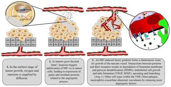
1.2. Tumor Control of Angiogenesis

In many ways, tumors can be considered functional organs as opposed to a group of aberrant cells. The tumor stroma includes mesenchymal-derived cells, inflammatory cells, and vascular cells, albeit in an irregular fashion that has been modified by the tumor to tailor to its survival needs [11][12]. Tumors are therefore capable of inducing angiogenesis by co-opting the same pro-angiogenic program. Small tumors devoid of vasculature are often observed in solid tumor types—their oxygen and nutrient demands being supplied by passive diffusion from nearby vessels [13]. However, as tumors grow beyond 2 mm2, the tumor core becomes increasingly hypoxic and the process of angiogenesis begins to fuel oxygen and nutrient demands [14]. This moment has been termed “the angiogenic switch” in which tumor cells respond to low oxygen perfusion by releasing many of the angiogenic factors represented in Figure 1 [15]. Cellular responses to low oxygen are primarily regulated by DNA-binding transcription factors known as hypoxia inducible factors (HIF). HIFs are heterodimeric proteins that consist of a constitutively expressed HIF-1ß subunit and an oxygen-regulating subunit (HIF-1α or HIF-2α) [16][17]. These alpha subunits are composed of an amino terminal basic Helix-Loop-Helix (bHLH) necessary for DNA binding to hypoxia response elements (HRE), transactivation domains (N-TAD and C-TAD) that are vital for activation of HIF target genes, PAS-A and PAS-B domains for protein-protein dimerization, and an oxygen-dependent degradation domain (ODDD). Redundancy in HIF-1α stabilization is evident as a secondary lysine residue within the ODDD can be acetylated by an acetyl transferase enzyme called arrest-defective-1 (ARD-1) to favour degradation of HIF-1α [18]. The expression of ARD-1 is decreased in hypoxia, resulting in stabilized HIF-1α under this condition [18].
Under normoxic conditions, the prolyl hydroxylase domain (PHD) uses oxygen as a rate-limiting substrate and iron as a cofactor to hydroxylate two proline residues within the ODDD [18]. Hydroxylated HIF-1α becomes associated with Von Hippel Lindau factor (pVHL) and elongins B and C, cullin-2 (cul-2) and rbx1 co-factors, forming a complex with E3 ubiquitin ligase activity (HIF-1α-VBC complex) [19]. However, under hypoxic conditions, HIF-1α is stabilized through limited PHD activity. This allows generation and accumulation of non-hydroxylated HIF-1α. Further, HIF-1α stability is controlled by ubiquitin ligases that are PHD enzymes themselves, as well as pVHL-interacting deubiquitinating enzyme (VDU2), which acts to destabilize ubiquitin ligases on HIF-1α [18]. Given the significantly short half-life of HIF-1α (<1 min in a perfused lung), it is constantly being degraded at physiological oxygen levels in normal cells and is subject to tight regulation should oxygen levels decline [20]. In contrast, the median oxygenation of an untreated tumor falls between approximately 0.3% and 4.2%, with most untreated tumors exhibiting median oxygen levels <2% [21]. This level of hypoxia triggers the release and stabilization of HIF-1α while also inducing oncogenic mechanisms that further derail the HIF pathway and make tumors less dependent on oxygen [21]. Tumor-induced mutations in the binding pocket of pVHL have been shown to disrupt HIF-1α interactions and thereby disassemble the E3 ubiquitin ligase (VEC) complex [22]. More directly, in lung cancer, TP53 mutants have been shown to exert a gain of function on HIF-1, leading to heightened expression of hypoxia-response genes [23]. HIF-1α is capable of binding directly to the tumor suppressor, favouring mouse double minute 2 homolog (Mdm2) ubiquitination and proteosomal degradation of HIF-1α, which is not possible in TP53 mutants or knockouts [24]. Several studies have shown that HIF expression is abrogated upon phosphoinositide 3-kinase (PI3K) pathway inhibition regardless of oxygen levels [25][26]. Similarly, HIF-1 is upregulated by AKT in human gastric cancer, breast cancer, and non-small cell lung cancer [27][28].

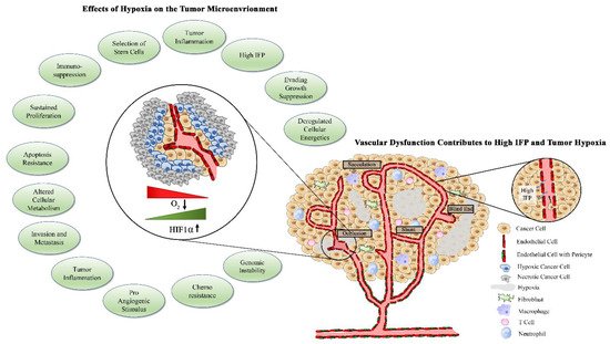
1.3. Factors Contributing to Tumor Vascular Dysfunction

Tumors initiate the angiogenic process through activation of multiple factors including the most prominent angiogenic ligand, vascular endothelial growth factors (VEGF), and its receptors including VEGFR2 [29][30][31]. The VEGF family of proteins includes VEGF-A, VEGF-B, VEGF-C, VEGF-D, VEGF-E, and placenta growth factor (PIGF) [31]. VEGF-C and VEGF-D are studied as regulators of lymphangiogenesis, while VEGF-A is commonly referred to simply as VEGF due to its dominant role in angiogenesis. VEGF undergoes alternative splicing, leading to several isoforms that differ based on heparin binding affinity, localization to the extracellular matrix, or diffusive potential. The VEGF gene is transcriptionally regulated in response to HIF, and its levels must be tightly controlled to prevent aberrant angiogenesis [31]. Due to their control over HIF, tumor cells release exaggerated levels of VEGF to the extracellular space in response to hypoxia [32]. High concentrations of VEGF surrounding endothelial cells select for excess tip cells, which then contribute to irregular branching and tortuous vascular networks. The basement membrane of tumor vessels, which serves as a physical barrier for cancer cell metastasis to surrounding tissues, is often absent or thin due to chemical degradation by tumor-derived proteases [33][34]. The monolayer of endothelial cells is often disorganized and cells are plagued with abnormal gene expression profiles, karyotypic abnormalities, and chromosomal instability [35][36][37]. Compared with normal endothelial cells, tumor endothelial cells contain four times the amount of total RNA, indicating enhanced gene expression. Indeed, tumor endothelial cells have enhanced expression of VEGFR-1 and -2 and are therefore more responsive to VEGF stimulation [38]. Recently, tumor endothelial cells have been shown to have enhanced expression of markers of angiogenesis and stemness such as CD61, CD105, Sca-1, CD34, CD90, and ALDH [39][40]. These expression profiles contribute to the escalated angiogenic potential of tumor endothelial cells compared with normal endothelial cells, which facilitates the aberrant vascular arrangement seen in tumors [41]. Extracellular factors such as VEGF, PMA, TGF-ß, and cytochalasin B, which are overexpressed in the tumor microenvironment, have been shown to impact fenestration formation in endothelial cells [42][43]. Given that these plasma membrane microdomains are vital for the exchange of solutes and water at the interface of tissue and vasculature, tumor endothelial cells are often more porous compared with normal counterparts [43]. Abnormal VEGF signaling in tumor endothelial cells also leads to downregulation of connexin expression, causing gap junction dysfunction, increasing vascular fenestrations, and increasing vascular permeability [44][45][46]. In fact, VEGF was initially identified based on its ability to increase vascular permeability and extravasation of plasma proteins, such as fibrinogen [47].
Pericytes are specialized smooth muscle cells that are recruited to mature and stabilized vessels through release of PDGF-ß by ECs [48]. Mice deficient in PDGF-ß signaling lack pericytes and succumb to micro hemorrhaging, demonstrating the importance of these cells for proper vascular function [48][49]. Signals secreted by pericytes maintain EC survival by leading to enhanced expression of BCL-w antiapoptotic protein [49]. Pericytes therefore also shelter normal vessels from anti-angiogenic therapies, allowing for tumor-targeted action of these agents. Hypoxia and downstream angiogenic factors released by tumor cells disengage pericytes from endothelial cells as the initial step to the formation of the nascent vascular sprout. Therefore, tumor-associated vessels are largely devoid of pericytes or demonstrate weak connections between pericytes and endothelial cells, contributing to an immature vascular phenotype and facilitating continued angiogenesis [50].

1.4. Abnormal Vasculature Results in Limited Treatment Delivery

The abnormalities of the tumor vasculature result in poor tissue perfusion, which poses a physical barrier to therapy delivery to tumors. Of the number of delivery and uptake impediments, elevated interstitial pressure (IFP) is considered to be the most significant barrier to therapy access to the tumor [51][52][53]. The etiology of IFP elevation is multifactorial and involves high vascular permeability and mechanical compression of lymphatic blood vessels [54][55]. Disrupted vascular morphology with reduced pericyte coverage is associated with a loss of endothelial cell junction integrity and an activated endothelium, resulting in vessels that are leaky and extravasate fluid into the tumor environment, thereby increasing pressure within the tumor [56][57]. Combined with solid stress, in which accumulation of cancer cells, stromal cells, cancer-associated fibroblasts (CAFs), and their associated extracellular matrix create high mechanical pressure within the tumor, IFP leads to a significant elevation in intratumoral pressure [58].
This high IFP causes a stasis in flow throughout the tumor, which results in tumor hypoxia and acidosis [59]. Elevated hypoxia as a consequence of high IFP is associated with poor outcome in cancer patients and is considered an early response marker for cancer therapeutics such as chemotherapy and radiation [60][61]. As another consequence of reduced perfusion and flow within the tumor, there is impediment of drug uptake and delivery within the tumor tissue [62]. With the elevated IFP, there is an attenuated transvascular osmotic pressure difference, resulting in impaired delivery of drugs throughout the tumor [63]. Even in tumors in which there is vascular heterogeneity, drugs will become concentrated in regions that have sufficient blood supply but will have limited migration to areas in which IFP is higher and vessel density is decreased [64]. Although IFP is often discussed in relation to the primary tumor, it is important to note that larger metastatic tumors also demonstrate elevated IFP and decreased drug uptake, potentially contributing to the development of drug-resistant metastatic disease.
While intra-tumoral treatment delivery decreases off-target toxicities, it fails to account for metastatic disease and has not led to significant survival benefit compared to systemic administration [65]. Clinical use of intra-tumoral drugs is also impractical for some tumor subtypes such as ovarian and pancreatic cancers, which are inaccessible through transdermal injection. In order to prove effective, systemic agents must not only navigate from the injection site to the tumor vasculature but must also gain access and disperse throughout a tumor, which is often plagued with impediments to this process, posing a therapeutic challenge [66]. The properties of the tumor microenvironment that pose issues for treatment delivery are depicted in Figure 2.
Figure 2. Tumor hypoxia activates several tumorigenic processes. Tumor vasculature has altered morphology, with reduced pericyte coverage. The immature tumor vessels are characterized by blind end shunts, torturous pathway, sacculations, decreased luminal size, and increased fenestrations. Excessively fenestrated vessels allow for fluid extravasation and increased interstitial fluid pressure (IFP) and facilitate intravasation and migration of metastatic tumor cells. Elevated IFP and disrupted tissue perfusion contribute to areas of acute and chronic hypoxia, which can activate numerous pro-tumorigenic processes.

2. Vascular Normalizing Agents as Adjuvants to Traditional Cancer Therapeutics

Normalized tumor vessels have also been shown to re-program many other aspects of the tumor microenvironment known to limit delivery of cancer therapies discussed earlier in this review, giving rise to the term ‘microenvironment normalization’ [67]. Anti-angiogenic drugs have opened new avenues for combination therapy. In a humanized murine model of colorectal adenocarcinoma, combination therapy with anti-PDGFR and anti-VEGFR tyrosine kinase inhibitors decreased IFP in tumors, allowing for enhanced delivery of taxol therapy [68]. Improved delivery of chemotherapy through vascular normalization in solid tumors has been extensively reviewed [69]. We and others have extended the utility of vascular normalization to enhancing the delivery and functionality of agents beyond traditional chemotherapy. The vascular shutdown typical of oncolytic viruses (OV) was prevented using thrombospondin type-1 repeats in a mouse model of advanced stage ovarian cancer [70]. This led to enhanced intratumoral trafficking of immune cell subsets, thereby improving immunotherapeutic success [67][70][71]. In addition to enhancing vascular perfusion and providing a conduit for immune cells, the enhanced oxygenation of tumors as a result of low-dose anti-angiogenic therapy has improved immune cell function and reprogramed immune cell subsets with greater anti-tumor capabilities [72]. Vascular normalizing therapies continue to be recognized for their oxygen-modulating function in sensitizing tumors to traditional therapies, which have often been met with resistance [73].

References

  1. Pereira, R.D.; de Long, N.E.; Wang, R.C.; Yazdi, F.T.; Holloway, A.C.; Raha, S. Angiogenesis in the Placenta: The Role of Reactive Oxygen Species Signaling. BioMed Res. Int. 2015, 2015, 814543.
  2. Dangat, K.; Khaire, A.; Joshi, S. Cross Talk of Vascular Endothelial Growth Factor and Neurotrophins in Mammary Gland Development. Growth Factors 2020, 38, 16–24.
  3. Kumar, P.; Kumar, S.; Udupa, E.P.; Kumar, U.; Rao, P.; Honnegowda, T. Role of Angiogenesis and Angiogenic Factors in Acute and Chronic Wound Healing. Plast. Aesthet. Res. 2015, 2, 243–249.
  4. Ucuzian, A.A.; Gassman, A.A.; East, A.T.; Greisler, H.P. Molecular Mediators of Angiogenesis. J. Burn Care Res. 2010, 31, 158–175.
  5. Yokota, Y.; Nakajima, H.; Wakayama, Y.; Muto, A.; Kawakami, K.; Fukuhara, S.; Mochizuki, N. Endothelial Ca2+ Oscillations Reflect VEGFR Signaling-Regulated Angiogenic Capacity in Vivo. eLife 2015, 4, e08817.
  6. Ghajar, C.M.; George, S.C.; Putnam, A.J. Matrix Metalloproteinase Control of Capillary Morphogenesis. Crit. Rev. Eukaryot. Gene Expr. 2008, 18, 251–278.
  7. Sauteur, L.; Krudewig, A.; Herwig, L.; Ehrenfeuchter, N.; Lenard, A.; Affolter, M.; Belting, H.G. Cdh5/VE-Cadherin Promotes Endothelial Cell Interface Elongation via Cortical Actin Polymerization during Angiogenic Sprouting. Cell Rep. 2014, 9, 504–513.
  8. Franco, C.A.; Jones, M.L.; Bernabeu, M.O.; Vion, A.C.; Barbacena, P.; Fan, J.; Mathivet, T.; Fonseca, C.G.; Ragab, A.; Yamaguchi, T.P.; et al. Non-Canonical Wnt Signalling Modulates the Endothelial Shear Stress Flow Sensor in Vascular Remodelling. eLife 2016, 5, e07727.
  9. Chen, Q.; Jiang, L.; Li, C.; Hu, D.; Bu, J.W.; Cai, D.; Du, J.L. Haemodynamics-Driven Developmental Pruning of Brain Vasculature in Zebrafish. PLoS Biol. 2012, 10, e1001374.
  10. Murakami, M. Signaling Required for Blood Vessel Maintenance: Molecular Basis and Pathological Manifestations. Int. J. Vasc. Med. 2012, 2012, 293641.
  11. Bremnes, R.M.; Dønnem, T.; Al-Saad, S.; Al-Shibli, K.; Andersen, S.; Sirera, R.; Camps, C.; Marinez, I.; Busund, L.T. The Role of Tumor Stroma in Cancer Progression and Prognosis: Emphasis on Carcinoma-Associated Fibroblasts and Non-Small Cell Lung Cancer. J. Thorac. Oncol. 2011, 6, 209–217.
  12. Murphy, K.J.; Chambers, C.R.; Herrmann, D.; Timpson, P.; Pereira, B.A. Dynamic Stromal Alterations Influence Tumor-Stroma Crosstalk to Promote Pancreatic Cancer and Treatment Resistance. Cancers 2021, 13, 3481.
  13. Folkman, J. Tumor Angiogenesis: Therapeutic Implications. N. Engl. J. Med. 1971, 285, 1182–1186.
  14. Hanahan, D.; Weinberg, R.A. The Hallmarks of Cancer. Cell 2000, 100, 57–70.
  15. Folkman, J.; Hanahan, D. Switch to the Angiogenic Phenotype during Tumorigenesis. Princess Takamatsu Symp. 1991, 22, 22.
  16. Comerford, K.M.; Wallace, T.J.; Karhausen, J.; Louis, N.A.; Montalto, M.C.; Colgan, S.P. Hypoxia-Inducible Factor-1-Dependent Regulation of the Multidrug Resistance (MDR1) Gene. Cancer Res. 2002, 62, 62.
  17. Wilkins, S.E.; Abboud, M.I.; Hancock, R.L.; Schofield, C.J. Targeting Protein-Protein Interactions in the HIF System. ChemMedChem 2016, 11, 773–786.
  18. Jeong, J.-W.; Bae, M.-K.; Ahn, M.-Y.; Kim, S.-H.; Sohn, T.-K.; Bae, M.-H.; Yoo, M.-A.; Song, E.J.; Lee, K.-J.; Kim, K.-W. Regulation and Destabilization of HIF-1 by ARD1-Mediated Acetylation Quitin-Proteasome Pathway (Salceda and Caro The Association of PVHL and HIF-1 under nor-Moxic Conditions Is Triggered by the Posttranslational. Cell 2002, 111, 709–720.
  19. Strowitzki, M.; Cummins, E.; Taylor, C. Protein Hydroxylation by Hypoxia-Inducible Factor (HIF) Hydroxylases: Unique or Ubiquitous? Cells 2019, 8, 384.
  20. Yu, A.Y.; Frid, M.G.; Shimoda, L.A.; Wiener, C.M.; Stenmark, K.; Semenza, G.L. Temporal, Spatial, and Oxygen-Regulated Expression of Hypoxia-Inducible Factor-1 in the Lung. Am. J. Physiol. Lung Cell. Mol. Physiol. 1998, 275, L818–L826.
  21. McKeown, S.R. Defining Normoxia, Physoxia and Hypoxia in Tumours—Implications for Treatment Response. Br. J. Radiol. 2014, 87, 20130676.
  22. Artemov, A.V.; Zhigalova, N.; Zhenilo, S.; Mazur, A.M.; Prokhortchouk, E.B. VHL Inactivation without Hypoxia Is Sufficient to Achieve Genome Hypermethylation. Sci. Rep. 2018, 8, 10667.
  23. Amelio, I.; Mancini, M.; Petrova, V.; Cairns, R.A.; Vikhreva, P.; Nicolai, S.; Marini, A.; Antonov, A.A.; le Quesne, J.; Baena Acevedo, J.D.; et al. P53 Mutants Cooperate with HIF-1 in Transcriptional Regulation of Extracellular Matrix Components to Promote Tumor Progression. Proc. Natl. Acad. Sci. USA 2018, 115, E10869–E10878.
  24. Ravi, R.; Mookerjee, B.; Bhujwalla, Z.M.; Sutter, C.H.; Artemov, D.; Zeng, Q.; Dillehay, L.E.; Madan, A.; Semenza, G.L.; Bedi, A. Regulation of Tumor Angiogenesis by P53-Induced Degradation of Hypoxia- Inducible Factor 1α. Genes Dev. 2000, 14, 34–44.
  25. Mohlin, S.; Hamidian, A.; von Stedingk, K.; Bridges, E.; Wigerup, C.; Bexell, D.; Påhlman, S. PI3K-MTORC2 but Not PI3K-MTORC1 Regulates Transcription of HIF2A/EPAS1and Vascularization in Neuroblastoma. Cancer Res. 2015, 75, 4617–4628.
  26. Zhong, H.; Chiles, K.; Feldser, D.; Laughner, E.; Hanrahan, C.; Georgescu, M.M.; Simons, J.W.; Semenza, G.L. Modulation of Hypoxia-Inducible Factor 1α Expression by the Epidermal Growth Factor/Phosphatidylinositol 3-Kinase/PTEN/AKT/FRAP Pathway in Human Prostate Cancer Cells: Implications for Tumor Angiogenesis and Therapeutics. Cancer Res. 2000, 60, 1541–1545.
  27. Lee, B.I.; Kim, W.H.; Jung, J.; Cho, S.J.; Park, J.W.; Kim, J.; Chung, H.Y.; Chang, M.S.; Nam, S.Y. A Hypoxia-Independent up-Regulation of Hypoxia-Inducible Factor-1 by AKT Contributes to Angiogenesis in Human Gastric Cancer. Carcinogenesis 2008, 29, 44–51.
  28. Stegeman, H.; Span, P.N.; Peeters, W.J.M.; Verheijen, M.M.G.; Grénman, R.; Meijer, T.W.H.; Kaanders, J.H.A.M.; Bussink, J. Interaction between Hypoxia, AKT and HIF-1 Signaling in HNSCC and NSCLC: Implications for Future Treatment Strategies. Future Sci. OA 2016, 2.
  29. Ferrara, N. Vascular Endothelial Growth Factor: Basic Science and Clinical Progress. Endocr. Rev. 2004, 25, 581–611.
  30. Claesson-Welsh, L.; Welsh, M. VEGFA and Tumour Angiogenesis. J. Intern. Med. 2013, 273, 114–127.
  31. Li, X.; Eriksson, U. Novel VEGF Family Members: VEGF-B, VEGF-C and VEGF-D. Int. J. Biochem. Cell Biol. 2001, 33, 421–426.
  32. Liang, L.; Yue, Z.; Du, W.; Li, Y.; Tao, H.; Wang, D.; Wang, R.; Huang, Z.; He, N.; Xie, X.; et al. Molecular Imaging of Inducible VEGF Expression and Tumor Progression in a Breast Cancer Model. Cell. Physiol. Biochem. 2017, 42, 407–415.
  33. Chang, J.; Chaudhuri, O. Beyond Proteases: Basement Membrane Mechanics and Cancer Invasion. J. Cell Biol. 2019, 218, 2456–2469.
  34. Zucker, S.; Vacirca, J. Role of Matrix Metalloproteinases (MMPs) in Colorectal Cancer. Cancer Metastasis Rev. 2004, 23, 101–117.
  35. Hashizume, H.; Baluk, P.; Morikawa, S.; McLean, J.W.; Thurston, G.; Roberge, S.; Jain, R.K.; McDonald, D.M. Openings between Defective Endothelial Cells Explain Tumor Vessel Leakiness. Am. J. Pathol. 2000, 156, 1363–1380.
  36. Hida, K.; Hida, Y.; Amin, D.N.; Flint, A.F.; Panigrahy, D.; Morton, C.C.; Klagsbrun, M. Tumor-Associated Endothelial Cells with Cytogenetic Abnormalities. Cancer Res. 2004, 64, 8249–8255.
  37. Schaaf, M.B.; Garg, A.D.; Agostinis, P. Defining the Role of the Tumor Vasculature in Antitumor Immunity and Immunotherapy Article. Cell Death Dis. 2018, 9, 115.
  38. Matsuda, K.; Ohga, N.; Hida, Y.; Muraki, C.; Tsuchiya, K.; Kurosu, T.; Akino, T.; Shih, S.C.; Totsuka, Y.; Klagsbrun, M.; et al. Isolated Tumor Endothelial Cells Maintain Specific Character during Long-Term Culture. Biochem. Biophys. Res. Commun. 2010, 394, 947–954.
  39. Sievert, W.; Tapio, S.; Breuninger, S.; Gaipl, U.; Andratschke, N.; Trott, K.R.; Multhoff, G. Adhesion Molecule Expression and Function of Primary Endothelial Cells in Benign and Malignant Tissues Correlates with Proliferation. PLoS ONE 2014, 9, e91808.
  40. Ohmura-Kakutani, H.; Akiyama, K.; Maishi, N.; Ohga, N.; Hida, Y.; Kawamoto, T.; Iida, J.; Shindoh, M.; Tsuchiya, K.; Shinohara, N.; et al. Identification of Tumor Endothelial Cells with High Aldehyde Dehydrogenase Activity and a Highly Angiogenic Phenotype. PLoS ONE 2014, 9, e113910.
  41. Mehran, R.; Nilsson, M.; Khajavi, M.; Du, Z.; Cascone, T.; Wu, H.K.; Cortes, A.; Xu, L.; Zurita, A.; Schier, R.; et al. Tumor Endothelial Markers Define Novel Subsets of Cancer-Specific Circulating Endothelial Cells Associated with Antitumor Efficacy. Cancer Res. 2014, 74, 2731–2741.
  42. Gatmaitan, Z.; Varticovski, L.; Ling, L.; Mikkelsen, R.; Steffan, A.M.; Arias, I.M. Studies on Fenestral Contraction in Rat Liver Endothelial Cells in Culture. Am. J. Pathol. 1996, 148, 2027–2041.
  43. Levick, J.R.; Smaje, L.H. An Analysis of the Permeability of a Fenestra. Microvasc. Res. 1987, 33, 233–256.
  44. Suarez, S.; Ballmer-Hofer, K. VEGF Transiently Disrupts Gap Junctional Communication in Endothelial Cells. J. Cell Sci. 2001, 114, 1229–1235.
  45. Nimlamool, W.; Andrews, R.M.K.; Falk, M.M. Connexin43 Phosphorylation by PKC and MAPK Signals VEGF-Mediated Gap Junction Internalization. Mol. Biol. Cell 2015, 26, 2755–2768.
  46. Thuringer, D. The Vascular Endothelial Growth Factor-Induced Disruption of Gap Junctions is Relayed by an Autocrine Communication via ATP Release in Coronary Capillary Endothelium. Ann. N. Y. Acad. Sci. 2004, 1030, 14–27.
  47. Dvorak, H.F.; Senger, D.R.; Dvorak, A.M. Fibrin as a Component of the Tumor Stroma: Origins and Biological Significance. Cancer Metastasis Rev. 1983, 2, 41–73.
  48. Xiang, D.; Feng, Y.; Wang, J.; Zhang, X.; Shen, J.; Zou, R.; Yuan, Y. Platelet-derived Growth Factor-BB Promotes Proliferation and Migration of Retinal Microvascular Pericytes by Up-regulating the Expression of C-X-C Chemokine Receptor Types 4. Exp. Ther. Med. 2019, 18, 4022–4030.
  49. Lindahl, P.; Johansson, B.R.; Levéen, P.; Betsholtz, C. Pericyte Loss and Microaneurysm Formation in PDGF-B-Deficient Mice. Science 1997, 277, 242–245.
  50. Franco, M.; Roswall, P.; Cortez, E.; Hanahan, D.; Pietras, K. Pericytes Promote Endothelial Cell Survival through Induction of Autocrine VEGF-Asignaling and Bcl-w Expression. Blood 2011, 118, 2906–2917.
  51. Díaz-Flores, L.; Gutiérrez, R.; Madrid, J.F.; Varela, H.; Valladares, F.; Acosta, E.; Martín-Vasallo, P.; Díaz-Flores, J. Pericytes. Morphofunction, Interactions and Pathology in a Quiescent and Activated Mesenchymal Cell Niche. Histol. Histopathol. 2009, 24, 909–969.
  52. Jain, R.K. Transport of Molecules in the Tumor Interstitium: A Review. Cancer Res. 1987, 47, 3039–3051.
  53. Libutti, S.K.; Tamarkin, L.; Nilubol, N. Targeting the Invincible Barrier for Drug Delivery in Solid Cancers: Interstitial Fluid Pressure. Oncotarget 2018, 9, 35723–35725.
  54. Griffon-Etienne, G.; Boucher, Y.; Brekken, C.; Suit, H.D.; Jain, R.K. Taxane-Induced Apoptosis Decompresses Blood Vessels and Lowers Interstitial Fluid Pressure in Solid Tumors: Clinical Implications. Cancer Res. 1999, 59, 59.
  55. Padera, T.P.; Stoll, B.R.; Tooredman, J.B.; Capen, D.; di Tomaso, E.; Jain, R.K. Pathology: Cancer Cells Compress Intratumour Vessels 1 11672. Nature 2004, 427, 695.
  56. Weis, S.M.; Cheresh, D.A. Pathophysiological Consequences of VEGF-Induced Vascular Permeability. Nature 2005, 437, 497–504.
  57. Nagy, J.A.; Dvorak, A.M.; Dvorak, H.F. Vascular Hyperpermeability, Angiogenesis, and Stroma Generation. Cold Spring Harb. Perspect. Med. 2012, 2, a006544.
  58. Stylianopoulos, T.; Martin, J.D.; Chauhan, V.P.; Jain, S.R.; Diop-Frimpong, B.; Bardeesy, N.; Smith, B.L.; Ferrone, C.R.; Hornicek, F.J.; Boucher, Y.; et al. Causes, Consequences, and Remedies for Growth-Induced Solid Stress in Murine and Human Tumors. Proc. Natl. Acad. Sci. USA 2012, 109, 15101–15108.
  59. Helmlinger, G.; Yuan, F.; Dellian, M.; Jain, R.K. Interstitial PH and PO2 Gradients in Solid Tumors in Vivo: High-Resolution Measurements Reveal a Lack of Correlation. Nat. Med. 1997, 3, 177–182.
  60. Simonsen, T.G.; Lund, K.V.; Hompland, T.; Kristensen, G.B.; Rofstad, E.K. DCE-MRI–Derived Measures of Tumor Hypoxia and Interstitial Fluid Pressure Predict Outcomes in Cervical Carcinoma. Int. J. Radiat. Oncol. Biol. Phys. 2018, 102, 1193–1201.
  61. Ferretti, S.; Allegrini, P.R.; Becquet, M.M.; McSheehy, P.M.J. Tumor Interstitial Fluid Pressure as an Early-Response Marker for Anticancer Therapeutics. Neoplasia 2009, 11, 874–881.
  62. Baish, J.W.; Netti, P.A.; Jain, R.K. Transmural Coupling of Fluid Flow in Microcirculatory Network and Interstitium in Tumors. Microvasc. Res. 1997, 53, 128–141.
  63. Wu, M.; Frieboes, H.B.; Chaplain, M.A.J.; McDougall, S.R.; Cristini, V.; Lowengrub, J.S. The Effect of Interstitial Pressure on Therapeutic Agent Transport: Coupling with the Tumor Blood and Lymphatic Vascular Systems. J. Theor. Biol. 2014, 355, 194–207.
  64. Jain, R.K.; Stylianopoulos, T. Delivering Nanomedicine to Solid Tumors. Nat. Rev. Clin. Oncol. 2010, 7, 653–664.
  65. Bender, L.H.; Abbate, F.; Walters, I.B. Intratumoral Administration of a Novel Cytotoxic Formulation with Strong Tissue Dispersive Properties Regresses Tumor Growth and Elicits Systemic Adaptive Immunity in in Vivo Models. Int. J. Mol. Sci. 2020, 21, 4493.
  66. Sriraman, S.K.; Aryasomayajula, B.; Torchilin, V.P. Barriers to Drug Delivery in Solid Tumors. Tissue Barriers 2014, 2, e29528.
  67. Mpekris, F.; Voutouri, C.; Baish, J.W.; Duda, D.G.; Munn, L.L.; Stylianopoulos, T.; Jain, R.K. Combining Microenvironment Normalization Strategies to Improve Cancer Immunotherapy. Proc. Natl. Acad. Sci. USA 2020, 117, 3728–3737.
  68. Kłosowska-Wardȩga, A.; Hasumi, Y.; Burmakin, M.; Åhgren, A.; Stuhr, L.; Moen, I.; Reed, R.K.; Rubin, K.; Hellberg, C.; Heldin, C.H. Combined Anti-Angiogenic Therapy Targeting PDGF and Vegf Receptors Lowers the Interstitial Fluid Pressure in a Murine Experimental Carcinoma. PLoS ONE 2009, 4, e8149.
  69. Chouaib, S.; Noman, M.Z.; Kosmatopoulos, K.; Curran, M.A. Hypoxic Stress: Obstacles and Opportunities for Innovative Immunotherapy of Cancer. Oncogene 2017, 36, 439–445.
  70. Matuszewska, K.; Santry, L.A.; van Vloten, J.P.; AuYeung, A.W.K.; Major, P.P.; Lawler, J.; Wootton, S.K.; Bridle, B.W.; Petrik, J. Combining Vascular Normalization with an Oncolytic Virus Enhances Immunotherapy in a Preclinical Model of Advanced-Stage Ovarian Cancer. Clin. Cancer Res. 2019, 25, 1624–1638.
  71. Shrimali, R.K.; Yu, Z.; Theoret, M.R.; Chinnasamy, D.; Restifo, N.P.; Rosenberg, S.A. Antiangiogenic Agents Can Increase Lymphocyte Infiltration into Tumor and Enhance the Effectiveness of Adoptive Immunotherapy of Cancer. Cancer Res. 2010, 70, 6171–6180.
  72. Tian, L.; Goldstein, A.; Wang, H.; Lo, H.C.; Kim, I.S.; Welte, T.; Sheng, K.; Dobrolecki, L.E.; Zhang, X.; Putluri, N.; et al. Mutual Regulation of Tumour Vessel Normalization and Immunostimulatory Reprogramming. Nature 2017, 544, 250–254.
  73. Goel, S.; Wong, A.H.K.; Jain, R.K. Vascular Normalization as a Therapeutic Strategy for Malignant and Nonmalignant Disease. Cold Spring Harb. Perspect. Med. 2012, 2, a006486.
More
Information
Subjects: Oncology
Contributor MDPI registered users' name will be linked to their SciProfiles pages. To register with us, please refer to https://encyclopedia.pub/register :
View Times: 527
Revisions: 2 times (View History)
Update Date: 17 Sep 2021
1000/1000
Video Production Service